
Abstract
The exact reconstruction of the tibial plateau and articular surface is the main operative aim in the treatment of tibial plateau depression fractures. For selected cases, a novel technique with the use of balloon tibioplasty in combination of bioabsorbable calcium phosphate cement is available. In this study, the first objective was to answer the question whether the clinical outcome parameters after balloon tibioplasty are comparable to open reduction procedures described in the literature. Secondly, we asked whether the cement absorption is safe in relation to adverse effects like osteolysis and measured the absorption ability during the bone conversion process in the proximal tibia bone. Eight patients (mean age 54 years; 4 males and 4 females) received the abovementioned surgical procedure. Mean follow-up period was 27 months. This study evaluated clinical outcome and radiological measured cement absorption within the postoperative course. Cement absorption was measured on X-rays and calculated based on the greatest extend on anterior–posterior and lateral view radiographs just after the operation on the latest available follow-up. WOMAC score showed a mean of 93. Radiologic absorption was 1/5 at a mean of 18 months. No osteolysis reaction was seen surrounding the cement. This far, promising clinical and radiological results have been shown with WOMAC scores comparable to the results of noninjured knees. The indication for this relatively new technique is restricted to isolated depression fractures. It is a useful tool to facilitate the reduction of select depressed tibial fractures. The radiologic absorption effect seems to be quite fast in bone remodeling and safe without any osteolysis or osseous reaction.
Keywords
Introduction
The exact reconstruction of the articular surface of the tibial plateau is of decisively importance in the treatment of tibial plateau depression fractures to prevent development of secondary arthritis of the knee. 1,2 The treatment of choice for tibial plateau depression fractures is open reduction and internal fixation. 3 The current operative standard procedure is the retrograde elevation of the depression zone via a tibial cortical window by means of a metal repositioning tamper, defect filling with autologous cancellous bone, and subsequent screw or plate osteosynthesis. 2,4
After encouraging results in the treatment of vertebral body fractures, a few years ago, the use of balloon tibioplasty was also described in tibial plateau fractures in cadaveric studies and some clinical case reports. Indication for balloon repositioning and tibioplasty includes depression fractures and combined depression fractures in the medial or lateral tibial plateau area. 5 –7
In recent literature, two experimental–biomechanical 8,9 and few cadaveric and clinical studies 10 –12 have shown good results for the short term in five patients. Furthermore, calcium phosphate cement augmentation has better mechanical stability than the use of cancellous bone. 13 Long-term results and experience are not yet available due to this recently described surgical technique.
To our knowledge, this study is the first describing clinical outcome parameters in depressed tibial fractures in combination with the radiologic absorption effect in bone remodeling.
Material and methods
In this retrospective analyzed study, eight patients were surveyed after tibial head depression fracture and undergoing balloon tibioplasty (Medtronic Inflate FX, Medtronic GmbH, Meerbusch, Germany) with calcium phosphate cement augmentation (Calcifix synthetic calcium phosphate paste, aap Implantate AG, Berlin, Germany) and fixation with minimally invasive compression screw and/or plate osteosynthesis (DePuy Synthes, Johnson & Johnson Medical GmbH, Umkirch, Germany) between 2014 and 2016 at the university orthopedic department.
Demographic distribution is shown in Table 1. Fracture type was classified regarding the AO classification.
Demographic data.

SD: standard deviation.
Localization and extent of the depression were visualized and discussed using X-ray (AP and lateral view), computed tomography (CT) for topography of the fracture, classification and planning of the surgical approach, and magnetic resonance tomography (MRI) for the occurrence of soft tissue injury, although the MRI did not influence the decision for using tibioplasty (Figure 1). The surgical procedure was then planned and the eligibility for balloon tibioplasty was analyzed. Only partial articular depression fractures of the tibial plateau are suitable for reconstruction of the joint surface with the aid of the described surgical technique of tibioplasty, therefore all other types of tibial plateau fractures were excluded from the study. All treated patients had an AO type 41-B depressed or split-depressed tibial plateau fracture (five cases subtype B2 and three cases B3). All patients were treated within 24 h between fracture and surgery.

Preoperative X-ray, MRI, and CT images of the left knee joint, anterolateral tibial head depression fracture type AO 41-B2.
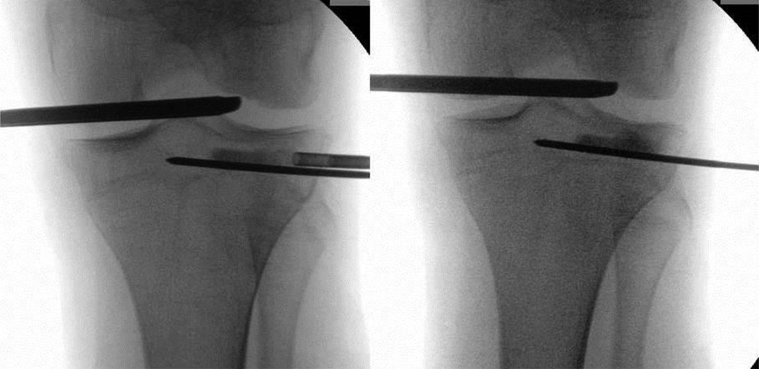
A knee joint arthroscopy was performed first to analyze the fracture gap as well as the articular tibial plateau impression (Figure 2). Then, the osteointroducer and trocar was placed from the medial or lateral side of the tibial plateau under fluoroscopic view and positioned 2–5 mm below the deepest point of the impression. 7 After predrilling and positioning a working drill, the balloon catheter was inserted. Radiopaque dots showed the position of the balloon under fluoroscopic view. Afterwards, the balloon was inflated step-by-step and reduction of the articular surface was performed. To ensure neither deviation of the balloon nor dislocation of the fracture occurred, both imaging and arthroscopic control of the reduction and articular surface were needed (Figure 3). To guarantee sufficient balloon inflation in a superior direction, a scoop cannula is part of the used instruments. If not available, a Kirschner wire can be drilled just distally of the inserted balloon catheter alternatively. The reduction maneuver was continued until the desired congruence of the articular surface was achieved. Finally, the balloon was slowly deflated. To maintain the reduction result during deflation, thin Kirschner wires were inserted through the fragment. Very quick deflation of the balloon may result in loss of reduction. After complete deflation, the balloon catheter was removed and the viscous calcium phosphate cement was introduced retrogradely into the cavity via 3 ml filled cement applicators (Figure 4). Two to three cement applicators were necessary per case. To avoid cement leakage, a step-by-step check was carried out with intraoperative radiographs. The cement applicator and tools were then removed. Within a period of 3–60 min, the calcium phosphate cement can be drilled with a drill at a low revolution (300 r min−1). The osteosynthesis was then finalized with compression screws (six cases) or a plate osteosynthesis (two cases) to prevent the fracture from secondary loss of reduction (Figure 5). All cases showed anatomic reduction. This calcium phosphate cementation technique of tibioplasty with the filling of the spongy cavity could have also be combined with autologous or allogeneic cancellous bone filling via a limited cortical window but wasn’t necessary in these cases.

Knee arthroscopy: View of the lateral compartment of the left knee joint. After lifting the lateral meniscus, a clearly palpable split and an impacted concave anterolateral tibial plateau appears.

Intraoperative radiographs, positioning of Kirschner wires to the distal lining, to avoid deviation of the balloon. After predrilling and positioning a working drill, inserting the balloon catheter and accurately positioning the balloon under control of X-ray marking points. Inflation of the balloon and reduction of the articular surface. Imaging and arthroscopic view of the reduction and articular surface.
Retrograde filling the spongious cavity with calcium phosphate cement.

Postoperative X-ray of the left knee joint of the same patient.
All patients had non-weight-bearing restriction for 6 weeks with early active mobilization of the knee under physiotherapy control, received follow-up at 6 weeks postoperatively including X-ray analysis, and were allowed full weight-bearing due to bony consolidation, afterwards. Osteosynthesis material removal was recommended at approximately 1 year postoperatively, but depending on patient’s specifications, the procedure was not performed in all cases.
All patients received clinical examination, KOOS-score and WOMAC-score evaluation, and analysis of the latest available X-ray regarding absorption of the calcium phosphate cement at the time of the latest follow-up in 2017–2018. X-rays were used for an orientating measurement, as there was no clinical indication for a CT scan (gold standard for volumetric measurements) at the time of follow-up. To measure distance in the anterior–posterior and lateral view X-rays of the knee, size was referenced to a 25 mm reference ball. The maximum cement penetration anterior–posterior and medial–lateral was measured on the immediate postoperative X-ray and on the latest available X-ray, related to the technique of the Response Evaluation Criteria In Solid Tumors (RECIST). 14 The difference in millimeters and absorption percentage were then calculated.
This anonymized retrospective study was registered with the number 17-180-0061 (IRB approval). Compliance with the STROCSS criteria is given. 15
Results
Eight patients received clinical examination at a mean of 27 months (standard deviation (SD) ±7) and radiological analysis of the latest X-ray after minimally invasive balloon tibioplasty with the use of bioabsorbable cement augmentation. Age at the time of surgery was at a mean of 54 years (SD ±17). Sex distribution was four males and four females (Table 1).
The clinical examination showed a mean maximum knee flexion of 139° (SD ±8°).
KOOS pain scores are shown in Table 2.
Clinical data and scores, and radiologic absorption.

SD: standard deviation.
WOMAC score showed a mean of 93 (SD ± 2).
The difference of longest cement penetration on anterior–posterior view radiographs of the treated knees directly postoperatively and at the time of the latest follow-up showed a mean of 7 mm (SD ±4 mm). The difference of longest cement penetration on lateral view radiographs of the treated knees directly postoperatively and at the time of the latest follow-up showed a mean of 6 mm (SD ±3 mm). Both radiographs therefore showed cement absorption rate of 20% and 23%, respectively, and new bone remodeling instead and reduced radiographic density of the bone cement. None of the cases showed osteolysis surrounding the bioabsorbable cement or a secondary dislocation, re-depression of the fragment, or secondary loss of reduction. All cases showed a normal fracture healing. None of the cases showed post-traumatic osteoarthritis.
Discussion
Balloon tibioplasty offers the possibility of a controlled and gentle reduction below the impression. Application of this minimally invasive technique, in comparison to conventional open surgical techniques, enables lifting more of the articular surface due to the relatively large balloon surface. The positioning of the tibioplasty balloon requires surgical precision. The balloon creates a well-defined bone cavity with a known volume. Fracture repositioning is ensured by augmentation with bioabsorbable calcium phosphate cement and optional osteosynthesis. Possible risks are cement leakage and secondary loss of repositioning.
In this survey, we were able to show good results after treating eight cases with isolated tibial depression fractures AO Type 41-B with the technique of balloon tibioplasty combined with the use of bioabsorbable cement and screw osteosynthesis. WOMAC score was at a mean of 93 after a mean follow-up of 27 months and cement absorption was measured to be approximately 1/5, supposed to increase in the following period.
Treatment of tibial plateau fractures is still a challenging surgical procedure and may result in articular incongruence, leading to secondary arthritis of the knee. Anatomic reduction of the articular surface is the main goal of the operation. Especially in elderly people, where fragility and osteoporosis may occur, the use of balloon tibioplasty may be an alternative and gentle technique to reduce tibial plateau fractures, although costs for the surgical procedure are higher. 16 In addition to that, minimal invasiveness and the ability of a symmetric augmentation for subchondral support are advantages of this new technique. Furthermore, neurological and vascular complication can be reduced, as open reduction at the depression area is not necessary. The reduction is gentler and homogeneously with less pressure peaks than with the classic reduction technique with instrument pushers. 8
There are only few studies available describing the technique of balloon tibioplasty and even less evaluating clinical outcome. To our knowledge, this study is the first one describing the bone remodeling and radiologic absorption effect in the follow-up time after reconstruction with bioabsorbable cement after tibial depressed fractures.
Many studies have evaluated conventional fracture reduction and osteosynthesis; however, as treated fracture types are very heterogenous, those results are hardly comparable to our collective. 17 –22
In 2016, Doria et al. described a retrospective case series of 28 patients: 14 cases received tibioplasty and 14 patients received conventional approach. 23 Postoperative radiological outcome in the treatment group was described as “maintained anatomic articular reduction,” which corresponds to our positive experience and results. Clinical data were described to be superior compared to the conventional approach, while clinical data were assessed using the Rasmussen Score System at 14 months postoperatively—probably comparable to good and excellent WOMAC scores after tibioplasty at a mean follow-up of 27 months in the present study.
Furthermore, even in case of multi-fragmentary impression fractures, an elevation of the joint surface is possible by repeated positioning and dilatation of the balloon as well as a final stabilization by means of a support plate osteosynthesis.
The use of absorbable cement augmentation has been reported in the treatment of depression fractures, filling bone defects and improving osteosynthesis in osteoporotic bone. 24 When bony consolidation is complete, the cement ideally should degrade. Several groups have developed absorbable calcium phosphate cements in the last decades, which have a similar chemical structure to the apatite of bone. However, degradation characteristics of the various available products show great variations. Clinical long-term studies have not yet been conducted so far. Whether these bone cement types fulfill the absorption requirements in clinical application is still not known. Our study provides first data in the tibial cancellous bone to degradation amount in the defined time of follow-up.
In the treatment of vertebral fracture surgery with bioabsorbable cement augmentation, a 2 vol% rate reduction was measured after a period of 12 months. 25
Extrapolating these values, Libicher et al. showed a far lower absorption rate compared to our results, although a different brand and type in a different skeletal area was used, which makes these results hard to compare.
Mauffrey et al. 6 surveyed possible minor and major complications in a case series of 20 patients. In some cases, balloon rupture, contrast agent leakage, cement leakage into the soft tissue, or intraoperative dislocation of the dorsal edge fragment occurred. In addition, the possibility of an intra-articular cement leakage, the failure of repositioning the depression zone, and the subsequent conversion to an open reduction and internal fixation technique have been discussed. In our study collective, none of these complications occurred.
There are several limitations to this study—at first its retrospective study design. Patients were treated depending on the expertise of one single surgeon, as well as on possible comorbidities and possibly other unknown criteria. One patient had a rheumatoid arthritis as comorbidity. Therefore, this study shows preliminary results of a relatively new developed surgical technique in selected tibial head depression fracture cases and might contain selection bias. Furthermore, the small number of patients in this study reduces explanatory power in comparison to other studies using a conventional surgical approach. This limits us to describe an effect dependent to age or gender. In addition, calcium phosphate cement absorption was only measured on two-dimensional X-rays; therefore, we were not able to calculate three-dimensional volume, which would be of interest for more precise results. For the later calculation, CT scans would have been necessary; they have not been performed for the lack of postoperative clinical indication and would therefore not be allowed due to regulations in clinical trials in Germany.
In the future, larger studies evaluating mid- or long-term outcome should be conducted to measure clinical outcome compared to conventional fracture repositioning and osteosynthesis techniques—Wang et al. described a promising study protocol for performing a randomized controlled study on this topic
26
Conclusion
Balloon tibioplasty with the use of bioabsorbable cement augmentation is a relatively new, helpful, minimally invasive procedure for closed reduction of tibial head depression fractures assisted in arthroscopic surgery.
We achieved excellent results and are able to report easy application and no occurrence of complications.
This far, promising clinical and radiological results have been shown with WOMAC scores comparable to the results of noninjured knees.
The radiologic measured absorption effect seems to be efficient, and good bone remodeling was shown.
To date, no randomized comparative studies have been reported that demonstrate a superiority of balloon tibioplasty compared to traditional fracture reduction and osteosynthesis. For this reason, the indication for this new procedure should be addressed closely and critically. Preoperative precise analysis of the fracture is essential. The indication for this novel technique is restricted to isolated depression fractures. In selected cases, however, according to the authors’ experience, it is a helpful and promising technique for the treatment of isolated tibial head depression fractures.
Footnotes
Declaration of conflicting interests
The author(s) declared no potential conflicts of interest with respect to the research, authorship, and/or publication of this article.
Funding
The author(s) received no financial support for the research, authorship, and/or publication of this article.