
Abstract
Purpose:
The purpose of this study is to compare biomechanical characteristics of tension band wiring using Kirschner wires (TBWKW), cannulated screws (TBWCS), and ring pins (TBWRP) for transverse fracture of the patella.
Methods:
A total of 48 polyurethane synthetic patellae were biomechanically tested. All patellae were osteotomized to create a transverse fracture. Each TBWKW, TBWCS, and TBWRP fixed 16 broken patellae. A specially designed fixation board simulated a knee with 90° flexion. Ten static tests and six dynamic tests were performed on each method. The static test is measuring maximum strength (N) during traction until breakage of the fixation. The dynamic test consisted of measuring the fracture gap (mm) after 10,000 repetitive loading cycles between 100 N and 300 N that simulated actual daily activity. A gap of 2 mm or more was defined as a failure in both tests.
Result:
The failure load was 438.6 ± 138.6 N, 422.2 ± 72.7 N, and 1106.8 ± 230.3 N for TBWKW, TBWRP, and TBWCS, respectively. TBWCS showed a statistically significant difference compared to TBWKW and TBWRP in the static test (p < 0.001). All the groups had no failure in the dynamic test. The mean fracture gap after completion of the dynamic test was 0.3267 ± 0.3395 mm, 0.2938 ± 0.2165 mm, and 0.0360 ± 0.0570 mm for TBWKW, TBWRP, and TBWCS, respectively (p = 0.044). The mean values in the dynamic test showed no statistical difference. There was a significant difference between TBWRP and TBWCS (p = 0.009), but others showed no difference with statistical significance.
Conclusion:
All three methods have sufficient stability at a daily activity. TBWCS showed a better failure load compared with TBWKW and TBWRP. TBWRP showed compatible mechanical characteristics with traditional tension band wiring. TBWRP could be an alternative method for TBWKW.
Introduction
Patella fracture is a fracture consisting 1% of all extremity fractures, and among them, transverse type (OTA/AO 34-C1) is the most common. 1 The purpose of operative treatment for patellar fracture is to restore anatomical reduction of the joint surface and to make a strong enough fixation to allow early passive range of motion exercise, which prevents intra- and periarticular fibrosis and helps articular cartilage healing. 2 –4 For this purpose, various fixation methods have been introduced. 4 –7
The most common type of fixation method used for transverse patella fracture is modified tension band wiring using two Kirschner wires (K wires) (TBWKW) across the fracture site with a figure of eight pattern cerclage wiring (Figure 1(a)). 3 Although there is a high union rate, various complications including protrusion and extrusion of the implant resulting in pain and wound problem have been reported. 8 Implant removal rate of TBWKW was up to 40%. 9 –11

Schematic drawing of the three tension band wiring. Tension band wiring using (a) K-wires, (b) ring pins, and (c) cannulated screws.
Tension band wiring using cannulated screw (TBWCS) was introduced to overcome these complications and is substituting TBWKW (Figure 1(b)). TBWCS generally showed superior results to TBWKW in biomedical and clinical study. 4,12 However, there are also some studies reporting inferior results with this method, and clinical outcome of TBWCS is still debatable. 11 Moreover, TBWCS has some innate problems including excessive bone loss, technical difficulty, fixation problem with small patella, and additional risk of fracture, 4,11 which can be a more important issue to consider in Asian population, whose patella is smaller than Caucasian population. 13
To overcome these problems, tension band wiring using ring pins (TBWRP) was introduced and showed a 100% union rate and no pin migration (Figure 1(c)). 14,15 TBWRP expanded its indication including olecranon fracture, clavicle fracture, ankle fracture, and AC separation with good clinical results. 16 Despite the increasing clinical use of TBWRP, there has been no mechanical test of the TBWRP until now. If the thickness of the ring pin and the K-wire is the same, the rigidity of the shaft may be similar. However, it cannot be concluded that the mechanical property of TBWKW and TBWRP is similar because of the thickness of the shaft and because the proximal part is different. Although K-wire was bent in TBWKW, the ring pins have a pinhole to passing through roll wire in TBWRP (Figure 1(a) and (c)). The structure difference of proximal part makes discrepancy in the method of holding the roll wire and blocking mechanism of the migration of the proximal fragment of fracture. A mechanical test of TBWRP is necessary for the final confirmation of the safety of this technique. The purpose of this study is to compare the mechanical characteristics of TBWKW, TBWCS, and TBWRP by performing static and dynamic biomechanical test.
Materials and methods
Preparation of specimen
We used polyurethane foam patellae as an experimental tool instead of a cadaver for the following reasons: (1) Patellar fractures mainly occur in relatively young and active people. 1,17 The available cadavers are usually elderly. Therefore, if there are not enough young cadavers, the results can vary from the actual situation due to osteoporotic bones. (2) Size and shapes of the individual patella can differ according to gender, age, and size of the body. Different shapes of the patella may not be suitable for obtaining a uniform result. (3) The latest polyurethane bone model composed of cortical and cancellous bone can simulate a structure similar to the normal bone, and it reported to be especially useful for comparing the fixation methods. 18 Besides, we can compare the results to other studies because previous several biomechanical tests already use polyurethane patella in a similar way. 8,19,20
This study was designed based on biomechanical studies of Wild et al., 8 Thelen et al., 19 and Dickens et al. 20 A total of 48 polyurethane foam patellae (Sawbones, Pacific Research Laboratories Inc., Vashon, Washington, USA) were used for the biomechanical test. The size of 25 × 3 mm2 transverse hole was generated on the central part of the patella to pass polyester band (Rib webbing, HS Korea, Seoul, Korea), which will play a role as quadriceps tendon and patellar tendon. After then, transverse fracture was made using 1-mm thick manual saw on the middle one-third of the patella (Figure 2). Fractured patella foams were reduced and fixed with the methods described below according to their groups (3 groups, 16 each).

The loops made of the polyester band were hung on proximal and distal fragments to reproduce extensor mechanism around the patella. The hole prevents a gap of the fracture due to the thickness of the nylon band.
TBWKW group
Two 2.0 mm K-wires (Solco Biomedical, Gyeonggi-do, Republic of Korea) were inserted perpendicular to the transverse axis of the patella and parallel to each other on its medial 1/3 and lateral 1/3. Through the hole generated before, two 25-cm polyester belts were passed. The 18G roll wire (Solco Biomedical) was passed around the K-wire, making vertical figure of eight wiring, and two knots were twisted on the distal part of K-wire and tightened. The proximal part of the K-wire was bent and cut, and impacted for the complete touch with the sawbone surface, completing the TBWKW fixation model.
TBWCS group
After reduction, two 1.0-mm K-wires (Solco Biomedical) were inserted perpendicular to the transverse axis of the patella on its medial 1/3 and lateral 1/3. Using drill bit, screw holes were generated, and two 4.0-mm cannulated screws (Asnis III Cannulated Screw System, Stryker Orthopaedics, Mahwan, New Jersey, USA) were inserted. Two 25-cm nylon bands were passed through the hole. The 18G roll wire was passed through the cannulated screw, making vertical figure of eight wiring, and K-wire at the distal tip of the screw was tightened, completing the TBWCS model.
TBWRP group
Surgical procedures were the same with TBWKW, except using 2.0-mm ring pin (Jaeil Medical, Seoul, Korea) instead of K-wire. Roll wire was passed through the ring of the pin, rounded making a figure of eight wiring, and tightened at the distal portion of the ring pin, completing the TBWRP model (Figure 3).

Application of tension band wiring with ring pin at sawbones patella. (a) Ring pins are inserted after the reduction of a patella fracture. (b) The steel roll wire of 18 gauge is passed through the hole of ring pins. (c) The roll wire was tightened after passing through distal of the ring pin and making a shape of eight. (d) The proximal part of the hole is cut without the need to bend like tension band wiring using K-wires.
After completion of the fixation in all groups, ends of the nylon bands were sewn together with industrial grade thread to complete proximal and distal loops.
Specimen positioning for mechanical test
Biomechanical test was performed using Instron material testing machine E3000 (Instron Engineering Corporation, Norwood, Massachusetts, USA) (Figure 4). Fixation board that can be fixed to the base of the Instron with a vertical rod was designed for this study and manufactured. On the base of the fixation board, fixation device for metal cable that can attach the lower part of the polyester band was built. And on the vertical rod, femur-mounting device, where femur polyurethane foam (Sawbones, Pacific Research Laboratories Inc.) will be fixed to act as distal femur, and pulley system, which will change the direction of the force generated by Instron, were built and metal cable was fixed to the Instron. Patella specimen was placed over the center of femoral condyle, where the patella contacts in native joint, and both polyester loops were connected to the superior and inferior cable. The angle of the polyester band was set to 90° because that degree is the most severe mechanical stress on patellofemoral joint. 21,22

Test setup. A specially designed fixation board was used to simulate the shape of the actual knee. Polyurethane foam patella fixed with a tension band ring was placed on the polyurethane femur using a polyester band. The length of the band was limited to 15 cm to reduce noise to result caused by a polyester band, and steel wires were used for an additional extension. Patella tendon bands were fixed to the bed, and quadriceps tendon bands were pulled by mechanical testing machine (Instron).
Biomechanical testing
Static test
Ten specimens of each fixation group were used for the static test. After mounting, preload of 100 N was applied, and tensile load was applied with a speed of 5 N/min. The definition of failure was when there is a failure point on a stress–strain curve or when fracture gap of 2 mm was observed, which was measured at the medial or lateral edge of the patella. Force at the failure was documented. Fracture gap was primarily observed with a ruler attached to the femoral condyle using magnification device, and for accurate measurement, digital ruler (Digital ABS AOS Caliper 0–150 mm, Digimatic, Rod, Neuss, Germany) was used for final decision of failure. During and after the test, mode of failure was observed.
Dynamic test
Six specimens of each fixation group were used for the dynamic test. After mounting, preload of 100 N was applied, and 100 N to 300 N was applied with the frequency of 5 Hz. After completion of 10,000 cycles, fracture gap was measured using digital ruler (Digital ABS AOS Caliper 0–150 mm, Digimatic, Rod) under 100 N, and the models were retrieved, and the medial and lateral fracture gap was measured using video measuring machine (VMQ100PK, TZTEK, Suzhou, China) (Figure 5). The definition of the gap of dynamic test was based on the mean of the values obtained from medial and lateral. The same as static test, a gap of 2 mm or more on both sides was defined as failure of fixation.

Measuring the gap of fracture by video measuring machine. The video measuring machine was used to measure the gap after the dynamic test of 10,000 cycles. The gaps were measured in 0.0001 mm units at the same position in all polyurethane patellae. The medial and lateral gaps were measured, and the average of the two was obtained. If a gap of 2.0 mm or more occurs, either medial or lateral was considered a fixed failure.
Statistical analysis
Statistical analysis was performed using SPSS Statistics version 23.0 (International Business Machines Corporation, New York, USA). One-way analysis of variance (ANOVA) and Dunnett’s T3 test were used for static testing, and Kruskal–Wallis and Mann–Whitney tests were used for dynamic testing. Statistical significance was defined when the p value was below 0.05 in ANOVA and Kruskal–Wallis test, and 0.017 in comparison of mean values for post hoc test between groups.
Result
Static test
All specimen except one specimen in TBWCS group failed due to a fracture gap of 2 mm. One specimen in TBWCS failed due to sudden breakage, which showed a failure point in stress–strain curve. The failure load was 438.6 ± 138.6 N, 422.2 ± 72.7 N, and 1106.8 ± 230.3 N for TBWKW, TBWRP, and TBWCS, respectively. TBWCS showed a statistically significant difference compared to TBWKW and TBWRP (p value < 0.001) (Table 1 and Figure 6).
Results of the static test.a

TTBW: traditional tension band wiring using Kirschner wires; TBWRP: tension band wiring using ring pins; TBWCS: tension band wiring using cannulated screws; ANOVA: analysis of variance.
a Data were reported as mean ± standard deviation (SD).
b p Value was calculated by one-way ANOVA.
c p Values were calculated by Dunnett T3.
d This value is statistically significant (<0.05).
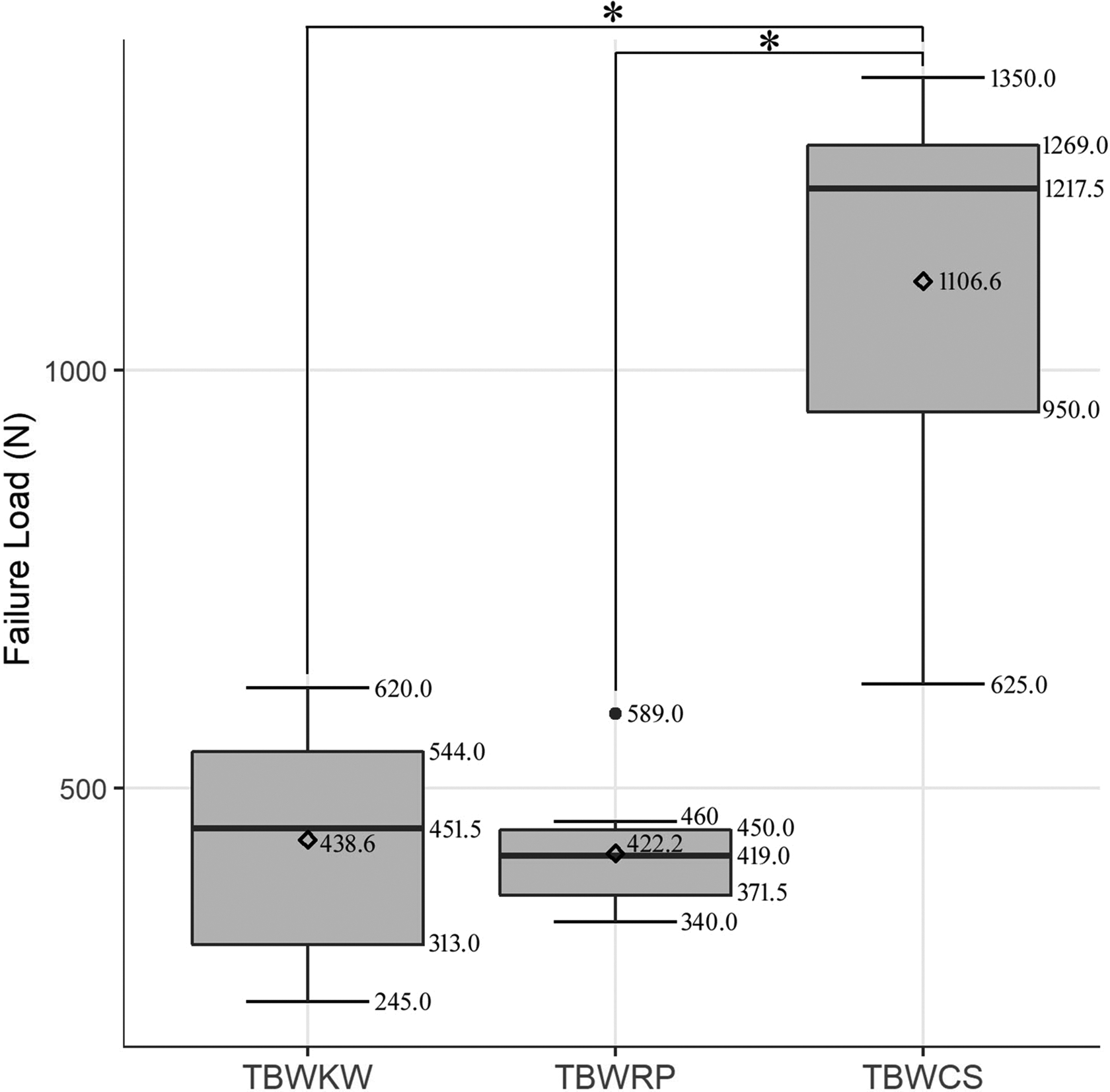
Box plot of failure load of the three tension band methods in a static test. TBWCS showed a statistically significant difference compared to TBWKW and TBWRP. *Significant differences. TTBW: traditional tension band wiring using Kirschner wires; TBWRP: tension band wiring using ring pins; TBWCS: tension band wiring using cannulated screws; TBWKW: tension band wiring using Kirschner wires.
Mode of failure showed difference between TBWKW, TBWRP, and TBWCS. In both TBWKW and TBWRP, slippage of distal portion of pin accompanied with loosening of the wire knot was the mode of failure. However, in the TBWCS group, head impaction was the mode of failure except for one specimen, which showed saw bone breakage.
Dynamic test
The mean fracture gap after completion of dynamic test was 0.3267 ± 0.3395 mm, 0.2938 ± 0.2165 mm, and 0.0360 ± 0.0570 mm for TBWKW, TBWRP, and TBWCS, respectively. All the groups had no failure, and the mean value showed no statistical difference. When comparing the mean values of the three groups with the Kruskal–Wallis test, it was found that there were some significant differences between groups (p = 0.044). When the comparison between two groups was compared by Mann–Whitney test, there was a significant difference between TBWRP and TBWCS (p = 0.009), but others showed no difference with statistical significance (Table 2 and Figure 7).
Results of the dynamic test.a

TTBW: traditional tension band wiring using Kirschner wires; TBWRP: tension band wiring using ring pins; TBWCS: tension band wiring using cannulated screws.
a Data were reported as mean ± standard deviation (SD).
b p Value was calculated by Kruskal–Wallis test.
c p Values were calculated Mann–Whitney test.
d This value is statistically significant.

Box plot of gaps of the three tension band methods in a dynamic test. There was a significant difference between TBWRP and TBWCS. *Significant differences. TTBW: traditional tension band wiring using Kirschner wires; TBWRP: tension band wiring using ring pins; TBWCS: tension band wiring using cannulated screws.
Discussion
This study is to identify the mechanical strength level of TBWRP compared to TBWKW and TBWRP. TBWRP was weaker than TBWCS in the static test but showed similar to TBWKW. In the dynamic test, a significant difference was only shown between TBWRP and TBWCS. Overall, TBWRP showed similar mechanical property to TBWKW in our study.
Our results show a same tendency to previous biomechanical tests for modified tension band wiring. Previous studies reported higher failure load in TBWCS compared to TBWKW for transverse patellar fracture in the static test. 8,12,23 The difference in the objective load between fixation methods can differ according to the study. Wild et al. 8 reported a failure load of TBWCS to be 1.6 times higher than TBWKW, but the result of this study was about 2.5 times higher in the TBWCS group. This difference is because of the difference in the definition of failure and the different angle of the stress applied to the patella. A statistically significant difference in failure load implies that TBWCS has higher fixation strength in the static test regardless of the definition of failure and the direction of a force. The dynamic test of this study indicates that with 10,000 times of cyclic stress, no failure occurred in all three methods. This is in accordance with the previous study by Thelen et al., 19 which showed no occurrence of a failure in TBWCS and TBWCS groups with the same force applied as this study was with 60° of flexion angle. However, the mean value of displacement was higher in our study, which implicates knee flexion of 90° will be likely to fail with higher probability compared to knee flexion angle of 60° if the same cyclic load is applied.
Superior biomechanical strength does not always guarantee a superior clinical result. Hoshino et al. 11 studied 448 patella fracture patients and reported that the failure rate in the TBWCS group was 7.5%, which was higher than TBWCS of 3.5%. Moreover, in the TBWCS group, there was a fracture of sawbones before reaching the failure fracture gap of 2 mm, and similar phenomenon was also reported in another biomechanical study by Baydar et al. 24 This may imply some concerns including higher risk of fracture due to excessive bone loss, which is suggested by previous reports. 12,25 Especially this can be a great problem for Asian, children, or female patients, whose patella is small. 13 Moreover, in senile patient whose strength of patella is weak due to osteoporosis, firm fixation with screw cannot be achieved or the excessively stiff screw can lead to bone fracture, which was reported by previous clinical study. 4
These clinical results indicate that the fixation strength required in a clinical situation is not that high, and modest fixation that can endure with daily activity with less bone loss is more important. The most of tension around the patella in daily activity is verified performing within 300 N in previous biomechanical tests. 12,23,25 Although there are some complications associated with migration of K-wire, TBWKW has been shown sufficient stability for a bone union. 26 In results of this study, TBWKW and TBWRP were proven enough clinical stability.
Although the TBWRP group showed similar fixation strength to TBWKW group, the clinical results of TBWRP are reported to be superior to TBWKW because of stationary implant. 26 Kim et al. 14 reported TBWRP to have 100% union rate with 0% pin migration with pin removal rate of 8.3%, which is lower than the previously reported outcome of MTBW. Kyung et al. 15 also reported 100% union rate and migration rate, which was statistically superior to TBWKW technique. The stability of our study and superior clinical outcomes demonstrate the usefulness of TBWRP in the transverse patella fracture. Moreover, the technique of TBWRP is relatively simple, almost identical to TBWCS, without additional procedure for K-wire bending, which is also an advantage of TBWRP.
There are some limitations to this study that needs to be mentioned. First, as the technique of TBWCS is recommended to position the thread into the cancellous bone, the result of static and dynamic test shown here may be an exaggerated value as the saw bone patella is composed of purely cortical part. However, the result of cadaveric study comparing TBWKW and TBWCS also reports higher load to failure in the TBWCS group, which is the same result with this study. 24 Second, interference of soft tissue cannot be put into consideration, which is an innate limitation of the mechanical test with sawbones. However, soft tissue issue can be another confounding factor of cadaver study, which cannot be controlled uniformly. Third, the pullout of K-wire or ring pin, which is clinically proven the most important difference between TBWKW and TBWRP, could not be reproduced with this experimental setting. We are considering to design a new experimental study to prove this phenomenon in the future.
Conclusion
TBWCS showed a better failure load compared with TBWKW and TBWRP. All three methods have sufficient stability at daily activity. TBWRP could be an alternative method for TBWKW because TBWRP showed compatible mechanical characteristics with TBWKW.
Supplemental Material
Supplementary - Biomechanical comparison of three tension band wiring techniques for transverse fracture of patella: Kirschner wires, cannulated screws, and ring pins
Supplementary for Biomechanical comparison of three tension band wiring techniques for transverse fracture of patella: Kirschner wires, cannulated screws, and ring pins by Kyung-Hag Lee, Yohan Lee, Young Ho Lee, Bong Wan Cho, Min Bom Kim and Goo Hyun Baek in Journal of Orthopaedic Surgery
Footnotes
Declaration of conflicting interests
The author(s) declared no potential conflicts of interest with respect to the research, authorship, and/or publication of this article.
Funding
The author(s) received no financial support for the research, authorship, and/or publication of this article.
Supplemental Material
Supplemental material for this article is available online.
References
Supplementary Material
Please find the following supplemental material available below.
For Open Access articles published under a Creative Commons License, all supplemental material carries the same license as the article it is associated with.
For non-Open Access articles published, all supplemental material carries a non-exclusive license, and permission requests for re-use of supplemental material or any part of supplemental material shall be sent directly to the copyright owner as specified in the copyright notice associated with the article.