
Abstract
Purpose:
The purpose of this study was to analyze the dynamic motion of the first carpometacarpal (CMC) ligaments on a three-dimensional (3-D) surface model and to examine the changes in the ligament lengths during the motion of the first CMC joint.
Methods:
Six fresh-frozen cadaver wrists were used to analyze the motion of the first CMC ligaments on a 3-D coordinate system using a digitizer. Four ligaments, namely, dorsoradial ligament (DRL), posterior oblique ligament (POL), superficial anterior oblique ligament (SAOL), and deep anterior oblique ligament (dAOL), were dissected and identified. Their attachments were digitized and represented on 3-D bone images. The distances between the ligament attachments of the first metacarpal and the trapezium, which were the ligament lengths, were measured during the extension–flexion and adduction–abduction of the first CMC joint.
Results:
Both the DRL and POL lengthened during flexion of the first CMC joint, and both the SAOL and dAOL lengthened during extension. Both the DRL and SAOL lengthened during adduction, and both the POL and dAOL lengthened during abduction. The DRL alone lengthened significantly at flexion and adduction when the first CMC joint was in dorsoradial dislocation.
Conclusions:
The lengths of four ligaments changed significantly during first CMC joint motion. This study suggested that the DRL contributes substantial stability to the first CMC joint, preventing dorsoradial dislocation. This 3-D information improves the knowledge and understanding of the function of individual ligaments and their roles in the stability of the first CMC joint.
Introduction
The first carpometacarpal (CMC) joint is a biconcave–convex saddle joint characterized by little bony support between the first metacarpal and the trapezium. 1 Therefore, the first CMC ligaments are thought to play an important role in the maintenance and stability of the first CMC joint. 2 –12 Currently, several studies on the anatomy of the first CMC ligaments have been reported in cadaveric dissections. 2,6,8 –10,13 –15 Presently, Bettinger et al. 2 and Nanno et al. 8 have described seven ligaments in examining the complex ligamentous anatomy of both the first metacarpal and the trapezium. They identified the superficial anterior oblique ligament (SAOL), the deep anterior oblique ligament (dAOL), the ulnar collateral ligament, the dorsoradial ligament (DRL), the posterior oblique ligament (POL) of the first CMC joint, the volar intermetacarpal ligament (vIML), and the dorsal intermetacarpal ligament (dIML) in the intermetacarpal joint. 2,8 Nevertheless, understanding of the primary stabilizer of the first CMC joint remains incomplete. It may be useful to analyze the dynamic motion of the first CMC ligaments and to evaluate the function and the role of each ligament in the first CMC joint.
In recent years, a few researchers used a method of modeling the paths of ligaments based on reconstructed three-dimensional (3-D) structures of the bones from computed tomography (CT) data of the first CMC joint and investigated the changes in length of the ligaments of the first CMC joint at certain thumb positions. 5,11 However, in their study, the origins and insertion sites of the ligaments were determined based only on previous anatomic information, and the individual variances in ligamentous anatomy were not considered. 5,11
At present, there has been little research analyzing the dynamic motion of the first CMC ligaments on a 3-D surface model of the first metacarpals and the trapezium with a combined approach of detailed dissection and digitization to evaluate the precise actual locations of the ligament attachments and ligament lengths of the first CMC joint. Nanno et al. 8 visually demonstrated qualitatively the anatomic 3-D attachment sites and paths of the first CMC ligaments and quantified their areas using a combination of meticulous dissection, CT imaging, and a 3-D digitization technique. By digitizing the anatomical 3-D description of the attachments and paths of the CMC ligaments, the distances between the ligament attachments of the first metacarpal and the trapezium, which were the lengths of the ligaments, were able to be measured during the motion of the first CMC joint.
The purpose of this study was to analyze the dynamic motion of the first CMC ligaments on a 3-D surface model and to examine the changes in their lengths during the motion of the first CMC joint using a 3-D digitization technique. This 3-D information was considered potentially helpful to improve the knowledge and understanding of the biomechanics of the first CMC joint and the function of individual CMC ligaments and their roles in joint stability.
Materials and methods
Six fresh-frozen cadaver upper extremities (three males, three pairs, with an age range of 62 to 69 years, average age = 64.3 years) were used in this study.
Two single half pins were inserted into both the distal radius and ulna to fix the extremities firmly to the digitizing platform (Figure 1). In addition, a 2-mm-diameter graphite rod was placed into the first metacarpal. A triad pin that had three 5-mm-diameter spheres with a 1.5 mm-diameter screw fixed in a cruciform arrangement on top was glued to the end of each rod, as markers to define the 3-D local coordinate system. Moreover, three 1.5-mm screws were placed into the trapezium, as the markers to define the 3-D local coordinate system.

Experimental setup for measuring the first CMC joint kinematics. Model of three-dimensional dynamic motion analysis of the first CMC ligaments. CMC: carpometacarpal.
In accordance with the techniques reported by Imaeda et al. 6 and Koff et al., 16 the neutral position of the CMJ was maintained by a load of 50 g for each of the six tendons: the extensor pollicis longus (EPL), the extensor pollicis brevis (EPB), the abductor pollicis longus (APL), the abductor pollicis brevis (APB), and the adductor pollicis (Add P) and the flexor pollicis longus (FPL). This constant 50-g load was applied to each tendon muscle unit to stabilize the first CMC joint. Each tendon was connected to a wire that looped around one of six freely moving pulleys.
To perform active motion of flexion–extension and abduction–adduction, the six tendons were pulled selectively by incrementally adding load of 0.5 pounds from 0 to 6 pounds. Specifically, the EPL and EPB were loaded during the extension of the first CMC joint, the FPL during flexion, the APL and APB during abduction, and the Add P during adduction.
The 3-D motions of the first metacarpal and trapezium were digitized manually on a 3-D coordinate system using Microscribe-3DX Digitizer (Immersion Corp., San Jose, CA, USA) and an original 3-D coordinate system software (Spider; developed by Orthopaedic Biomechanics Laboratory, the University of Texas Medical Branch, Galveston, TX, USA) during the passive motion of flexion–extension and abduction–adduction of the first CMC joint. 8
Moreover, the CMC ligaments of all the six specimens were meticulously dissected using loupe magnification. Four CMC ligaments, namely, the DRL, POL, SAOL, and dAOL, were identified. The soft tissues were excised to distinguish the accurate sites of ligament attachment. Those ligaments were carefully removed from the first metacarpal and trapezium, and their attachments were marked using an oil-based color marker. 8
Furthermore, the making of the 3-D digitized surface models with the ligament attachments was performed according to the methods reported by Nanno et al. 8 The ligament attachments and whole bone surfaces were digitized manually and represented on 3-D images of the bones using a MicroScribe-3DX Digitizer and an original 3-D surface reconstruction software (Spider; developed by Orthopaedic Biomechanics Laboratory, the University of Texas Medical Branch).
Furthermore, we represented three paths of each ligament that connected three points on the manually selected ligament attachment sites of both the first metacarpal and the trapezium using the Spider software (Figure 2). These three paths were chosen to represent the radial (r), central (c), and ulnar (u) parts of the ligaments. The ligament length was determined as the length of the straight line between the ligament attachments of the first metacarpal and the trapezium on a 3-D surface model. The 3-D dynamic motion analysis of the first CMC ligaments was performed on a 3-D coordinate system using the Spider software. The changes in the ligament lengths of the three paths of each ligament were calculated during extension–flexion and adduction–abduction of the first CMC joint. Their ligament lengths were also compared among three paths of each ligament. Additionally, the rates of change of the ligament length in four ligaments were compared at varied positions of the first CMC joint.

Measurement of the distances of four ligaments from the ligament attachments of the first metacarpal to those of the trapezium (in case of the DRL). The three fibers’ paths were chosen to represent the radial, central, and ulnar parts of the ligaments. 1MC: the first metacarpal; Tm: the trapezium; DRL: dorsoradial ligament; POL: posterior oblique ligament.
All analyses were performed by SPSS 21.0J. A nonpaired t-test was used to analyze the differences of each ligament length at 20° extension, 10°flexion, 25° adduction, and 5° abduction position to the neutral position of the first CMC joint, respectively. The ligament lengths of four ligaments at five positions of the CMC joint were expressed as absolute lengths and as percent changes compared with those at neutral position. Furthermore, the ligament lengths of each ligament were statistically compared among three paths at five positions of the first CMC joint with paired t-test, respectively. The p values of <0.05 were considered statistically significant for the motion differences.
Results
The lengths of four CMC ligaments changed significantly during extension–flexion and adduction–abduction of the first CMC joint.
We measured the distance of the centroid (c) of each ligament attachment between the first metacarpal and the trapezium on a 3-D surface model as the length (C) of the ligaments. 1. Change in the lengths (C) of ligaments during extension–flexion of the first CMC joint
Both the DRL and POL lengthened on the flexion of the first CMC joint, and both the SAOL and dAOL lengthened on the extension of the joint (Figure 3).

Changes in the length of four ligaments during extension–flexion motion of the first CMC joint. CMC: carpometacarpal; DRL: dorsoradial ligament; POL: posterior oblique ligament; SAOL: superficial anterior oblique ligament; dAOL: deep anterior oblique ligament.
Especially, at 20° extension position of the first CMC joint, both the SAOL and dAOL lengthened significantly, while the POL loosened significantly, compared at the neutral position, respectively (p < 0.05; Table 1). The length rates were 29.5 and 22.1%. The DRL loosened relatively at 20° extension of the first CMC joint than at the neutral position, but it did not reach significance.
The lengths of four ligaments at five positions of the first CMC joint.a

CMC: carpometacarpal; DRL: dorsoradial ligament; POL: posterior oblique ligament; SAOL: superficial anterior oblique ligament; dAOL: deep anterior oblique ligament.
aValues are given as mean mm (SD mm); [%] Percent stretch from neutral of four ligaments to four positions of the first CMC joint, negative numbers reflect shortening from the neutral position.
b p < 0.05.
Conversely, at 10° flexion of the first CMC joint, only the DRL lengthened significantly, compared to at the neutral position. The length rate was 21.9%.
2. Change in the lengths of ligaments during adduction–abduction of the first CMC joint
Both the DRL and SAOL lengthened on the adduction of the first CMC joint, and both the POL and dAOL lengthened on the abduction of the joint (Figure 4).
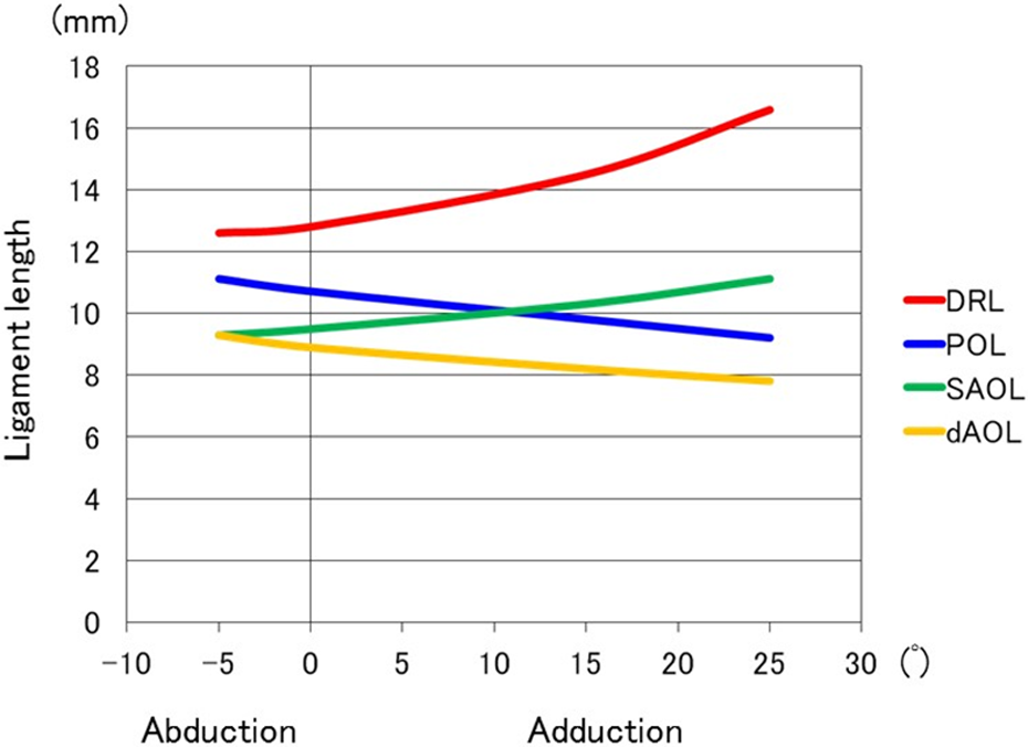
Changes in the lengths of four ligaments during adduction–abduction motion of the first CMC joint. CMC: carpometacarpal; DRL: dorsoradial ligament; POL: posterior oblique ligament; SAOL: superficial anterior oblique ligament; dAOL: deep anterior oblique ligament.
Especially, at 25° adduction of the first CMC joint, only the DRL lengthened significantly, compared to at the neutral position (p < 0.05). The length rate was 29.7%.
There were no significant differences in ligament lengths of the POL, SAOL, and dAOL during adduction–abduction motion of the first CMC joint, respectively. Consequently, only the DRL lengthened significantly at flexion and adduction when the first CMC joint was in dorsoradial dislocation (p < 0.05). On the other hand, the dAOL was taut at both extension and abduction with the first CMC joint in dorsoradial dislocation. Furthermore, the dAOL was the shortest among all four ligaments during all motions of the first CMC joint, except for the POL at extension position.
3. Change in the lengths of the three paths of each ligament during extension–flexion and abduction–adduction
We compared among three ligament lengths (C), (R), and (U), which were the distances of the radial (r), center (c), and ulnar (u) edge of each ligament attachment between the first metacarpal and the trapezium on a 3-D surface model.
There were no statistically significant differences of four ligaments among three paths of each ligament (Table 2).
The lengths of three paths of each of four ligaments at five positions of the first CMC joint.a
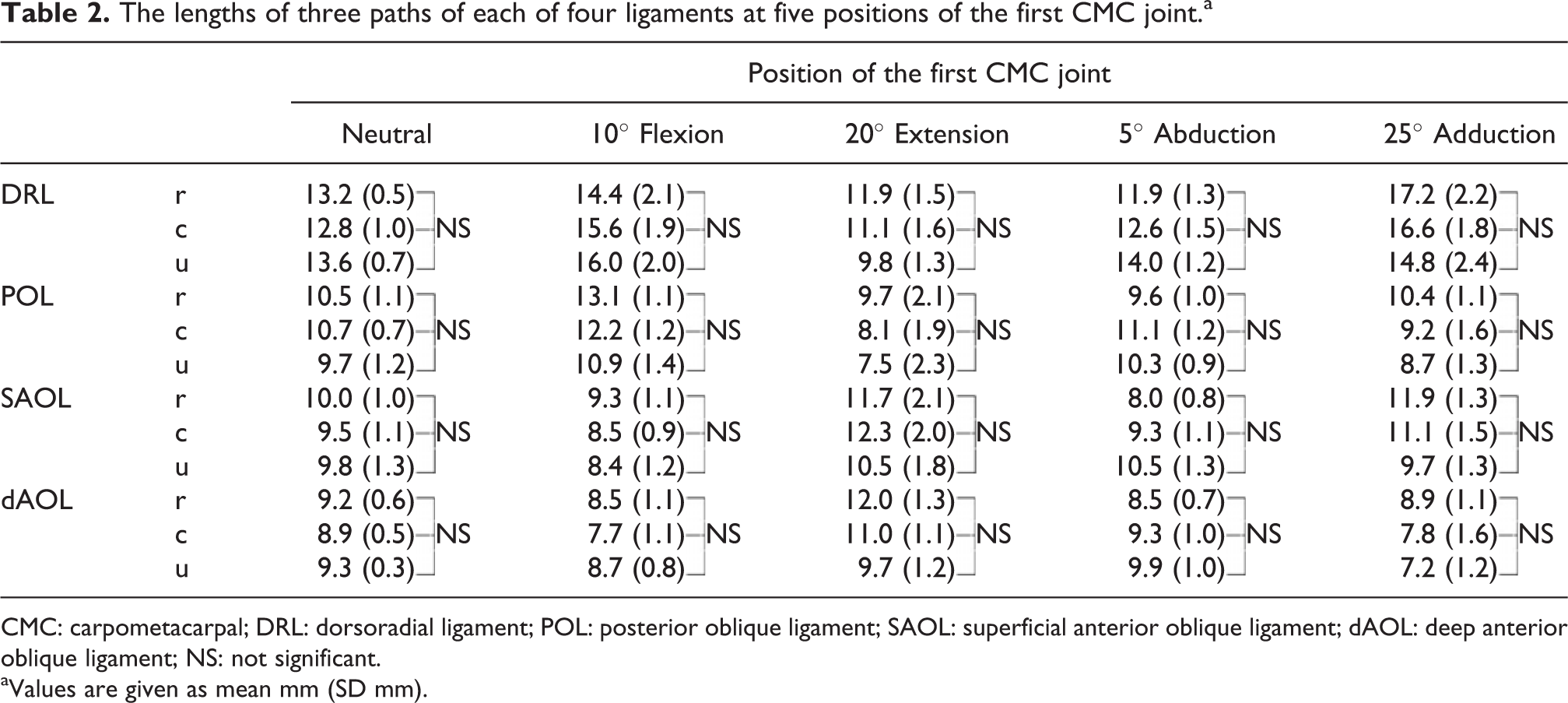
CMC: carpometacarpal; DRL: dorsoradial ligament; POL: posterior oblique ligament; SAOL: superficial anterior oblique ligament; dAOL: deep anterior oblique ligament; NS: not significant.
aValues are given as mean mm (SD mm).
Discussion
There have been a number of researches on the ligament anatomy of the first CMC joint in cadavers. 2,6,8 –10,13 –15 Especially, some anatomical studies identified the roles of various ligaments in stabilizing the first CMC joint. 2 –12 Earlier articles have mentioned that the dAOL also plays an important role in the stability of the first CMC joint. 6,9,10,13 Currently, increasing evidence from anatomical cadaver studies reveals the DRL to be the primary stabilizer of the first CMC joint. 3,5,7,8,11 –15 Despite a considerable increase in knowledge about the CMC ligaments, there have been some inconsistencies in the understanding of the efficacy of these ligaments for the stability of the first CMC joint. Therefore, a 3-D dynamic motion analysis of the first CMC ligaments may provide useful information about the role of each ligament on the stability of the first CMC joint.
Recently, a few researchers have investigated in vivo the 3-D kinematics of the first CMC joint and the changes in the ligament lengths noninvasively at several thumb positions using CT bone registration techniques. 5,11 However, these techniques involved no direct visualization of the ligaments. 5,11 Moreover, they manually defined the points of both the insertion and the origin of the ligaments on 3-D bone models using information from previously published descriptions of cadaveric dissection. 5,11 Therefore, they could confirm neither the precise ligament attachments of the first CMC ligaments nor anatomical variations of the ligaments.
Nanno et al. 8 have reported in detail the precise anatomical 3-D description of the attachments and paths of the CMC ligaments using a combination of meticulous dissection, CT imaging, and a 3-D digitization technique. The distance between each ligament attachment of the first metacarpal and the trapezium on a 3-D surface model, which was defined as the length of the ligaments, could be measured using a 3-D digitization technique. The current study was the first to combine precise 3-D dynamic motion analysis and manual digitization of ligament attachment location to quantify the changes in the ligament lengths of the first CMC ligaments during motion of the first CMC joint and to determine the role of each ligament for the stability of the first CMC joint. Consequently, it was found that both the DRL and POL lengthened during flexion of the first CMC joint and both the SAOL and dAOL lengthened during extension of the joint. Additionally, both the DRL and SAOL lengthened during adduction of the first CMC joint and both the POL and dAOL lengthened during abduction of the joint.
Halilaj et al. 5 also evaluated the changes in the lengths of the DRL and AOL during the first CMC motions using in vivo kinematics data. They indicated that the AOL was slack in most of the range of CMC joint motion, whereas the DRL was in tension in positions that involve joint flexion and adduction, similar to our findings. 5 Moreover, they suggested that the DRL plays an important role in the maintenance and dorsoradial stability of the first CMC joint. 5 However, they have not examined separating the AOL to the SAOL and dAOL. 5 We confirmed that the change in the dAOL was different from that in the SAOL.
On the other hand, Tan et al. 11 examined in vivo five ligament lengths (DRL, POL, dAOL, vIML, and dIML) during flexion, adduction, or opposition motion of the first CMC joint using previous anatomic information. They reported that both the DRL and POL lengthened, while the dAOL loosened at flexion and opposition positions of the first CMC joint. 11 Additionally, the POL lengthened at the abduction position of the joint. 11 In the current study, similar results were obtained. However, they had not examined the ligament lengths at either extension or adduction positions of the first CMC joint. 11 Moreover, they investigated the ligament lengths only at certain fixed positions of the first CMC joint without examining them consecutively during the first CMC joint motion. 11 In the current study, only the DRL lengthened significantly at both flexion and adduction positions of the first CMC joint. We considered that the DRL was stretched most tensely in a combined flexion and adduction position, that is, the dorsoradial dislocation of the first CMC joint. Moreover, the DRL was always the longest ligament among four ligaments during all motion of the first CMC joint. The length rates of the DRL were statistically 122 and 130% at thumb flexion and adduction, respectively, compared to at the neutral position. These findings suggest that the DRL must contribute substantial stability to the first CMC joint preventing dorsoradial dislocation.
Conversely, the dAOL is situated at a mirror image position to the DRL. The current study demonstrated that the dAOL was taut at both extension and abduction, which was the reposition position for dislocation of the first CMC joint. Furthermore, the dAOL was the shortest among all four ligaments during all motion of the first CMC joint. Moreover, the current study indicated that there was no significant difference in ligament lengths of the dAOL during all motion of the first CMC joint. This information suggests that the dAOL acts as a pivot point for the circumduction motion of the first CMC opposition. It is thus thought that the dAOL may play only a minor role in the stability of the first CMC joint. Our findings revealed significant differences of changes in the ligament length between the DRL and AOL and support the findings of the most recent studies 3,5,7,8,11 –15 that have stated the functional importance of the DRL.
In the current study, we also compared the ligament lengths among three paths, which were the central (c), radial (r), and ulnar (u) edge of each ligament during extension–flexion and abduction–adduction. Consequently, there were found to be no statistically significant differences in four ligaments among three paths of each ligament. Therefore, it would be reasonable that the distance of the central (c) path between the ligament attachment sites of both the first metacarpal and the trapezium was selected among three paths of each ligament for the dynamic motion analysis of the first CMC ligaments.
The present study had several limitations. First, a small number of specimens was used. A large number of specimens would better clarify precisely variations in the lengths of the first CMC ligaments. Second, the models in this study may not reproduce accurately in vivo kinematics, similar to any other biomechanical study that uses specimens. However, using techniques reported in the previous studies, 6,16 we were able to analyze the dynamic motion of the first CMC joint. And, finally, the mean age of our specimens was comparatively high, which may have had an impact on ligament integrity.
Conclusion
The current study has demonstrated the value of a combined approach of detailed dissection and digitization to analyze the 3-D dynamic motion of the first CMC ligaments and to examine the changes in the lengths of the ligaments during the motion of the first CMC joint. This 3-D information improves the knowledge and understanding of the anatomy and biomechanics of the first CMC ligaments and both the function of individual ligaments and their roles in joint stability. Especially, this study suggests that one of the mechanisms responsible for the dorsoradial dislocation of the first CMC joint is excessive traction of the DRL and that the DRL is the primary stabilizer in the first CMC joint. This should also help in the precise assessment of treatment for the fracture–dislocation of the first CMC joint when performing ligament repair and reconstruction.
Footnotes
Acknowledgment
The authors gratefully acknowledge the help of Steven F. Viegas, MD, William L. Buford, PhD, Rita M. Patterson PhD, and Clark R. Andersen, MS, for their assistance with this study, and L. Frumson for his help in the manuscript preparation.
Declaration of conflicting interests
The author(s) declared no potential conflicts of interest with respect to the research, authorship, and/or publication of this article.
Funding
The author(s) received no financial support for the research, authorship, and/or publication of this article.