
Abstract
The authors present a case report of a rare upper extremity mycotic aneurysm in a 36-year-old male with multiple comorbidities, including diabetes, end-stage renal disease, and prior infections. Initially, the patient presented with ischemic symptoms in the right small finger and underwent successful surgical debridement of the radial and ulnar arteries. Subsequently, the patient suffered a cardiac arrest five weeks postoperatively and underwent radial arterial line placement, which led to worsening vascular symptoms in the affected extremity. A second surgery revealed a diagnosis of mycotic aneurysm of the distal ulnar artery. The diseased artery was resected and reconstructed using vein grafts. Following reconstruction, the patient recovered with restored perfusion and resolution of pain. This case highlights the clinical course of diagnosing upper limb mycotic aneurysm and reconstruction of upper limb arteries, stressing the importance of clinical suspicion and timely surgical intervention.
Introduction
Mycotic aneurysms are rare presentations, resulting from arterial injury combined with microbial seeding of a vessel wall, leading to destruction of the media and elastic lamina.1,2 Staphylococcus aureus is the most common pathogen, followed by Salmonella and Pseudomonas species. 1 Infection-induced inflammation activates matrix metalloproteinases, weakening the vessel wall and predisposing to rupture, thrombosis, or distal ischemia.2,3 There are no specific diagnostic criteria for mycotic aneurysms. A thorough evaluation involving blood tests and imaging can help characterize the condition and guide management.1,4 Prolonged antibiotic therapy is needed, with definitive management requiring surgery to prevent complications such as aneurysmal rupture, sepsis, thrombosis, and distal limb ischemia.2,5
Case Report
Patient Introduction
A 36-year-old right-hand–dominant male with end-stage renal disease, type 1 diabetes, and chronic foot infection presented with two weeks of progressive pain, paresthesia, and discoloration of the right small finger. Examination revealed fingertip ulceration without infection. Radial and brachial pulses were palpable; however, the ulnar pulse was absent. Computed tomography angiography (CTA) and magnetic resonance angiography (MRA) demonstrated severe arterial calcification, with 50% distal radial stenosis, and no flow in the ulnar artery.
Given his comorbidities, a conservative approach was initially taken. Chemical sympathectomy via botulinum toxin was attempted for ischemic pain but failed. The patient wished to then pursue limb salvage.
Initial Operation–Adventitial Stripping of the Radial and Ulnar Arteries
In the main operation room, exploration revealed heavily calcified radial and ulnar arteries. Both vessels underwent adventitial stripping over 5 cm (Figure 1), with small finger debridement. Postoperatively, pain resolved completely and doppler signals were present in both arteries.
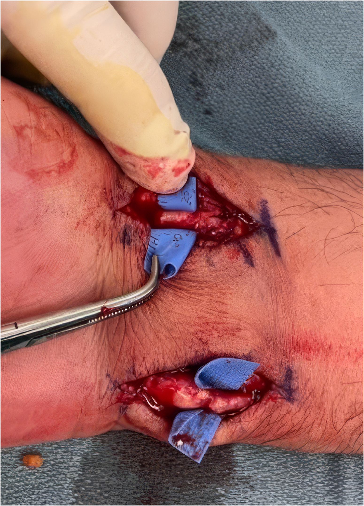
Adventitial stripping of the radial and ulnar arteries.
Unfortunately, five weeks later, the patient suffered a cardiac arrest that required cardiopulmonary resuscitation and insertion of an arterial line into their right radial artery at the wrist. Two months later, necrosis of the small and index fingertips developed. Angiography revealed poor distal radial and ulnar flow but a patent deep palmar arch, prompting the decision for bypass reconstruction using a vein graft.
Second Operation (Six Months Following)
During re-exploration, multiple fluid-filled sacs encased the ulnar artery (Figure 2). Rupture of these encapsulated structures released purulent discharge, with cultures positive for Streptococcus species (Figure 3). Histopathologic findings showed the ulnar artery was thickened and hemorrhagic over a 7 cm segment, with above 90% luminal occlusion. There was destruction of the tunica media and internal elastic lamina, dense neutrophilic infiltration, and intraluminal bacterial colonies, findings consistent with a mycotic aneurysm (Figure 4).
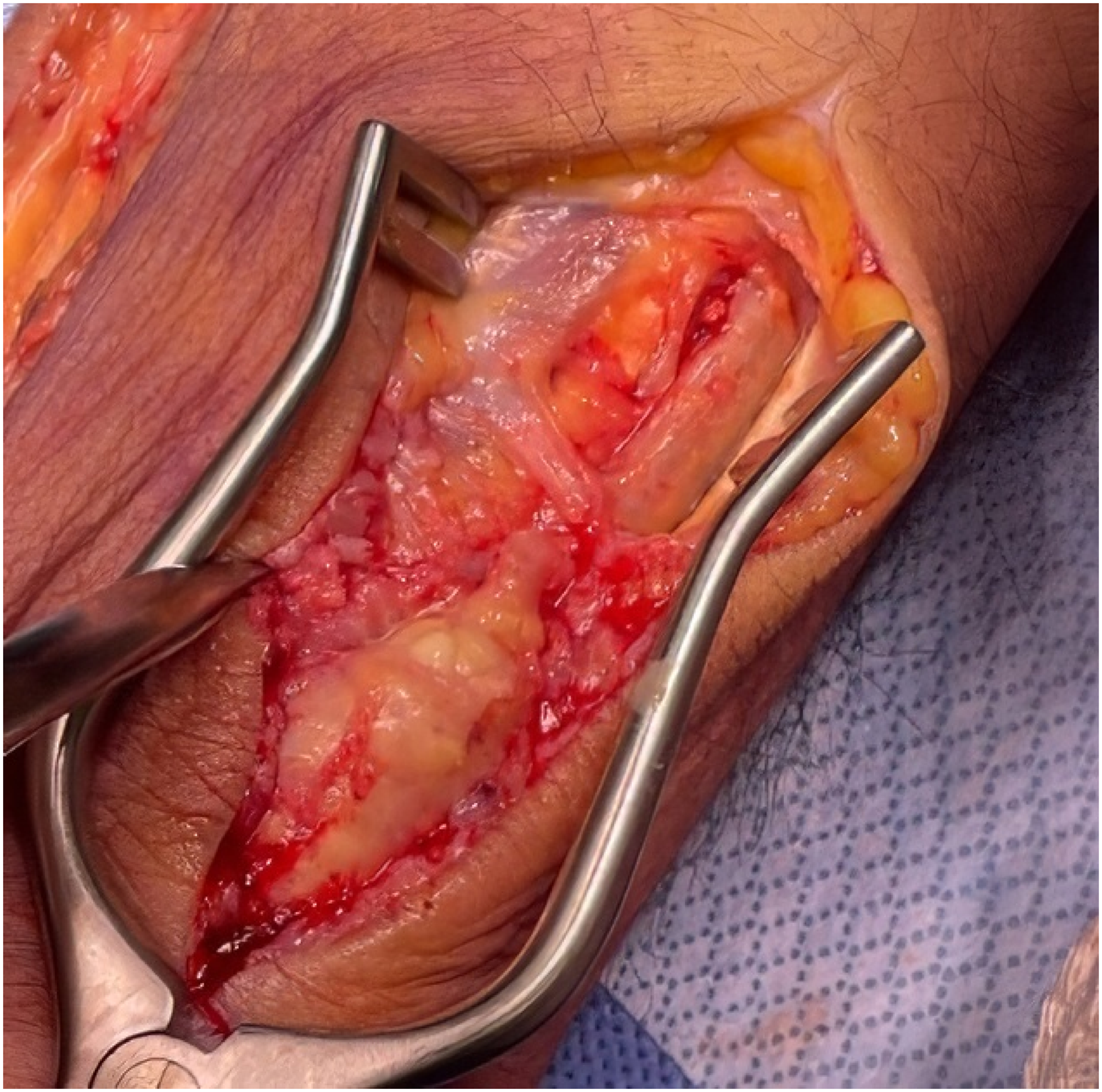
Ulnar artery encapsulated structures.
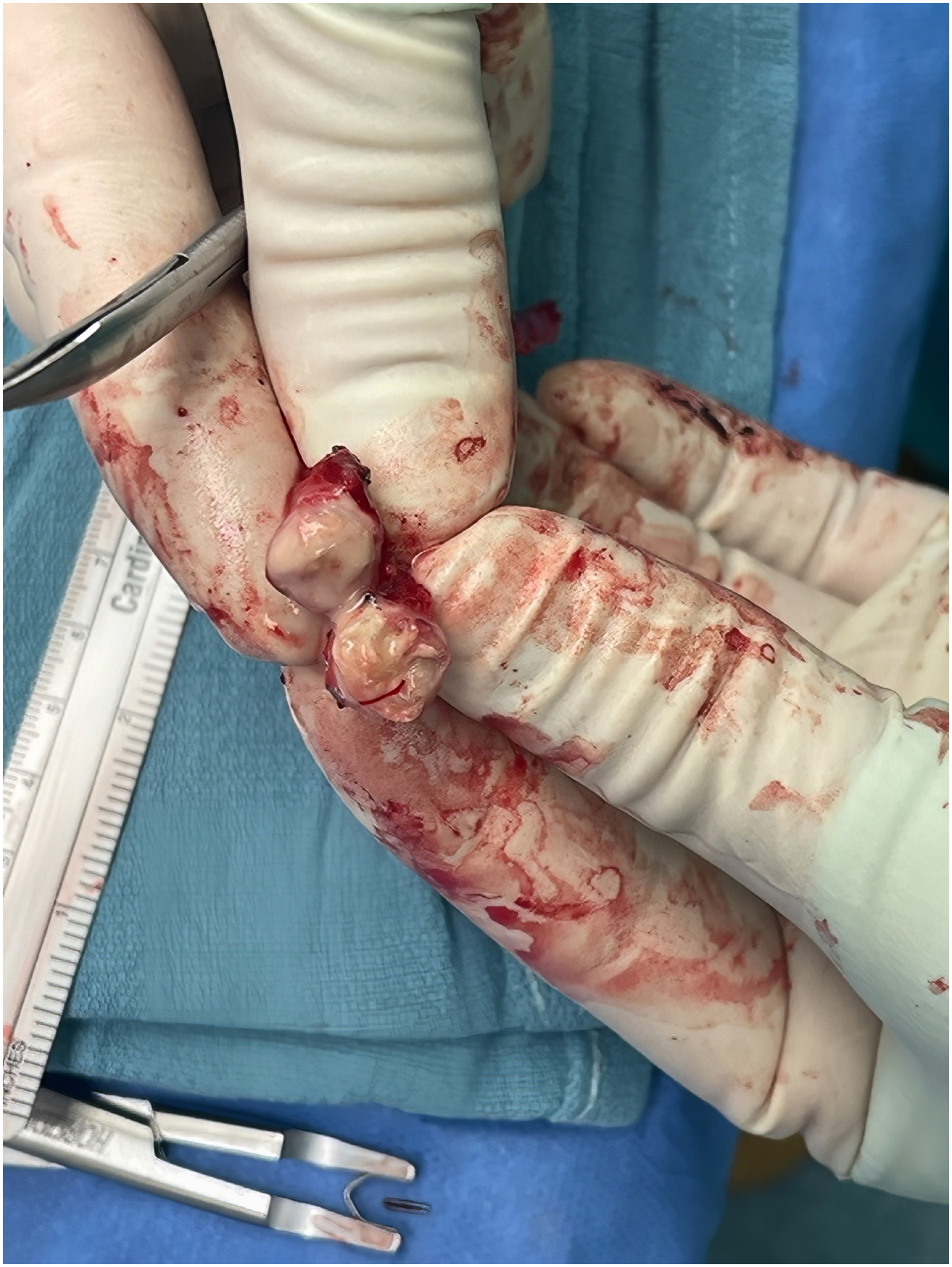
Encapsulated structures from ulnar artery.
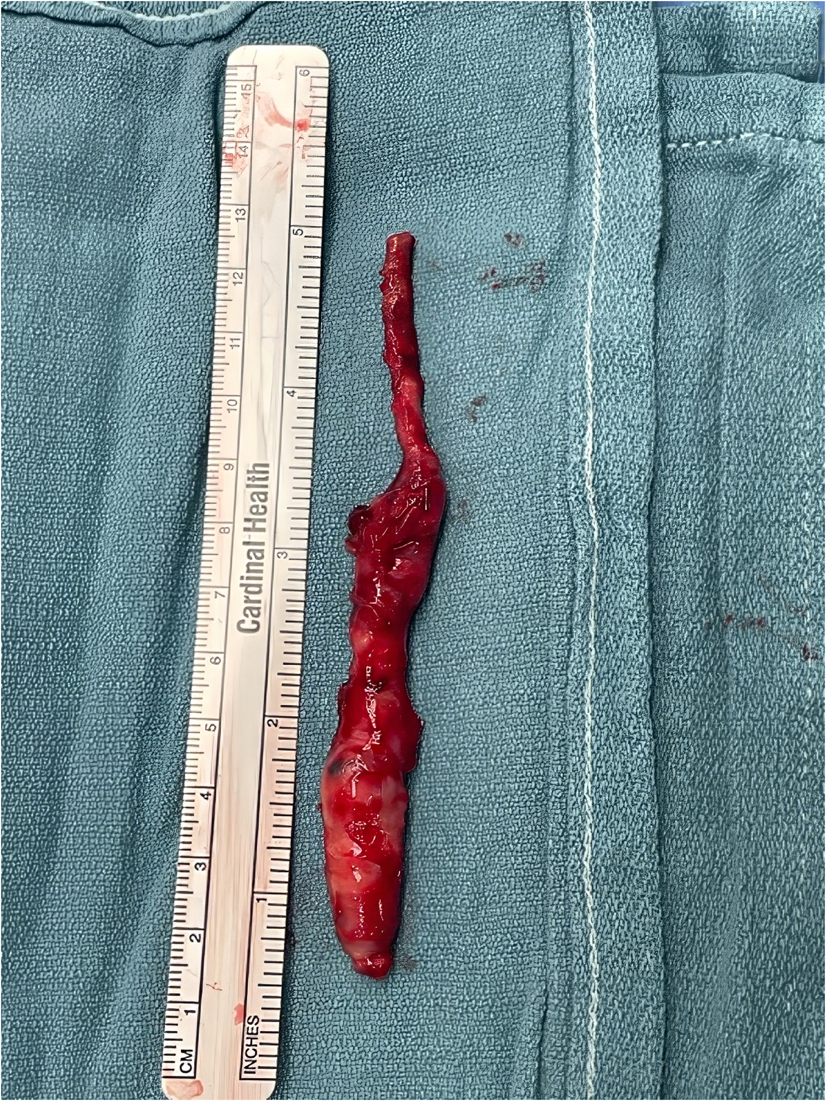
Ulnar artery mycotic aneurysm.
The diseased arterial segments were resected, and interpositional vein grafts from the foot were anastomosed to the superficial and deep palmar arches. The patient recovered uneventfully and was discharged several days later. At two-month follow-up, perfusion was restored to all digits with full resolution of pain.
Discussion
Mycotic aneurysms are relatively uncommon, accounting for 0·7%–3% of all aortic aneurysms and rarely occur in the upper extremities. 6 The combination of arterial trauma and hematogenous bacterial seeding played a central role in the subsequent formation of the mycotic aneurysm.
This case highlights the complexity of diagnosis and treatment, being noteworthy due to the placement of an arterial line following the initial procedure, combined with chronic foot infections that likely precipitated bacterial seeding of previously injured vessels. Clinical suspicion and imaging were crucial to operative planning, but the definitive diagnosis was only confirmed intraoperatively. Prompt surgical debridement and bypass reconstruction achieved source control and limb salvage.
Conclusion
Upper extremity mycotic aneurysms are rare but serious infections requiring timely recognition and surgical management. Patient comorbidity can significantly contribute to the development of mycotic aneurysms and subsequently exacerbate vascular compromise requiring reconstruction. Surgical intervention, including resection and bypass grafting, was required to successfully restore perfusion and allow limb salvage.
Footnotes
Contributory Roles
Declaration of Conflicting Interests
The authors declared no potential conflicts of interest with respect to the research, authorship, and/or publication of this article.
Funding
The authors received no financial support for the research, authorship, and/or publication of this article.
Consent Statement
Informed consent was obtained from the participant included in the study regarding both the publication of their information and images.