
Abstract
Orthopedic implant-associated infections, primarily caused by biofilm-forming Staphylococcus aureus, pose significant clinical challenges. These infections often lead to implant failure, prolonged antibiotic treatments, and an increased risk of revision surgeries, emphasizing the need for effective biofilm-resistant implant materials. In this study, we present a dual-functional titanium screw (Ti-S) grafted with chitosan (Cs), a biocompatible polymer known for its osteogenic and antimicrobial properties while maintaining mechanical integrity. The chitosan-modified titanium screw (Cs-Ti-S) was prepared via chemical immobilization to enhance resistance to biofilm formation while promoting osseointegration and preserving biomechanical integrity. Biomechanical testing confirmed that chitosan modification did not compromise mechanical performance, as Cs-Ti-S exhibited a torsional yield strength of 1.70 ± 0.00 Nm compared to 1.76 ± 0.05 Nm for unmodified titanium screws (Un-Ti-S), and an axial pullout force of 68.66 ± 14.36 N for Cs-Ti-S versus 70.33 ± 9.71 N for Un-Ti-S. Micro-scratch tests revealed similar hardness values (1.26 ± 0.03 GPa for Cs-Ti-S vs. 1.40 ± 0.07 GPa for Un-Ti-S) and scratch resistance, ensuring surface durability. Gene expression analysis showed upregulated β1-integrin on Cs-Ti-S at 24 h post-infection, indicating improved osteoblast adhesion. Scanning electron microscopy (SEM) analysis confirmed significantly reduced bacterial biofilm formation on Cs-Ti-S. Moreover, the combination of povidone-iodide (PI) treatment on Cs-Ti-S surfaces significantly inhibited biofilm formation over 7 days, unlike Un-Ti-S, which retained significant adhesion. These results suggest chitosan grafting as a scalable, non-antibiotic strategy to enhance antimicrobial resistance and osseointegration.
Introduction
Internal fixation devices, such as wires, staples, nails, screws, plates, and absorbable pins, are essential in orthopedic surgery for stabilizing fractures and facilitating bone healing after fractures or osteotomy procedures. This technique involves the surgical implantation of various fixation devices, including plates, screws, nails, rods, and wires, with the choice of implant and method depending on the fracture’s location and characteristics.1–12 Among these devices, Ti screws are particularly favored for their superior strength-to-weight ratio, excellent corrosion, low-modulus of elasticity, high fatigue and wear resistance, and bioinert nature.13–24 These fixation devices offer many advantages, such as providing stability, allowing early mobilization, and reducing the risk of malunion.25–30 However, complications like cortical porosity, stress shielding, delayed union, and refractures can arise, emphasizing the importance of careful device selection and design to ensure optimal healing outcomes.25,31–40
A critical complication associated with internal fixation devices is implant-associated infection, primarily driven by biofilm formation.41–46 The treatment for biofilm formation adds an extra financial burden to the patient’s healthcare costs and increases recovery time. These infections occur in 1%–2% of primary replacements and 3%–5% of revisions, with some reports indicating rates as high as 20%. The primary pathogens responsible for periprosthetic infections are Staphylococcus epidermidis and Staphylococcus aureus, accounting for approximately 70% of cases.47–53 Biofilm formation on orthopedic implants occurs in several stages, beginning with bacterial adherence, followed by accumulation and the development of structured communities encased in a protective glycocalyx.52,53 This biofilm not only facilitates bacterial survival and communication but also confers resistance to antibiotics and immune responses, making such infections extremely difficult to treat.54–58
To mitigate these challenges, implant surface modifications aimed at enhancing osseointegration while reducing bacterial colonization have been explored.59–61 Various surface coatings, including hydroxyapatite (HA),62–64 poly (methyl methacrylate) (PMMA),65–67 arginine-glycine-aspartate (RGD) peptides,68–71 and bone morphogenetic proteins (BMPs),72–74 have shown promise in promoting tissue integration. However, these materials lack inherent antimicrobial properties and remain vulnerable to infection. Antimicrobial coatings such as ionic silver75,76 and biodegradable gentamicin-hydroxyapatite77,78 have also been investigated, but concerns over cytotoxicity and antibiotic resistance limit their long-term clinical utility.79,80
Chitosan, a natural polymer derived from chitin, offers a compelling alternative due to its dual functionality—supporting osteogenesis81–84 and exhibiting antimicrobial85–89 activity. When applied to titanium implants, chitosan enhances osteoblast proliferation and cell adhesion, promoting robust osseointegration.90–93 Simultaneously, it reduces bacterial adhesion, including methicillin-resistant Staphylococcus aureus (MRSA), thereby lowering infection risk.94–96
Despite the promising biological properties of chitosan, its application to load-bearing orthopedic implants, particularly titanium screws, has not been thoroughly investigated. Most existing studies on chitosan-modified implants focus on flat or non-load-bearing surfaces, such as plates or coatings on dental implants, where mechanical demands are relatively low. However, orthopedic screws play a unique biomechanical role—they must sustain dynamic axial, torsional, and shear loads while maintaining anchorage in bone tissue.97–100 Applying a surface coating like chitosan raises critical concerns: Will it alter the screw’s mechanical strength, torque resistance, or stability under physiological conditions? These questions remain largely unanswered, creating a barrier to clinical translation.
Moreover, while chitosan exhibits inherent antimicrobial and osteoconductive properties, its efficacy against robust biofilms formed by pathogens such as Staphylo-coccus aureus and MRSA may be limited under certain clinical conditions. In this study, an adjunctive strategy using post-fabrication washing of Cs-Ti-S surfaces with PI, a broad-spectrum antiseptic, was implemented to enhance bacterial resistance. This method avoids potential alterations in chitosan’s molecular structure and mechanical integrity that may arise from direct chemical interaction with iodine-based compounds. PI has been shown to interact electrostatically with the amino groups in chitosan, potentially disrupting the charge balance and altering film stability and bioactivity.101–103 When applied as a surface rinse, PI may form a transient antimicrobial layer without compromising chitosan’s coating adherence or mechanical properties. This wash-based technique may thus offer a clinically viable and scalable strategy for enhancing implant sterility without sacrificing osseointegration capacity.
This integrated evaluation of both mechanical and biological performance represents a significant advancement in the design of infection-resistant orthopedic implants. By demonstrating that a bioactive, antimicrobial coating can be applied to load-bearing implants without impairing their function, this study bridges a critical translational gap. The findings may inform future strategies in orthopedic device engineering, reduce infection-related revision surgeries, and ultimately improve clinical outcomes in patients undergoing internal fixation.
Materials and methods
Screw preparation
Cancellous titanium screws with a diameter of 4 mm and a length of 10 mm (Cat. No. 406.010) were purchased from Synthes® USA. Dopamine hydrochloride, Tris hydrochloride (TRIS-HCL), glutaraldehyde (25%), and low molecular weight chitosan (75%–85% deacetylated) were obtained from Sigma-Aldrich. The screws were ultrasonically cleaned sequentially in acetone, absolute ethanol, and deionized (DI) water for 10 min each, then dried under a vacuum. These prepared samples were referred to as Un-Ti-S, where “Un” denotes uncoated.
To fabricate titanium screw with chitosan, the Un-Ti-S samples were first treated with 48% sulfuric acid (H2SO4) solution at 60°C with constant stirring for 3 h. The samples were then thoroughly washed with DI water and dried under vacuum, resulting in SA-Ti-S, where “SA” denotes sulfuric acid-treated. The SA-Ti-S screws were immersed in a 5 mg/mL solution of dopamine hydrochloride in 10% (v/v) 0.1 M TRIS-HCL aqueous buffer at room temperature for 12 h, followed by overnight treatment in 3% glutaraldehyde at 4°C. Finally, the screws were immersed in a 0.5% (w/v) chitosan solution prepared in 1% acetic acid at room temperature for 18 h. This concentration provided optimal solubility and viscosity to ensure uniform coating without aggregation, while the mildly acidic environment effectively protonated chitosan’s amine groups for strong surface adhesion. The 18-h soaking duration was chosen based on reports that chitosan adsorption typically plateaus within this timeframe, promoting stable film formation while minimizing the risk of over-deposition or degradation that can occur with extended incubation. 104 After rinsing with DI water and drying under vacuum, the screws were referred to as Cs-Ti-S, where “Cs” denotes chitosan-coated.
Characterization of screws
Surface characterization
The surface morphology of both Un-Ti-S and Cs-Ti-S screws were analyzed with a FEI Quanta 350 SEM. High-resolution SEM imaging was performed at multiple regions of each screw, with particular focus on the crest and valley areas of the thread surfaces to assess localized surface features. For the Cs-Ti-S group, special attention was given to evaluating the distribution, uniformity, and adherence of the chitosan coating across the surface. Images were analyzed for changes in surface roughness, coating continuity, and any evidence of aggregation or defects in the chitosan layer. Comparative analysis between Un-Ti-S and Cs-Ti-S screws allowed for qualitative assessment of topographical differences and provided visual confirmation of the surface modifications.
Biomechanical characterization
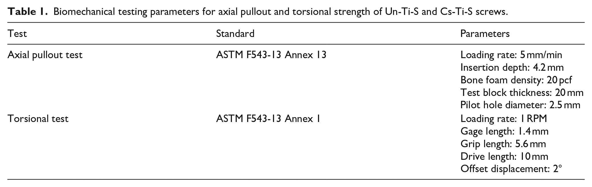
Biomechanical characterization was conducted to evaluate the mechanical integrity of the Un-Ti-S and Cs-Ti-S. Two standardized mechanical tests were performed according to ASTM F543-13 protocols: an axial pullout test (Annex 13) and a torsional strength test (Annex 1), following parameters summarized in Table 1. In short, for the axial pullout test, screws were inserted into 20 pcf density synthetic bone foam block to a depth of 4.2 mm, using pilot home of 2.5 mm diameter. The pullout force was applied at a constant loading rate of 5 mm/min until failure, as illustrated in Figure 1(a). For the torsional test, each screw was subjected to rational force at a rate of 1 RPM, with a gauge length of 1.4 mm and grip length of 5.6 mm. The drive length was maintained at 10 mm, and the offset displacement was limited to 2° as shown in Figure 1(b). These parameters ensured consistent evaluation of the screw’s resistance to axial and torsional loads under conditions mimicking clinical fixation scenarios.
Biomechanical testing parameters for axial pullout and torsional strength of Un-Ti-S and Cs-Ti-S screws.

Photographs illustrating the biomechanical testing setup and samples after testing: (a) axial pullout strength testing setup, (b) torsional strength testing setup, (c) post-test Un-Ti-S sample, and (d) post-test Cs-Ti-S sample.
Micro-scratching test of the modified surface
To assess surface mechanical integrity, a micro-scratch test was performed to evaluate the scratch resistance and surface hardness of the material. Due to the curved and threaded geometry of the screws, direct testing on screws surfaces was not feasible. Instead, a flat titanium foil was used to simulate the screw surface conditions. Specifically, unmodified titanium foil and chitosan-modified titanium foil were prepared and subjected to micro-scratch testing according to ASTM G171-03 guidelines. This approach allowed for standardized and reproducible measurements of surface hardness and coating adhesion, which are representative of the material behavior observed on screw surfaces.
Gene expression
To explore the molecular response of osteoblastic cells to bacterial infection and Cs-Ti-S surfaces, quantitative real-time PCR (qRT-PCR) was employed as a high-sensitivity method to measure gene expression relevant to osseointegration and apoptosis. Osteoblastic cells adhered to both Un-Ti-S and Cs-Ti-S, under infected and uninfected conditions, were analyzed to identify transcriptional changes critical for implant integration and immune response modulation.
Prior to RNA collection, extracellular bacteria were selectively removed through a 100 µg/mL gentamicin treatment for 1 h, ensuring that only intracellular or adherent bacterial effects influenced the gene expression profile. 105 Identical antibiotic treatment was applied to all conditions to maintain experimental consistency. Adherent osteoblastic cells were lysed directly on the Ti foil surfaces, and total RNA was extracted using the GeneJET RNA Purification Kit (Thermo Scientific, USA), following the manufacturer’s protocol. RNA concentration and purity were assessed using UV/Vis spectrophotometry (Beckman Coulter DU-730) at 260/280 nm to ensure sample quality.
Subsequently, complementary cDNA was synthesized from high-purity RNA using the High-Capacity cDNA Reverse Transcription Kit (Applied Biosystems, USA). Equal volumes of cDNA from each sample were used for TaqMan-based qRT-PCR, conducted using the 7500 Fast Real-Time PCR System (Applied Biosystems, USA) and Universal PCR Master Mix. Target genes included:
β1-integrin (Hs00559595_m1) and α5-integrin (Hs01547673_m1)—Critical mediators of osteoblast adhesion to extracellular matrix and titanium surfaces, strongly associated with early-stage osseointegration and bone remodeling.106,107
TNFRSF10B (Hs00366278_m1)—A death receptor linked to apoptotic pathways, indicative of cellular stress and immune signaling post-infection. 108
TNFRSF11A (Hs00921372_m1)—A key regulator of osteoclastogenesis and bone resorption, relevant for bone remodeling balance. 109
GAPDH (Hs02758991_g1)—Used as a housekeeping gene for normalization.110,111
These gene targets were selected based on their established roles in the cellular response to implant materials and infection. All the specific genes were purchased from Applied Biosystem, USA, and the expression levels were normalized to GAPDH and interpreted relative to baseline (uninfected, Un-Ti-S). This approach allowed for precise quantification of osteogenic and apoptotic gene expression, offering mechanistic insight into how chitosan surface modification and bacterial infection influence the biological processes essential for successful implant integration.
Biofilm formation
SEM was used to examine the morphology and distribution of bacterial biofilm on the screw samples. The samples were either washed with a 0.35% PI (10%) solution (referred to as washed Un-Ti-S or washed Cs-Ti-S) or left unwashed (unwashed Un-Ti-S or unwashed Cs-Ti-S). After incubating the samples for 1, 3, and 7 days at 37°C, following initial bacterial seeding (106–107 CFU/mL), the growth medium was discarded, and the screw samples were washed with PBS before being air-dried. The samples were then fixed with 2.5% glutaraldehyde and prepared for SEM imaging using a standard protocol involving washing a graded series of ethanol (30%, 50%, 70%, 90%, and 100%) to remove any water content from the samples and prevent distortion during SEM imaging. 112 The samples were immersed in each ethanol solution for approximately 10–15 min to ensure thorough dehydration. After dehydration, the samples were air-dried to remove any residual ethanol. Finally, the samples were sputter-coated with a thin layer of gold to enhance conductivity and prevent charging effects during SEM imaging. This coating process involved placing the samples in a vacuum chamber and applying a fine, even layer of gold, which allows for high-resolution imaging of the biofilm morphology and distribution on the screw surfaces.
Force between the bacteria/biofilm and the screw surface
To further evaluate the strength of bacterial adhesion to the Ti surface, a mechanical detachment assay was performed using ultrasonication and vortexing. This approach provided an indirect assessment of biofilm-surface interaction strength. Screw samples that had been incubated under identical bacterial culture condition (without PI treatment) were subjected to ultrasonication at 40 KHz for 10 min, followed by a 10-second vortex, to apply a uniform detachment force. Post-treatment, the screws were immediately fixed and analyzed via SEM to quantify the remaining surface-bound bacteria.
Cell culture
Staphylococcus aureus (ATCC 6538) was cultured overnight in tryptic soy broth (TSB) at 37°C. The bacterial concentration was determined by performing serial dilutions of the culture, followed by spreading 100 µL for each dilution onto a TSA plate. After incubation at 37°C for 24 h, colony-forming units (CFUs) were counted to estimate the bacterial concentration.
Osteoblastic cells, SaOS-2 (ATCC number: HTB-85), were cultured in DMEM (Dulbecco’s Modified Eagle Medium) supplemented with 10% fetal bovine serum (FBS), 1% penicillin/streptomycin, and 1% essential amino acids, following the manufacturer’s guidelines. The cells were maintained in a humidified incubator at 37°C with 5% CO2. Once the cells reached 80%–90% confluency, they were detached using 0.25% trypsin-EDTA solution and centrifuged. The cell pellet was resuspended in a fresh medium, and the cells were counted and seeded for subsequent experiments.
Statistical analysis
All experiments were performed using four biologically independent replicates (n = 4) per condition and time point to ensure data reliability and reproducibility. Quantitative results are presented as mean ± standard deviation (SD), representing the central tendency and variability across replicates. Prior to analysis, data were assessed for normality and homogeneity of variances using the Shapiro–Wilk test and Levene’s test, respectively. For pairwise comparisons between experimental groups, unpaired two-tailed Student’s t-tests were used if the assumptions of normality were met. In cases where normality could not be assumed, the non-parametric Mann–Whitney U test was employed. Where applicable, multiple comparisons were corrected using the Bonferroni method. All statistical comparisons were designed to evaluate the impact of chitosan surface modification, bacterial infection, and their combined effect on outcomes such as mechanical performance, gene expression, and cellular behavior. A p-value of less than 0.05 was considered statistically significant, indicating a high level of confidence that the observed differences were not due to random variation.
Results
Characterization of screws
Surface characterization
The surface morphology of Un-Ti-S and Cs-Ti-S was evaluated using SEM to confirm successful surface modification and examine localized topographical changes. The SEM images revealed clear distinctions between the two groups, supporting the effectiveness of the chitosan coating process (Figure 2).

SEM images of the screw surfaces for Un-Ti-S (A, a1, a2) and Cs-Ti-S (B, b1, b2). Panels a1 and b1 depict the valley regions of the respective screws, while panels a2 and b2 illustrate the crest regions of the screw threads for the respective.
In the Un-Ti-S group, the Ti screws surfaces exhibited a consistently rough texture across both the valley (Figure 2(a1)) and crest (Figure 2(a2)) regions of the thread. This roughness likely resulted from the acid etching and cleaning process, but lacked any additional surface treatment. In contrast, the Cs-Ti-S group demonstrated a markedly different surface profile. Importantly, region-specific differences were observed with the Cs-Ti-S group. The valleys of the threads (Figure 2(b1)) appeared rougher and more densely coated, while the crest regions (Figure 2(b2)) showed a smoother surface with a thinner, but still continuous, layer of chitosan. This variation in coating morphology suggests that the topography of the screw influenced the distribution and thickness of the chitosan layer, with recessed regions possibly retaining more coating during the immersion and drying process.
Bio-mechanical characterization
The biomechanical characterization of Un-Ti-S and Cs-Ti-S was evaluated through torsional and axial pullout testing, and the results are summarized in Table 2. This assessment aimed to determine whether the chitosan coated impacted the mechanical performance of the screws under clinically relevant loading conditions.
Comparative biomechanical properties of Un-Ti-S and Cs-Ti-S.

Values are presented as mean ± standard deviation.
No statistically significant differences were found (p > 0.05).
In the torsional tests, both screw types exhibited comparable mechanical behavior. The maximum average torque was 2.36 ± 0.11 Nm for both Un-Ti-S and Cs-Ti-S screws, indicating no reduction in torque-bearing capacity due to surface modification. The torsional yield strength was also similar between groups (1.76 ± 0.05 Nm for Un-Ti-S vs 1.70 ± 0.00 Nm for Cs-Ti-S). Additionally, the breaking angle showed a slight increase in Cs-Ti-S (103.33 ± 5.77°) compared to Un-Ti-S (93.33 ± 5.77°), suggesting that the modified screws tolerated a higher degree of twist before failure. The maximum torque displacement followed a similar trend, with Cs-Ti-S showing a modest increase (93.66 ± 4.04°) versus (87.66 ± 8.08°) for Un-Ti-S. Torsional stiffness remained statistically unchanged between the groups (0.21 ± 0.02 Nm/deg for Un-Ti-S and 0.19 ± 0.01 Nm/deg for Cs-Ti-S).
For the axial pullout test, the average pullout strength was 70.33 ± 9.71 N for Un-Ti-S and 68.66 ± 14.36 N for Cs-Ti-S, indicating that chitosan surface modification had no statistically significant effect on the screw’s anchorage in synthetic bone material. Overall, these results confirms that chitosan immobilization did not compromise the mechanical integrity of the Ti screws in either torsional or axial loading modes, supporting its clinical viability for load-bearing orthopedic applications.
Micro-scratching test of the modified surface
The result from the micro-scratch demonstrated that the chitosan coating did not significantly alter the mechanical surface properties of the titanium substrate. The mean hardness of the Un-Ti-S samples was 1 a.40 ± 0.07 GPa, while the Cs-Ti-S samples exhibited a slightly lower hardness of 1.26 ± 0.03 GPa. Although there was a minor reduction in hardness following chitosan immobilization, the difference was not statistically significant (p > 0.05), suggesting that the coating process preserved the underlying mechanical integrity of the material.
In terms of scratch resistance, the average scratch width-used as a proxy for surface deformation under constant load-was measured at 165.41 ± 3.60 µm for Un-Ti-S and 174.23 ± 2.00 µm for Cs-Ti-S. The slightly wider scratch width observed in the Cs-Ti-S group is consistent with the presence of a softer chitosan layer on the titanium surface. However, this variation also did not reach statistical significance, further supporting the mechanical compatibility of the coating. Although representative micrographs of scratch tracks were not available due to equipment constraints, the testing was performed under standardized conditions in accordance with ASTM G171-03, and the quantitative data provide a reliable assessment of surface mechanical behavior. To support interpretation, a schematic representation of the micro-scratch test setup has been included to illustrate the principle of the testing procedure (Figure 3). Future studies are planned to incorporate profilometry or high-resolution imaging (e.g. optical microscopy or SEM) for direct visualization of surface deformation. Collectively, these findings confirm that chitosan coating does not adversely impact the surface hardness or scratch resistance of titanium, reinforcing its potential use in load-bearing orthopedic implants.

Schematic representation of the micro-scratch test setup used to evaluate surface mechanical properties. A conical stylus applies a controlled normal load to the Ti surface while traversing from left to right, generating a scratch track. The resulting scratch width and depth provide quantitative measures of surface hardness and resistance to deformation.
Gene expression
To assess the impact of surface modification and bacterial infection on osteoblastic gene regulation, quantitative gene expression analysis was conducted at 2 h and 24 h post-infection. The focus was placed on key markers involved in cell adhesion, bone modeling, and apoptosis, specifically: β1-integrin, α5-integrin, TNFRSF10B, and TNFRSF11A.
At both 2- and 24-h post-infection, β1-integrin and α5-integrin mRNA were detected across all experimental groups, reflecting baseline osteoblastic adhesion activity. Notably, at 24 h, β1-integrin expression was significantly upregulated in cells cultured on Cs-Ti-S compared to both Un-Ti-S and uninfected controls (p < 0.05; Figures 4 and 5). This enhanced expression indicates a time-dependent osteogenic response, suggesting that the chitosan-modified surface promotes integrin-mediated adhesion and early-stage osseointegration, particularly under inflammatory stress. In contrast, α5-integrin expression remained stable across all groups and time points, with no statistically significant differences ob-served (p > 0.05), indicating that while α5-integrin supports basal cell adhesion and osteogenic function, it may not be as dynamically regulated in response to surface modification or infection.

Fold change in gene expression relative to the Un-Ti-S at 2 h post-infection. Gene expression levels of (a) β1-integrin and (b) α5-integrin are shown.

Fold change in gene expression relative to Un-Ti-S at 24 h post-infection. Gene expression levels of (a) β-integrin and (b) α5-integrin are presented.
Expression of TNFRSF10B, a gene associated with apoptotic signaling and cellular stress responses, was consistently below the detection threshold in all samples. Its minimal expression implies that the infection conditions used in this study did not trigger significant activation of extrinsic apoptotic pathways in osteoblastic cells. Similarly, TNFRSF11A, a regulator of osteoclastogenesis and bone remodeling, was detected at low levels with no significant variation across groups, suggesting limited activation of bone resorption signaling at the tested time points. Both genes were expressed so little by the cells that the genes could not be detected within the limitations of the experiment.
Taken together, these findings indicate that chitosan surface modification exerts a measurable influence on the transcriptional response of osteoblastic cells, particularly enhancing β1-integrin expression in the later stages of infection. This supports the hypothesis that chitosan coatings may promote osseointegrative signaling pathways even under bacterial challenge, without inducing apoptotic stress responses. These results provide molecular evidence for the biocompatibility and functional relevance of Cs-Ti-S surfaces in implant applications.
Biofilm formation
Biofilm formation on the screws surfaces unwashed with PI
Biofilm development on the surface of unwashed Un-Ti-S and Cs-Ti-S screws was assessed via SEM at Days 1, 3 and 7 following bacterial exposure. Figure 6 illustrates the comparative surface colonization on both screw types. Prior to imaging, samples were gently rinsed with PBS to remove loosely attached planktonic bacteria, leaving only adherent microbial communities for visualization. The screws at all the experiment period showed a higher number of bacteria on the thread areas compared to the head and neck region of the screws. Thus, the bacteria and the biofilms on the thread regions only were considered for comparison purposes.

Biofilm formation on unwashed Un-Ti-S (A, C, c) and Cs-Ti-S (B, D, d) surfaces. On Day 7, the Un-Ti-S surface (C, c) is completely engulfed by a dense extracellular matrix, indicating extensive biofilm development, whereas the Cs-Ti-S surface (D, d) shows only sparse bacterial aggregates, with no mature biofilm formation.
On Un-Ti-S, early signs of bacterial attachment were visible on Day 1, with microcolonies adhering along the thread surface (Figure 6(A)). As the incubation period progressed, bacterial colonization increased markedly. By Day 3 and more prominently on Day 7, the threads were densely coated with mature biofilm structure composed of large, confluent bacterial clusters embedded in a well-developed extracellular polymeric matrix (Figure 6(C, c)).
In contrast, Cs-Ti-S screws without PI treatment exhibited significantly lower levels of bacterial colonization. At Day 1, no observable bacterial attachment or biofilm formation was detected on the chitosan-coated threads (Figure 6(B)). By Day 7, only a few isolated bacterial aggregates were seen (Figure 6(D)), and even at higher magnification (Figure 6(d)), there was no evidence of a mature biofilm matrix or widespread surface colonization. These findings demonstrate that chitosan surface modification alone inhibits early bacterial adhesion and biofilm development over a 7-day period.
Biofilm formation on the screws surfaces washed with PI
To assess the synergistic anti-biofilm effect of surface coating and antiseptic treatment, Un-Ti-S and Cs-Ti-S were treated with PI and subjected to identical biofilm evaluation via SEM. Figure 7 presents the results at Days 1, 3, and 7 post-infections.

Biofilm formation on Un-Ti-S and Cs-Ti-S surfaces after povidone-iodine (PI) washing over a 7-day period, observed by SEM at both low (50 μm) and high (5 μm) magnifications. Panels A–C show Un-Ti-S surfaces on Days 1, 3, and 7, respectively, with corresponding high-magnification views in panels a–c. Panels D–F show Cs-Ti-S surfaces on Days 1, 3, and 7, respectively, with corresponding high-magnification views in panels d–f. The Un-Ti-S surface exhibited significantly denser biofilm and a higher bacterial count on Day 3 (B, b) and Day 7 (C, c) compared to the Cs-Ti-S surface, which displayed much lower bacterial presence on Day 3 (E, e) and Day 7 (F, f). These observations suggest that PI treatment, in combination with the chitosan modification on Cs-Ti-S, effectively reduced bacterial adhesion and biofilm formation over the 7 days.
On PI-treated Un-Ti-S, bacterial adhesion was initially suppressed compared to unwashed. SEM image from Day 1 (Figure 7(A, a)) showed only a few scattered bacteria with no evident biofilm. However, by Day 3 (Figure 7(B, b)), bacterial accumulation increased, with visible clusters forming along the thread surfaces. By Day 7, these clusters developed into a visible biofilm with extracellular matrix deposition (Figure 7(C, c)).
Remarkably, Cs-Ti-S treated with PI demonstrated robust resistance to bacterial colonization throughout the 7-day observation period. On Day 1, SEM imaging revealed very limited bacterial adhesion (Figure 7(D, d)). Even by Days 3 and 7 (Figure 7(E, e) and (F, f)), the surface remained mostly free of bacterial buildup, with no significant biofilm formation observed. Only sparse and isolated bacterial cells were noted, with no organized matrix or aggregation, with a density much lower than unwashed. These results indicate that the combination of chitosan surface modification and PI treatment effectively inhibits biofilm initiation and progression, providing superior protection against microbial colonization over time.
Adhesion force between the bacterial/biofilm and the screw surface
The results revealed a clear difference in bacterial adhesion between the two surfaces. On Un-Ti-S screws, SEM images (Figure 8(A, a)) showed that a substantial number of bacteria remained firmly attached to the thread surface even after sonication and vortexing, indication strong bacterial-surface interactions. These observations suggest that the native Ti surface provides favorable adhesion sites that support more robust bacterial attachment.
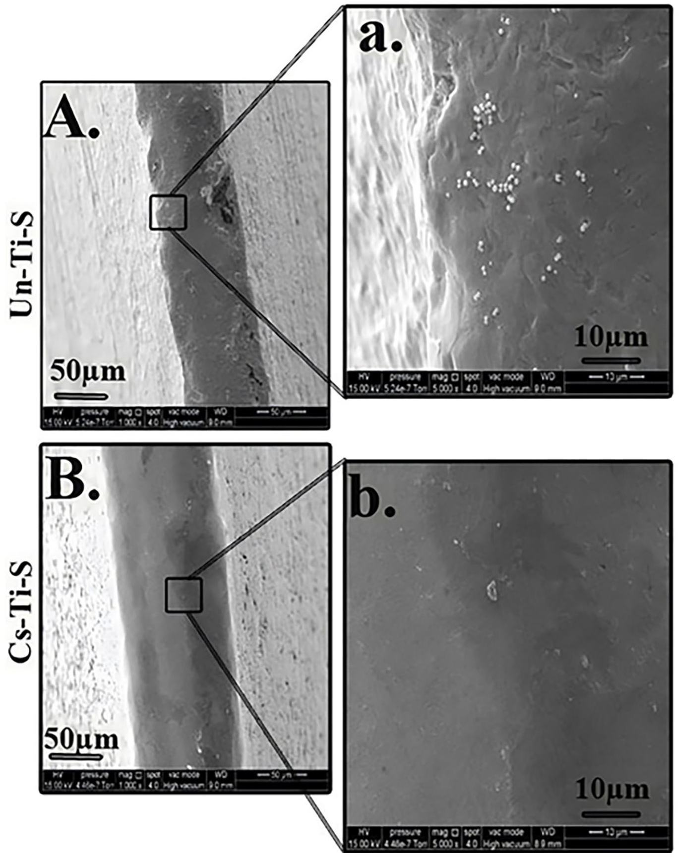
SEM of Un-Ti-S and Cs-Ti-S surfaces after sonication. A significantly higher number of bacteria was found to be present on the Un-Ti-S surface (A, a) after sonication in comparison to the Cs-Ti-S surface (B, b).
In contrast, Cs-Ti-S screws exhibited a drastically different response to the same detachment treatment. SEM images (Figure 8(B, b)) revealed minimal bacterial retention, with only a few scattered bacterial cells visible. The sonication force effectively dislodged the biofilm from the Cs-Ti-S surface, indicating a significantly weaker adhesion strength between bacteria and the chitosan-modified substrate. These findings strongly support the anti-adhesive properties of chitosan coatings and reinforce the evidence that chitosan modification not only reduces bacterial colonization but also weakens bacterial adhesion, making surface-bound biofilms more susceptible to physical removal. This is a critical advantage in clinical settings where mechanical debridement or lavage is used to manage implant-associated infections.
Discussion
This study presents a comprehensive investigation into the mechanical, antimicrobial, and biological performance of Cs-Ti-S + PI as a dual-functional strategy to enhance orthopedic implant outcomes. Compared to Un-Ti-S, the modified screw demonstrated promising improvements in biofilm resistance and osteogenic signaling while preserving essential biomechanical properties, making it a strong candidate for infection-resistant, load-bearing orthopedic applications.
Surface characterization confirmed uniform chitosan coating with preserved surface topography. In our earlier work, we characterized the surface morphology of chitosan-immobilized titanium (SA-Cs-Ti) using SEM.93,113 The images revealed that the chitosan coating conformed to the underlying titanium substrate, resulting in a roughened surface with visible porosity. This porous structure is advantageous as it can enhance osteoblast adhesion and proliferation, contributing to improved osseointegration. In the current study, SEM analysis of the chitosan-coated titanium screws corroborated these findings, showing a uniformly distributed chitosan layer with micro-scale porosity. The porous morphology is consistent with our previous observations and supports the potential of the chitosan coating to facilitate cellular interactions and integration with bone tissue. The same two studies also quantitatively demonstrated that chitosan coatings significantly reduce both bacterial adhesion and invasion on titanium surfaces. For example, in a co-culture model mimicking postoperative infection, chitosan-coated titanium exhibited substantially lower bacterial adhesion, with approximately 2,233 ± 681 CFU compared to 5367 ± 1662 CFU on uncoated titanium after 4 h. Additionally, live/dead fluorescence staining was employed to visualize and quantify the reduction of viable bacteria on chitosan-modified titanium, providing qualitative insights into biofilm integrity and spatial distribution. These results confirm that chitosan immobilization can disrupt biofilm maturation and reduce microbial viability, thereby lowering the risk of implant-associated infection.
Furthermore, while precise measurements for coating thickness were not conducted in this study, findings from others indicate that chitosan coatings applied via similar methods typically exhibit thicknesses ranging from 1 to 10 mm, depending on the specific application parameters. 114 Future studies will aim to precisely quantify the coating thickness using cross-sectional imaging techniques, which will offer more detailed insight into the structural characteristics of the chitosan layer. Although FTIR data were not obtained in the current work, we recognize its importance in confirming chemical composition and coating integrity. Therefore, FTIR analysis is planned in future investigations to validate the presence of chitosan and to assess potential interactions with the titanium substrate.
Biomechanical testing showed that Cs-Ti-S maintained comparable torsional yield strength, pullout force, and hardness relative to Un-Ti-S, indicating that the coating process did not impair mechanical performance. These results are critical because any surface modification intended for load-bearing implants must preserve core functional stability under physiological stress. At the molecular level, β1-integrin expression was significantly upregulated on Cs-Ti-S at 24 h post-infection. As a key transmembrane receptor involved in osteoblast attachment to the extracellular matrix and implant surfaces, increased β1-integrin expression suggests enhanced focal adhesion formation and osteogenic signaling, indicative of early-stage osseointegration. The stable expression of α5-integrin and the absence of detectable TNFRSF10B expression further support the cytocompatibility of Cs-Ti-S, indicating no activation of apoptosis pathways under inflammatory conditions.
In the current study, SEM analysis further validated these effects, showing that Cs-Ti-S surfaces substantially inhibited bacterial adhesion and biofilm development compared to Un-Ti-S. When combined with PI lavage, Cs-Ti-S remained free of dense biofilms over a 7-day period, in contrast to Un-Ti-S, which developed substantial bacterial accumulation. Even after standardized mechanical disruption via ultrasonication, Cs-Ti-S retained significantly fewer adherent bacteria than Un-Ti-S, indicating both reduced biofilm strength and improved cleanability of the modified surface.
Conclusion
This study highlights the potential of chitosan grafting as a dual-functional strategy for orthopedic implant applications by addressing two major clinical challenges: biofilm-associated infections and impaired osseointegration, while also not compromising the biomechanical integrity. Although our study showed clear evidence that biofilm formation on the untreated Ti surface is unavoidable once contaminated, Cs-Ti-S demonstrated strong antimicrobial efficacy, significantly reducing bacterial adhesion and biofilm development, especially when combined with PI treatment, while maintaining biomechanical integrity. Notably, upregulation of β1 integrin suggests enhanced osteoblast adhesion and osseointegration, reinforcing the biological compatibility of the modification. These findings underscore the scalability and clinical relevance of chitosan as a non-antibiotic, biocompatible coating for infection-resistant implants. Future work should explore long-term in vivo performance and elucidate the molecular mechanisms behind chitosan’s osteogenic and antimicrobial effects to support its clinical translation.
Footnotes
Ethical considerations
This study did not involve human participants, human data, human tissue, or animals. Therefore, ethical approval was not required.
Funding
The author(s) disclosed receipt of the following financial support for the research, authorship, and/or publication of this article: This work was supported by the National Institute of Arthritis and Musculoskeletal and Skin Diseases (NIAMS) of the National Institutes of Health (NIH) [R21AR065625], National Science Foundation (NSF; OIA-1849206) and Microfree Bio Technology LLC.
Declaration of conflicting interests
The author(s) declared no potential conflicts of interest with respect to the research, authorship, and/or publication of this article.
Data availability statement
The data supporting the findings of this study are available within the article.