
Abstract
After anastomosis of sutures or pins, the restoration of intestinal barrier function can avoid several complications, such as tissue damage and inflammation. Our previous studies demonstrated the feasibility of biodegradable magnesium (Mg) pins as novel anastomosing implants to spontaneously absorb in the body, avoiding secondary removal surgery and long-term inflammation. However, the effect of Mg pins on the intestinal tight junction barrier is rarely investigated. In this study, we conducted high-purity Mg pins inserted in the intestine of rats and prepared Mg extracts cultured intestinal epithelial cell line to investigate the biological effect on the intestinal barrier associated with tight junction protein expression. We discovered that the concentration of released Mg ions over 1.7 mM was the critical threshold, above which mRNA expression of intestinal tight junction and cell apoptosis were affected considerably. Results of the immunohistochemical analysis revealed that Mg functions to stimulate ZO-1, caspase-3, occluding, and claudin-3 expressions. We offer new insight into the effectiveness of biodegradable Mg materials as the next generation of intestinal anastomosis pins, which effectively filters toxins as well as bacteria, and reduces inflammation.
Introduction
Metallic implants, an important component of biomaterials, facilitate the healing or replacement of damaged tissues. 1 The metallic implants, including titanium, stainless steel and cobalt-chromium-based alloys, have been widely used in clinical applications due to their good mechanical strength and high fracture toughness. However, a predominant limitation of these non-degradable biomaterials is their tendency to release toxic metallic ions during corrosion or wear,2,3 resulting in inflammatory cascades that compromise biocompatibility or even cause tissue damage.4,5 A biodegradable material that can spontaneously degrade after completing a mission could reduce foreign body reactions and avoid secondary surgery to reduce this risk. Particularly, biodegradable magnesium (Mg) is approved by Conformite Europeenne (CE) as bone screws and coronary stents due to its superior elastic modulus, excellent fracture toughness, 6 and compressive yield strength similar to natural bone. Variable levels of intestinal bifidobacteria have been related to an inflammatory response caused by Mg deficiency. 7
Intestinal anastomosis is a common procedure in abdominal surgery in which metallic anastomotic devices assist intestinal healing. In our previous works, the Mg pins exhibit good biocompatibility for intestinal anastomosis with the healing of intestinal tissues. Moreover, the Mg pins demonstrated anti-inflammation and anti-tumor biofunctions.8,9 The recovery of barrier function after intestinal healing is important for maintaining normal intestinal permeability, allowing for the exchange of solutes and fluids between the lumen and tissues. 10 Additionally, intestinal barrier function specifies the role played by mucosa and the extracellular barrier components, including pathogens, mucus, antigens, toxins and bacteria, to prevent pro-inflammatory molecules from permeating into the mucosal tissues and the circulatory system of the human body. 11
Tight junctions (TJs) are functions of protein complexes that form intercellular boundaries between the plasma membranes of endothelial and epithelial cells. Epithelial tight junctions are involved in maintaining the intestinal barriers and regulating the permeation of ions, nutrients, and water. Additionally, some TJ-associated proteins, such as occludin and claudin-3, play an important role in the dynamic behavior of cytoplasmic and transmembrane proteins of tight junctions. 12 Numerous clinical studies demonstrate that the intestinal TJ barrier is important in intestinal disease pathogenesis. TJ barrier disruption would increase paracellular permeability, activating the mucosal immune system and subsequently inducing sustained inflammation and tissue damage. 13 Most previous reports focused on the biocompatibility and bio-function of Mg materials, 14 but the effect of Mg materials on the intestinal TJ barrier has yet to be studied.
This study aims to analyze the effect of different concentrations of high-purity magnesium (HP-Mg) extracts on IEC-6 cells by examining apoptosis percentage and TJ-associated protein expression in vitro. After inserting HP-Mg pins into the small intestinal wall of rats, we examined TJ-associated protein expression 1 week later using immunohistochemical analysis.
Methods
Cell culture
The intestine epithelial cell line IEC-6 was cultured in dulbecco’s modified eagle medium (DMEM, Sigma Aldrich) with 10% fetal bovine serum (HyClone) and 1% penicillin and streptomycin (Sigma Aldrich) at 37°C with 5% CO2. The cells in the logarithmic phase of growth were used for the experiment. The study design has been approved by the ethics committee of Affiliated Wuxi No. 2 People’s Hospital of Jiangnan University.
Mg extract preparation
In the present study, the HP-Mg (purity = 99.99%) discs and pins used were provided by Suzhou Origin Material and Medical Technology Co. Ltd. Before the experiment, all samples were ultrasonically cleaned with acetone, 100% ethanol, and 75% ethanol. Compared to the preparation of nanocomposite,15–18 Mg extracts were prepared following ISO 10993-5, and the surface area of HP-Mg discs (diameter = 11.3 mm and height = 2.0 mm) was 1.25 cm2/mL to the extraction medium ratio. ICP-AES was applied to examine Mg ion concentration (Table 1). Pin samples were deliberately inserted into the rat’s small intestinal wall (diameter = 0.5 mm and length = 3 mm).
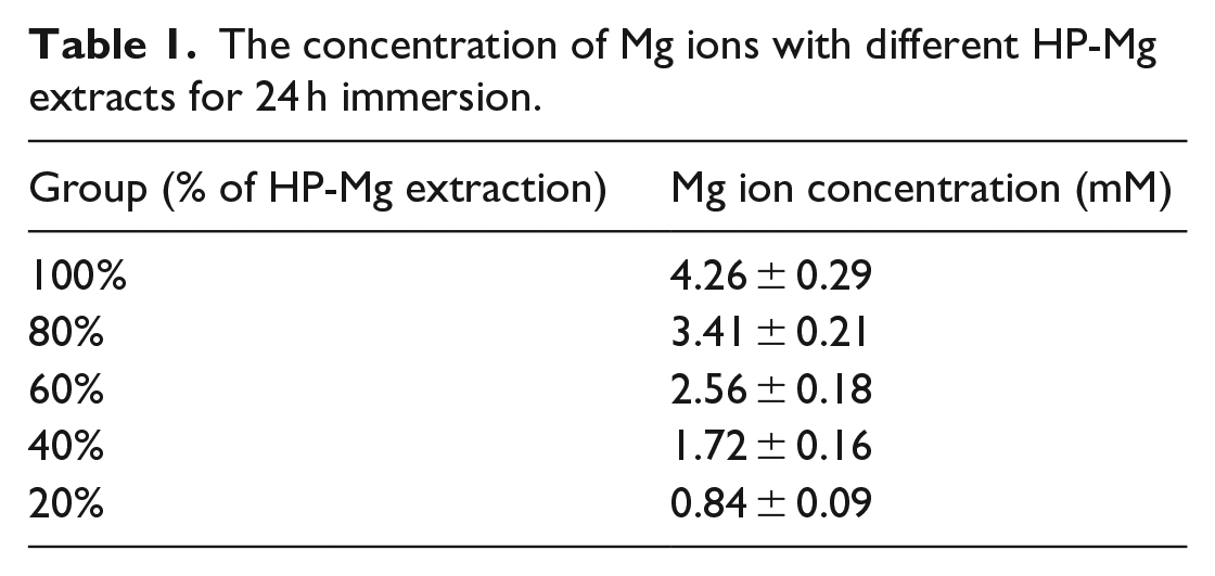
The concentration of Mg ions with different HP-Mg extracts for 24 h immersion.
Reverse transcription-polymerase chain reaction
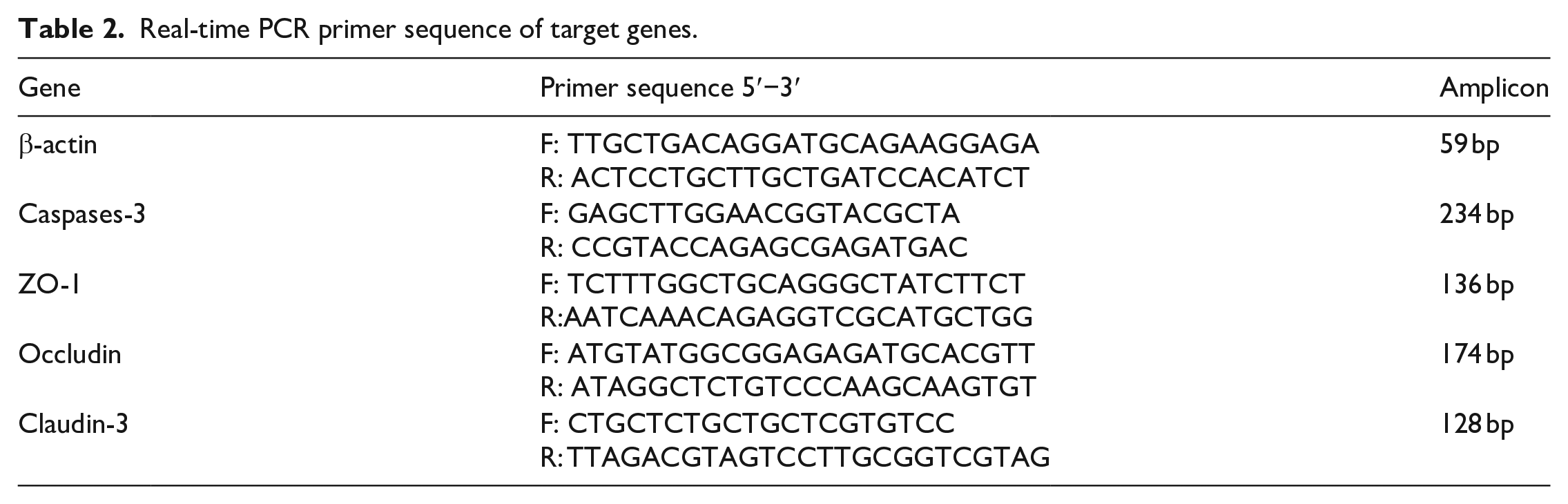
IEC-6 cells were co-cultured with HP-Mg extract gradient concentrations (20%, 40%, 60%, 80%, and 100%) for different duration (12 and 24 h), respectively. IEC-6 cells cultured with a normal medium were used as a control group. The primers for qRT-PCR assay of caspase-3, ZO-1, occluding, and claudin-3 were designed and synthesized by Sangon Biotech (Shanghai, China). The primer sequences are illustrated in Table 1. RT-qPCR was conducted using a quantitative real-time amplification system (Takara, Dalian, China). The mRNA isolation and RT-qPCR follow the description in the previous study. 19 The primer sequences are illustrated in Table 2.
Real-time PCR primer sequence of target genes.
Western blotting
IEC-6 cells were cultured in different concentrations of Mg extracts for 12 or 24 h. The protein expression levels were assessed using western blot analysis. Occludin antibody (cat#Ab167161, dilution 1:1200), TJ-associated proteins antibody ZO-1 (Ab190085; dilution 1:1000), claudin-3 (Ab214487; 1:1000), and caspase-3 (Ab32351; 1:1000) were purchased from Abcam. IEC-6 cells were lysed in RIPA buffer to obtain total cell lysates. In each lane, 20 μg of protein were separated using SDS-PAGE from each sample. The gels were transferred to polyvinylidene fluoride membranes (Merck Millipore) and immunoblotted with the aforementioned antibodies, followed by incubation with HRP-conjugated monoclonal goat anti-rabbit and donkey anti-goat secondary antibodies. β-actin monoclonal antibody (Abcam, cat#ab115777) was used as the internal control. The band signal was visualized using ECL (Merck Millipore).
Cell apoptosis analysis
The details followed the previously described method. 9 The apoptotic rate of IEC-6 cells cultured in gradient concentration of Mg extracts was measured using flow cytometry. IEC-6 cells were plated on the six-well plate at a density of 2 × 10⁵ cells in the cellular apoptosis assay. The cells were trypsinized, centrifuged, and washed in triplicate with PBS before being re-suspended in 250 μL binding buffer at the concentration of 1 × 10⁶ cells/mL. Each cell group was stained with 5 µL Annexin V (1 mg/mL) (eBioScience, USA) at RT for 15 min. Afterward, the cells were stained with propidium iodide (1 mg/mL) for 5 min. The apoptotic rate of IEC-6 cells cultured in gradient concentration of Mg extracts was measured using flow cytometry. Each test was repeated three times independently and separately.
Animal experiments
Fourteen adult male SPF Sprague Dawley rats (bodyweight 269 ± 24.1 g) were randomly divided into two groups (n = 7 in each group). All the rats were kept under standardized temperatures, with free access to food and water. The anesthesia and surgery procedures were described in a previous study. 20 Subsequently, the tools used in the experiment were sterilized using 29 kGy of 60-Co radiation. HP-Mg pins were embedded in the small intestinal wall of the 7 rats in the HP-Mg group. In contrast, Ti pins were implanted into the seven rats in the control group. The full-thickness suture was conducted, and the strengthening examination was performed in the seromuscular layer to prevent possible bleeding. Blood was collected from the orbit, and Mg ions concentration was measured by Automatic Analyzer (7600-030, HITACHI, Japan).
IHC analysis
When the rats were sacrificed, their small intestine tissues were removed and fixed in 10% buffered formaldehyde. After deparaffinization and rehydration, the IHC sections were observed with primary antibodies against occludin (cat#Ab167161, dilution 1:200, Abcam, UK), ZO-1 (Ab190085; dilution 1:150, Abcam), caspase-3 (Ab32351; 1:150, Abcam), and claudin-3 (Ab214487; 1:200, Abcam). Biotinylated immunoglobulin was used as a secondary antibody, and DAB was employed as a chromogen.
Statistical analysis
All the data were presented as mean ± Standard deviation (SD). The one-way ANOVA test was applied to analyze the results.
Results
RT-PCR quantification of caspase-3, ZO-1, occludin, and claudin-3 mRNA expression
Rats had HP-Mg pins implanted in their small intestinal wall. As shown in Figure 1, the concentration of Mg ions in the blood of rats after Mg implantation was 3.6 ±0.7 mM, a significant increase compared to the control group of 1.1 ± 0.1 mM. A previous study investigated HP-Mg pins’ mechanical and degradable properties, demonstrating that they served as an effective anastomosis material. 9 Figure 2 presents relative mRNA levels of caspase-3, ZO-1, occluding, and claudin-3 in the IEC-6 cells co-cultured with HP-Mg extraction for 12 and 24 h. Caspase-3 and occludin expressions up-regulated with increasing Mg extract concentration. Conversely, ZO-1 and claudin-3 protein expressions were down-regulated with an increased tendency of Mg extracts. The critical threshold was the 40% concentration of HP-Mg extracts (released Mg ion concentration was 1.72 ± 0.16 mM). Above this value, TJs protein the mRNA expression displayed significant variation. We performed a western blotting assay to determine caspase-3, occludin, ZO-1, and claudin-3 protein expressions in the IEC-6 cells and to examine the possible changes in TJ-associated protein expression induced by the various concentration of Mg extracts. We discovered that the 40% concentration of HP-Mg extracts (1.72 ± 0.16 mM) is the threshold value, consistent with qRT-PCR analysis. However, a concentration of HP-Mg extracts higher than 40% could induce an altered expression pattern of TJ-associated proteins, whereas a concentration lower than 40% exhibits a negligible effect on TJ protein expression. Figure 3 illustrates that caspase-3 and occludin expressions are up-regulated at Mg extract higher concentrations (above 40%), while ZO-1 and claudin-3 protein expressions are down-regulated at Mg extract higher concentrations.

The concentration of Mg ions in rats’ blood.
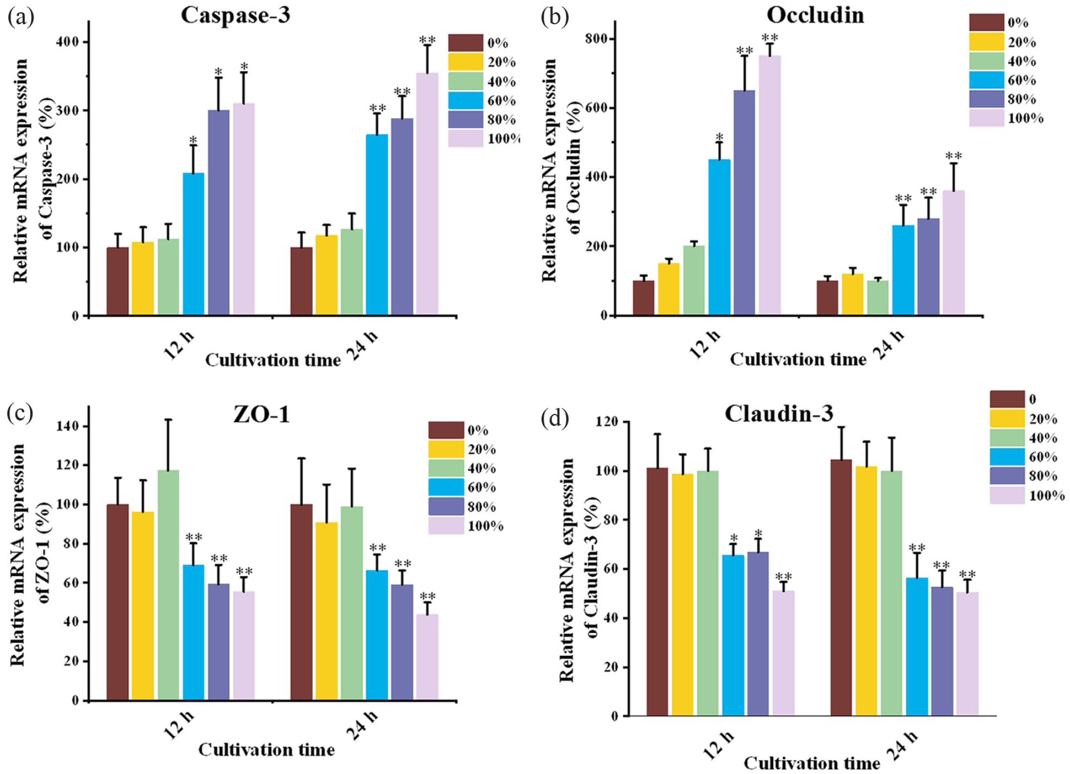
The relative mRNA levels of TJ-associated proteins in IEC-6 cells were cultured in the extraction media for 12 and 24 h: (a) Caspase-3, (b) ZO-1, (c) Occludin, and (d) Claudin-3. (GraphPad Prism 9 software was used for data processing. Values are expressed as the mean ± standard deviation or standard error as indicated. Differences between groups were evaluated using one-way analysis of variance. p < 0.05 was considered to indicate a statistically significant difference compared with 0% group. Similarly, **p < 0.01).

Western blotting assay of TJs protein expression: (a) TJ-associated proteins expression in 12 h group and (b) TJ-associated proteins expression in 24 h group.
The ratio of apoptotic cells with different concentrations Mg extracts group
We performed an apoptosis assay using flow cytometry to evaluate the effect of exposure to Mg extracts on the apoptosis of IEC-6 cells. Figure 4 presents that apoptotic IEC-6 cell percentage is significantly enhanced in high-concentration Mg extracts (60%, 80%, and 100%) than in low-concentration Mg extracts (0%, 20%, and 40%). We demonstrate that a 40% concentration of Mg extracts is a critical value because a higher concentration of Mg extracts results in a higher percentage of apoptotic IEC-6 cells.

The effect of different concentrations of Mg extracts on the apoptosis of IEC6 cells after 12 h (a) and 24 h (b) treatment. The corresponding statistical results of apoptotic rate after 12 h (c) and 24 h (d) treatment. (p < 0.05 was considered to indicate a statistically significant difference compared with the 0% group. Similarly, **p < 0.01).
Immunohistochemical analysis of caspase-3, ZO-1, occludin, and claudin-3
Immunohistochemical analysis revealed that HP-Mg pins up-regulated caspase-3 and claudin-3 expressions while down-regulated occludin and ZO-1 expression. Figure 5 indicates the IHC images of caspase-3, claudin-3, occludin, and ZO-1 expressions. The caspase-3 and occludin expression levels were improved after the co-culture with HP-Mg extracts, while claudin-3 and ZO-1 were suppressed.

Immunohistochemical analysis of caspase-3, claudin-3, occludin, and ZO-1 in surgery and mucosa areas.
Discussion
Mg and its alloys have excellent mechanical properties, good biocompatibility and low toxicity as a new medical implantable material. 21 Magnesium is an essential nutrient that plays an important role in nerve, muscle, bone, and heart function, as well as most metabolic processes in the human body, such as protein synthesis and enzyme activation. 22 Mg implant material can be developed for short-term or temporary implants due to its similar elastic modulus to humans (approximately 45 GPa) and degradability. 2 Magnesium deficiency easily decreases TJ-associated protein expression and affects cell barrier function. 7 The recovery of the intestinal barrier and absorption functions depends on healing intestinal tissue structure. Therefore, detecting the expression of intestinal function-related factors is critical for evaluating implant material biocompatibility.
Our previous study reported that the residual HP-Mg staples were 89.7% of their original weight 30 days after surgery, demonstrating that high-purity magnesium could gradually degrade in the rat’s intestine. Moreover, our study revealed the suppressive effect of high-purity Mg pins on the inflammatory response in rectal anastomoses. 23 Nevertheless, its effect on intestinal TJ barrier function should be evaluated accurately to promote the use of HP-Mg pins in intestinal tract reconstruction surgery. According to previous study results, we aim to ascertain the effect of differentiated HP-Mg extract concentration on TJ-associated protein expression in IEC-6 cells to shed light on its effect on intestinal integrity and provide a theoretical basis for the clinical use of high-purity Mg pins in anastomosis.
Claudins are involved in the maintenance of selective permeation of intercellular ions and solutes. Caspase-3, as the central role played in the cascade reaction of caspases, is one of the TJ-associated proteins in the gut, and is involved in cellular apoptosis. Therefore, claudins and caspase-3 are important indicators for evaluating intestinal barrier function.24,25 Occludin is a 65 kDa integral plasma membrane protein located at the tight junctions and involved in cytokine-induced mediation of TJs para-cellular permeability barrier, preventing macromolecule transport through the tight junction. 26 It is well-established that TJ-associated protein expression pattern correlates closely with intestinal barrier function. 27 An aberrant TJ-associated protein expression improves the odds of anastomotic leakage after gastrointestinal surgery. In addition to the influential factors of biocompatibility that might affect the healing after anastomosis, other factors, such as cytotoxicity, cell apoptosis and intestinal barrier function, are also vital to ensure the safety and efficacy of HP-Mg pins in clinical practice.
The present study demonstrates that the concentration of Mg ions about 1.7 mM is the threshold for TJ-associated protein expression and cellular apoptosis. When the concentration of Mg extracts was greater than 40%, the apoptosis rate of IEC-6 cells was enhanced with a higher concentration of Mg extract co-culture. A relatively higher concentration of Mg extracts affected TJ-associated protein and mRNA expressions. TJ-associated protein expressions contribute to maintaining the functional integrity of the intestinal mucosal barrier. A similar result was discovered in vivo that the concentration of Mg ions approximately 3.6 mM is beneficial to the intestinal barrier function. It is shown in immunohistochemical staining that expresses the differences between the groups near and far from Mg implants in TJs expression levels at different sites are obvious (Figure 5). Although individual proteins could not reliably obtain changes in the effect of Mg ion concentration on their expression, the effect of Mg ion on TJs in animal tissues was consistent with cytological data overall. It is also significant that cells are more prone to necrocytosis at low concentrations and for short periods of Mg induction. On the contrary, increasing concentration made cells more prone to apoptosis, which is consistent with the change in caspase-3 expression. This further suggests that the changes in caspase-3-mediated cell tight junctions will be in response to Mg ions.
Besides, previous researchers anticipated that Mg degradation in vivo would subsequently lead to extensive diffusion of magnesium ions into the surrounding tissues. 28 These suggest that HP-Mg has an important influence on anastomotic healing. Mg ion diffusion induces ECM accumulation and synthesis, stimulating anastomotic healing. 29 Magnesium plays a critical role in innate immune regulation that involves NF-κB activation and cytokine production. 30 Additionally, hydrogen gas, a product of magnesium degradation, can be used in treating inflammation, tumors and intestinal injury methods. 31 The gradually released hydrogen gas will be accumulated around the adjacent tissues, subsequently rendering a protective effect on the epithelial cells.
Conclusions
The present research demonstrates that Mg ion concentration is essential for intestinal barrier function after tissue healing. When the concentration of Mg extracts is above 1.72 ± 0.16 mM, it induces an aberrant TJ-associated protein expression and promotes cellular apoptosis. Therefore, using an appropriate corrosion rate of Mg pins should be considered to avoid the release of excessive Mg ions resulting in reduced intestinal barrier function.
Footnotes
Author contributions
Ting Shan did experiments and wrote the manuscript. Jun Yan revised the manuscript. Xiaonong Zhang provided the advice. Yigang Chen designed this study and provided the funding.
Declaration of conflicting interests
The author(s) declared no potential conflicts of interest with respect to the research, authorship, and/or publication of this article.
Funding
The author(s) disclosed receipt of the following financial support for the research, authorship, and/or publication of this article: This work was supported by the Double 100 Talent Project for young and middle-aged people (HB2020031) and Precision Medicine Projectof Wuxi Health Committee (J202109).