
Abstract
With limited availability of auto- and allografts, there is increasing demand for alternative bone repair and regeneration materials. Inspired by a mimetic approach, the utility of producing engineered native protein scaffolds is being increasingly realized, demonstrating the need for continued research in this field. In previous work, we detailed a process for producing mineralized collagen scaffolds using tendon to create collagen templates of highly aligned, natively crosslinked collagen fibrils. The process produced mineral phase closely matching that of native bone, and integration of mineral with the collagen template was demonstrated to be easily controlled, allowing scaffolds to be mechanically tuned. In the current study, we have extended this work to investigate how variation in the mineralization level of these scaffolds affects the osteogenic response of pre-osteoblastic cells. Scaffolds were produced under three treatment groups, where collagen templates underwent 0, 5, or 20 mineralization cycles. Scaffolds in each treatment group were cultured with MC3T3-E1 cells for 1, 7, or 14 days. Morphologic assessment under SEM indicated decreased attachment to the mineralized scaffolds, supported by DNA results showing a significant drop between culture days 1 and 7 for mineralized scaffolds only. For adherent cells, increasing scaffold mineralization also delayed cell spreading. While mineralization presented a barrier to cell coverage of scaffolds, it increased osteogenic activity, with cells on the mineralized scaffolds showing significantly greater alkaline phosphatase activity and osteocalcin production. Understanding how increasing collagen mineralization effects pre-osteoblast function may enable design of more advanced mineralized collagen scaffolds for bone repair and regeneration.
Introduction
Serving as the current gold standard bone repair material, autografts offer many advantages over alternative materials, including inherent biocompatibility and osteogenic potential.1,2 Availability of autograft tissue remains a strong limitation for its use however, along with donor site morbidities and extended time under anesthesia for tissue harvesting. While allografts are available in larger quantities, supply remains limited and concerns regarding disease transmission exist.1,3 Alternative materials, including various synthetic and natural biopolymers, are under widespread investigation to fulfill the increasing clinical demand for bone scaffolds and to avoid limitations associated with graft use. 4 With the potential to use these scaffolds for a wide variety of defect treatments that require different levels of load-bearing ability, methods for creating mechanically tailored scaffolds are required, and an understanding of how such changes impact cellular response developed.
Bone grafts for internal fixation may be indicated for use in some fractures to bridge bone ends and provide stability to the fracture site in an attempt to facilitate repair and regeneration. The specific healing processes that occur following surgical intervention are directed in part by the origin of bone graft material selected and bone type, that is, cortical or cancelous.5,6 Remodeling of autologous bone grafts proceeds similarly to normal physiological bone remodeling due to the presence of preserved mesenchymal stem cells (MSC) and osteogenic proteins.7–9 This process begins by apposition of bone, including initial osteoclast resorption followed by graft vascularization and fibroblast ingrowth.5,6 Osteoblasts fill resorption pits created by the osteoclasts with osteoid, which eventually becomes mineralized and subsequently remodeled.5,10 Complete resorption and replacement of the autograft with new bone typically occurs within 6–12 months but may take years if the graft is derived from dense cortical bone.6,11 A subset of autograft tissue includes bone marrow aspirate (BMA). BMA contains many similar osteogenic factors as bone autografts and is often used along with other graft materials as an extender. 8
Grafts foreign to the recipient host include xeno- and allografts, including demineralized bone matrix, which is available in fresh, frozen, or freeze-dried forms. 8 Due to the foreign nature of these grafts and depending on processing techniques, the initial stages of remodeling may involve undesirable immune responses. 11 As a result, integration of allografts or xenografts may be delayed in comparison to autografts. 12 For example, Zhang et al. 13 compared the use of autograft to allograft for instrumented atlantoaxial fusions in 32 pediatric patients. Although similar fusion rates were observed with both grafting materials, fusion time was 3 months longer when allografts were used. 13 Interestingly, the overall incidence of surgery-related complications was significantly higher for the autograft group, including a seroma formation and pelvic fracture associated with the donor site. 13 In a comparative study by Shibuya et al. 14 of 61 foot and ankle surgery patients, xenograft incorporation into human bone was found to be slower than incorporation of autografts and allografts. For xenografts, while supply is significantly less constrained compared to allografts, processing methods, including decellularization and sterilization techniques vary greatly, which may result in variable clinical outcomes.5,15
Materials being explored as graft substitutes include various synthetic and natural polymers, bioactive glasses, minerals, ceramics, and many composite preparations thereof. 8 Cellular responses to these graft substitutes vary depending on their chemical composition, structure, and mechanics. 16 While some graft alternatives have been shown to allow progressive healing similar to that occurring with graft use, healing typically progresses more slowly due to a general lack of osteoinductive potential. 11 To accelerate repair and remodeling, various biological factors have been incorporated into graft substitutes. These include bone morphogenetic proteins (BMP), fibroblast growth factors (FGF), vascular endothelial growth factors (VEGF), platelet-rich plasma (PRP), and many bioinorganic ions such as zinc, magnesium, cobalt, and strontium.11,17 Biologic incorporation within graft substitutes may improve the rate of bone formation and remodeling, however their use comes with increased cost and possible post-operative complications, including heterotopic bone growth and oncogenic concerns.8,11,17,18
Aside from collagen, the extracellular matrix (ECM) of bone contains various proteins involved in important osteogenic processes; more than 200 of which have been reported to date. 1 Some of the major non-collagenous proteins (NCP) found within bone include osteonectin, osteocalcin (OCN), fibronectin, osteopontin, and bone sialoprotein.1,19 Of these, OCN in particular has been widely used as a late stage biomarker for osteogenic differentiation. 1 OCN is thought to bind bone mineral with its N-terminal domain, contributing toward the rate of mineral crystal growth or maturation, while aiding in the regulation of bone matrix production via its C-terminal domain.20,21 Another widely used marker for osteogenic differentiation is alkaline phosphatase (ALP), otherwise known as tissue non-specific alkaline phosphatase (TNAP). 22 ALP is expressed early on in the development of bone and decreases as development progresses. 22 ALP is thought to play a role in promoting collagen mineralization through its hydrolysis of pyrophosphate (PPi), a mineralization inhibitor. 23 Additionally, NCPs and various ions and small molecules, such as magnesium and citrate, are thought to alter crystallization kinetics and promote the intrafibrillar mineralization of collagen during bone formation and repair. 24 To mimic the role of these NCPs, polyanionic macromolecules of varying molecular weights (MW) are frequently used during mineralization, including poly(acrylic acid) and poly(aspartic acid) (pAsp).4,24
Our laboratory recently developed a process for producing mineralized collagen scaffolds that feature highly aligned and natively crosslinked collagen fibrils combined with a mineral phase closely matching that of native bone. 25 We subsequently demonstrated that the mineral content of these scaffolds can be easily controlled during production, allowing tuning of a scaffold’s resulting mechanical properties, both in terms of macroscale flexural modulus and nanoscale collagen fibril stiffness. 26 In the present study, we make use of our developed scaffold production process to investigate how variations in the mineral content of aligned, natively crosslinked collagen scaffolds affect the osteogenic activity of cells. Our results provide new insights into how variations in scaffold mineralization may alter the functional response of osteoblasts.
Materials and methods
An overview of the experimental design is shown in Figure 1.

Overview of the experimental design. Collagen sheets underwent three different treatments (0, 5, or 20 mineralization cycles) with samples from each treatment cultured for three durations (1, 7, or 14 days) yielding nine sample groups for analysis.
Collagen acquisition and initial processing
Common digital extensor (CDE) tendons were dissected from the forelimbs of steers aged 2–3 years, which were killed for food at a local abattoir. Tendons were stored immediately in phosphate-buffered saline (PBS) solution at
Decellularization was conducted following a method previously described by Woods and Gratzer.
27
Briefly, tendons were first exposed to a hypotonic 10 mM tris buffer, containing serine and metalloprotease inhibitors, along with 1% penicillin/streptomycin and 1% amphotericin B for 36 h at room temperature. Tendons were then moved to a high saline 50 mM tris buffer containing 1% Triton X-100 along with antibiotics and protease inhibitors for 48 h at room temperature, followed by Hanks’ buffer rinse and wash in a DNase/RNase solution. This was followed by a 48 h, room temperature soak in 1% Triton X-100 solution with 50 mM TRIZMA base and antibiotics. Finally, the tendon samples were rinsed in PBS with antibiotics and stored in a similar solution at
Decellularized tendons were cut into 2-cm-long segments, mounted on a steel block using optimal cutting temperature compound (Fisher Healthcare, Ottawa, ON), frozen in liquid nitrogen, and cryosectioned longitudinally using a Leica SM2000R sliding microtome to produce 200-µ
Alternate soaking mineralization
Mineralization of the phosphorylated collagen sheets was performed following the method of Grue and Veres.
25
Using a stainless steel mesh basket, sheets were first soaked in 200 mL ddH2O before soaking in 200 mL of a magnesium (0.01 M) and citrate (0.02 M) doped calcium solution (CaCl2/MgCl2, 0.2 M) within a temperature-controlled water bath at
Cell culture
MC3T3-E1 osteoblast precursor cells derived from mouse calvaria (Sigma-Aldrich, MO, USA) were cultured in
Visualization of cell morphology on scaffolds
Two scaffolds from each treatment group (0, 5, and 20 mineralization cycles) were examined under SEM following 1, 7, and 14 days of culture. Scaffolds were removed from the culture plates and transferred to fresh 24-well plates and fixed in 2.5% electron microscopy-grade glutaraldehyde in PBS for 1 h at room temperature under constant agitation, followed by rinsing in ddH2O and dehydration in graded ethanol. Scaffolds were then critical point dried, mounted on SEM stubs using carbon tape, coated with gold-palladium, and examined at 5 kV, 10
Sample groups for biochemical analyses
Biochemical analyses for dsDNA content, ALP activity, and OCN content were undertaken. For each of the three culture durations (1, 7, and 14 days), the tests were performed on n = 6 scaffolds from each treatment group (0, 5, and 20 mineralization cycles). In addition to these 54 scaffolds, an additional six scaffolds per treatment group were prepared and cultured for 14 days without the addition of AA. As a no-cell control, analyses were also performed on a single scaffold for each of the nine treatment/culture duration combinations that was prepared and maintained under the same conditions, but in the absence of cells. For each scaffold, biochemical measurements were made in duplicate and then averaged.
Cell proliferation
DNA, measured via Quant-IT™ PicoGreen® dsDNA assay kit (Invitrogen-Thermo Fisher Scientific, Berlin, Germany) was used as a proxy for the number of cells present on scaffolds.31,32 At days 1, 7, and 14, scaffolds were transferred to a new 24-well plate, washed with sterile PBS, and incubated in TrypLE Express reagent (Invitrogen-Thermo Fisher Scientific, Berlin, Germany) for 30 min. Cell-TrypLE suspension was drawn off and centrifuged at 3000 rpm for 15 min followed by resuspension of cells in sterile PBS. The cells were then lysed through three freeze-thaw cycles followed by further centrifugation at 3000 rpm for 15 min to remove cellular debris. From the supernatant, 50
Cellular dsDNA measurement was performed according to the kit manufacturer’s instructions. Briefly, lambda DNA standards were prepared by eight-point serial dilution of a 2
ALP activity
To evaluate early osteogenic differentiation of the MC3T3-E1 cells, ALP activity was assessed using an alkaline phosphatase, diethanolamine detection kit (Sigma-Aldrich, MO, USA).
Cells lysates were used from the previously described proliferation assay. ALP measurements were performed according to manufacturer’s instructions. Briefly, 5
OCN content
OCN content was measured from the cell lysate using a mouse osteocalcin ELISA kit (MyBioSource, CA, USA). Briefly, 100
Statistics
Numerical data are presented as mean ± SD. DNA, ALP, and OCN results were analyzed using full-factorial two-way ANOVA, performed on rank transformed data to improve normality. This was followed by nonparametric Kruskal-Wallis tests and then Wilcoxon tests between individual pairs. Differences with p ≤ 0.05 were considered statistically significant.
Results
Cell morphology and scaffold ultrastructure
The response of MC3T3-E1 pre-osteoblast cells to culture on aligned collagen fibril scaffolds following 0, 5, or 20 alternate soaking mineralization cycles was assessed using SEM (Figure 2). Cells on the unmineralized scaffolds exhibited mostly unidirectional cytoplasmic extension well-aligned with the direction of collagen fibrils after 1 day of culture (Figure 2(a)). By day 7, the elongated cells had started to spread laterally (Figure 2(b)). By 14 days, the cells had become confluent (Figure 2(c)).

SEM images of MC3T3-E1 cells cultured on collagen scaffolds of varying mineral content, achieved via 0, 5, or 20 cycles of alternate soaking mineralization: (a–c) on unmineralized collagen scaffolds, cells had elongated by day 1, had begun to spread at day 7, and had reached confluence at day 14, (d–f) on scaffolds treated with five mineralization cycles, cells elongated and spread, but spreading did not occur to the same extent as in controls, and (g–i) on scaffolds treated with 20 mineralization cycles, cells showed little spreading after 14 days.
Cells on the five cycle mineralized scaffolds exhibited similar unidirectional cytoplasmic extension after 1 day of culturing, however the direction of elongation was more random due to the partial coverage of underlying collagen fibrils by mineral (Figure 2(d)). After 7 days of culture, cells appeared more elongated and fusiform compared to cells on the unmineralized scaffolds (Figure 2(e) vs (b)). By 14 days, the cells had spread laterally taking on a polygonal morphology (Figure 2(f)) appearing similar to those on the unmineralized scaffolds after 7 days of culture (Figure 2(b)).
Cells grown on the 20 cycle mineralized scaffolds exhibited similar elongation patterns as cells grown on the five cycle scaffolds after 1 day of culture but with cellular extensions more commonly observed (Figures 2(g), 3(a), and (d)). By day 7, cells began to spread laterally, similar to what was observed on the five cycle mineralized scaffolds, with a small number of cells taking on a polygonal morphology (Figures 2(h) and 3(e)). At 14 days, cell appearance had not changed appreciably compared to at 7 days (Figures 2(i) and 3). Cells grown on the mineralized scaffolds exhibited close association with the underlying mineral phase at all timepoints, with cells gradually becoming more embedded and integrated with the mineral as culture time increased (Figure 3).

SEM images of MC3T3-E1 cells cultured on mineralized collagen scaffolds: (a) five cycle mineralized collagen scaffold showing close association between cells and mineral after 1 day of culture, (b) five cycle mineralized scaffold after 7 days of culture showing cells partially covered by mineral, (c) five cycle mineralized scaffold after 14 days of culture, (d) 20 cycle mineralized collagen scaffold showing similar cell-mineral interactions following 1 day of culture, (e) 20 cycle mineralized scaffold after 7 days of culture, and (f) after 14 days of culture, some cells on scaffolds from the 20 mineralization cycle group were almost entirely covered by mineral.
Observed at all timepoints, cells on both the control and mineralized scaffolds possessed microvilli on their surfaces along with evidence of ECM production after 7 days (Figure 4), indicating increased activity during the culturing period. 33
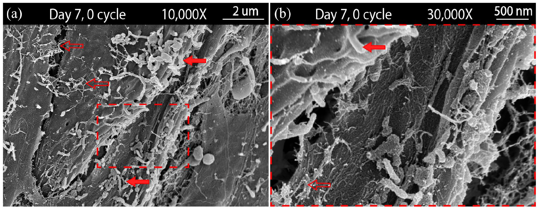
SEM images of MC3T3-E1 cells cultured on an unmineralized collagen scaffold for 7 days: (a) cells showing surface microvilli (solid red arrows) and evidence of ECM deposition (hollow red arrows) and (b) magnified region of (a). Both features, microvilli and ECM deposition, were also seen for the mineralized scaffolds.
Cell proliferation
On unmineralized scaffolds, DNA content did not change over 14 days of culture (Figure 5(a)). With SEM observations having shown monolayer coverage of the unmineralized scaffolds by 14 days (Figure 2(c)), the DNA and SEM results together suggest that surface coverage was largely achieved via cell spreading over the 14 days culture duration.

(a) DNA measurements indicated that cell numbers remained consistent on unmineralized (0 cycle) scaffolds, while initially decreasing and then remaining constant for mineralized scaffolds (5 and 20 cycle), (b) ALP activity normalized to DNA concentration showed increases for the mineralized scaffolds only, with a greater increase after 14 days of culture for the 20 cycle group compared to the 5 cycle group, and (c) OCN content normalized to DNA concentration similarly showed increases only for the cells cultured on mineralized scaffolds. After 14 days of culture, levels of OCN for the 20 cycle group nearly reached significance over the 5 cycle group with p = 0.0552. Bars represent differences between treatment groups for a given culture time while letters represent differences between culture times for a given treatment group.
On mineralized scaffolds, DNA content dropped significantly within the first 7 days of culture, and then remained unchanged to 14 days (Figure 5(a)). This was consistent with SEM observations, showing a sparsity of cells after 7 days of culture on the 5 and 20 cycle mineralized scaffolds compared to the unmineralized scaffolds (Figure 2(e), (h) vs (b)).
Cell differentiation
The potential for differentiation of the MC3T3-E1 pre-osteoblast cells on both unmineralized and mineralized collagen scaffolds was assessed by measuring ALP activity and OCN production.
ALP enzyme activity is an early biomarker of osteoblast differentiation.34,35 On unmineralized scaffolds, ALP activity did not change over the 14 days of culture (Figure 5(b)). On mineralized scaffolds, ALP activity increased significantly for both the 5 and 20 cycle groups within the first 7 days of culture (Figure 5(b)). While ALP activity did not change between 7 and 14 days of culture for the five cycle group, a significant increase was observed for the 20 cycle group (Figure 5(b)).
To ensure that the freeze-thaw cycling used to lyse cells didn’t negatively impact the ability to measure ALP activity, a test was done comparing the kit-provided enzyme control to the same enzyme control after undergoing 3X freeze-thaw cycles. No difference was found between ALP activity of the fresh enzyme controls versus those that experienced freeze-thaw cycling (p = 0.9889; n = 9).
OCN production is thought to occur during the later stages of osteoblast differentiation. 35 On unmineralized scaffolds, OCN content did not increase during the 14 days of culture (Figure 5(c)). OCN content increased significantly for the five cycle group within 7 days of culture followed by a decrease when measured at day 14 (Figure 5(c)). For the 20 cycle group, OCN content was unchanged after 7 days of culture, but increased significantly after 14 days of culture, nearly reaching significance over the five cycle group (Figure 5(c)).
Discussion
Visualization of cell morphology on scaffolds
Cell morphology is closely tied to a wide variety of cellular processes, including cell growth, cytoskeletal organization, cell differentiation, and gene expression. 36 For example, MSCs possessing a flattened and well-spread morphology are more likely to differentiate into osteoblasts and contribute to osteogenesis compared to cells more rounded in appearance, which are thought to become adipocytes.37–39 This has been shown by Somaiah et al. 39 where MSCs were cultured on either uncoated tissue culture treated plastic or type I collagen coated tissue culture plates. Cells grown on the uncoated plastic were found to exhibit high levels of adipogenic differentiation, while the collagen coated surfaces enhanced cell proliferation and spreading, and promoted osteogenic differentiation. 39
In the current study, cells cultured on unmineralized collagen fibril scaffolds exhibited a flattened morphology and gradual cell spreading over time, with neighboring cells coming into contact and forming a confluent monolayer within 14 days (Figure 2(a)–(c)). Cells grown on the mineralized scaffolds showed a more elongated morphology along with overall lesser cell density (Figure 2(d)–(i)). Cells cultured on the 5 cycle scaffolds appeared more polygonal-like after 14 days of culture compared to cells on the 20 cycle scaffolds (Figures 2(f), (i), and 3). Cells also appeared more elongated and spindle-like on the 20 cycle scaffolds as compared to the 5 cycle and unmineralized groups (Figure 2). This resultant cell morphology may indicate less of an osteoblastic phenotype or may be a result of the highly mineralized surface of these scaffolds which may have required the cells to spread from mineral peak-to-peak, bridging topographical gaps or pores, resulting in a “stretched out” morphology. Cells were also observed within pores of the 20 cycle scaffolds with some extending under mineral clusters (Figure 3(d)–(f)).
These morphological results, including cell flattening and spreading, are similar to those found by Cheng et al. 36 who cultured MC3T3-E1 cells on poly-dopamine functionalized graphene oxide substrates. Following 7 days of culturing, cells appeared flattened and well-spread, a trend similarly found in our study for cells grown on 0 and 5 cycle scaffolds and 20 cycle scaffolds to some extent. The authors also reported the resultant morphology of the MC3T3-E1 cells to be a product of the underlying surface morphology and structure. 36 For example, cells grown on bare glass exhibited reduced attachment, spreading, growth, and fewer microvilli and pseudopods than cells grown on the graphene oxide substrates. 36 This phenomenon of cells morphologically responding to underlying substrates was found in our study where cells favored an initial elongated morphology in the direction of underlying collagen fibrils on the unmineralized scaffolds (Figure 2(a)).
Cellular production of ECM was observed to begin on all scaffolds following 7 days of culture (Figure 4). This observation resembles what was found by Quarles et al. 40 where extracellular matrix deposition by MC3T3-E1 cells was found to peak after 7 days of culture. In addition to ECM production, cells appeared to possess numerous microvilli on their surfaces (Figure 4), pointing to heightened activity. 33 These findings are similar to what was observed by Cheng et al., 36 where an increase in microvilli and ECM formation occurred following 7 days of culturing MC3T3-E1 cells.
Cell proliferation and differentiation
dsDNA content was found to remain stable throughout the 14 days of cell culturing on the unmineralized collagen scaffolds (Figure 5(a)). This is similar to what was found by Nijsure et al., 41 where the number of human osteosarcoma cells remained constant on unmineralized aligned-collagen scaffolds over 7 days of culturing. dsDNA content for mineralized scaffold groups was observed to decline after 7 days of culture (Figure 5(a)). The increased relative cellular proliferation associated with unmineralized scaffolds compared to mineralized scaffolds may be due in part to the increased protein surface area available for cell adhesion. Pre-osteoblasts express the ability to adhere to collagen through RGD-dependent integrin binding. 42 This binding affinity is likely reduced when collagen fibrils are covered with mineral, which may result in a relative increase in cellular proliferation on unmineralized collagen scaffolds. In addition to this, osteogenic differentiation was observed to be greater for cells grown on the mineralized scaffolds compared to cells cultured on the unmineralized control scaffolds (described below) (Figure 5(a) and (b)). It is known that the proliferation of MC3T3-E1 cells decreases during periods of differentiation, thus possibly accounting in-part for the lack of proliferation on mineralized scaffolds. 40 The decrease in dsDNA content observed for mineralized scaffold groups following 7 days of culture may indicate a degree of cell detachment or death on these scaffolds compared to the stationary dsDNA levels found on unmineralized scaffolds (Figure 5(a)).
ALP is considered a prominent biomarker of early-stage bone formation.35,43 In bone, ALP is a homodimeric glycoprotein enzyme thought to act as an inhibitor to the inhibitor of hydroxyapatite formation and growth, PPi, through its ability to cleave PPi into inorganic phosphate.43,44 ALP contains a calcium-binding domain site in addition to a region capable of binding matrix proteins, including type I collagen, and is found associated with the membranes of osteoblast secreted matrix vesicles in bone.43,45
After 1 day of culturing, ALP activity was similar for all treatment groups (0, 5, and 20 cycles) (Figure 5(b)). After 7 days of culture, cells on mineralized scaffolds exhibited significantly greater ALP activity than cells grown on unmineralized scaffolds; a trend that continued after 14 days of culturing (Figure 5(b)). This increase in ALP activity observed for cells cultured only on mineralized collagen scaffolds could indicate increased early stage osteogenic differentiation for these cells. 35 This trend is consistent with results found by Xu et al. 46 where MSCs expressed increasing levels of ALP activity over 14 days of culturing, beginning on day 7 when grown on bioglass-collagen scaffolds. Further, Ngiam et al. 47 compared the ALP activity of osteoblasts when cultured on either a PLGA/collagen scaffold or a mineralized PLGA/collagen scaffold. After 7 days of culturing, ALP activity was significantly greater for cells grown on the mineralized scaffolds compared to the unmineralized scaffolds. 47
OCN is one of the most abundant NCPs found within bone.48,49 Produced mainly by osteoblasts, OCN is a 5.6 kDa secreted protein that in its carboxylated form is one of the two main
OCN content for cells cultured on the unmineralized scaffolds remained low for the 14 days of culture, while significant increases occurred for the mineralized scaffolds (Figure 5(c)). In addition to the previously mentioned ALP results, increases in OCN content indicate that mineralized scaffolds better promoted osteogenic differentiation. Similar findings were reported by Chou et al. 53 who compared MC3T3-E1 growth and osteoblastic differentiation on PLGA and apatite-coated PLGA scaffolds. In that work OCN content increased for the mineral coated group compared to the uncoated group after 4 weeks of culture. 53 In another study by Lee et al., 54 the effect that mineralization had on the osteogenic activity of collagen-glycosaminoglycan scaffolds was evaluated using MSCs. It was found that mineralization of the scaffolds increased the expression of OCN after 14 days of culture, suggesting that mineralization may help cue cellular osteogenic differentiation. 54 Beyond the 14 days of culture tested in the current study, how cells might continue to respond to the scaffolds following osteoblastic differentiation remains to be determined, as does scaffold susceptibility to dissolution at longer time points and its effect on cellular response through ion release and changes in substrate mechanics.
The mechanical properties of substrate materials are critical factors in defining the resultant morphology and differentiation of adherent cells. 55 For example, Khatiwala et al. 56 observed increased proliferation, migration, and eventual osteoblastic differentiation of pre-osteoblast cells with increasing stiffness of collagenous hydrogels. From our previous work, progressive mineralization of collagen fibril templates by alternate soaking was shown to increase individual collagen fibril stiffness and macroscale scaffold flexural modulus in proportion to the number of mineralization cycles complete. 26 In addition to differences in cellular response that may have resulted from topographical differences between mineralized scaffolds, the current study may indicate that the increase in scaffold mechanics moving from 5 to 20 mineralization cycles is sufficient to enhance osteogenic differentiation of pre-osteoblasts, as measured by ALP activity and OCN content.
As is also demonstrated by the current work, there is a disadvantage to increased scaffold mineralization, with occlusion of collagen fibril surfaces by mineral appearing to significantly reduce cell adhesion. Considering how best to design future mineralized collagen scaffolds given these disparate effects of increasing mineralization may lead to the creation of new scaffolds with enhanced repair and regeneration potential.
Footnotes
Declaration of conflicting interests
The author(s) declared no potential conflicts of interest with respect to the research, authorship, and/or publication of this article.
Funding
The author(s) disclosed receipt of the following financial support for the research, authorship, and/or publication of this article: This work was funded by a grant to SPV from the Natural Sciences and Engineering Research Council of Canada (NSERC). BG thanks Research Nova Scotia (RNS) for providing graduate stipend support. We acknowledge the support of the Canada Foundation for Innovation, the Atlantic Innovation Fund, and other partners which fund the Facilities for Materials Characterization, managed by the Clean Technologies Research Institute, Dalhousie University.