
Abstract
Purpose:
This study aims to evaluate the antibacterial properties of 304 Cu-bearing stainless steel (SS) with different Cu contents (0, 2.5, 4.5 wt.%) against oral biofilms of Streptococcus mutans (S. mutans), Streptococcus sanguinis (S. sanguinis), and their mixture.
Methods:
Bacterial biofilms on the surface of 304-Cu SS were characterized by plate counting, 4′, 6-diamidino-2-phenylindole (DAPI) staining with aid of sanning electron microscopy (SEM) and 2, 3-bis (2-methoxy-4-nitro-5-sulfophenyl)-2H-tetrazolium-5-carboxanilide inner salt (XTT). In addition, the inhibition zone method was also employed to evaluate the antibacterial properties of 304-Cu SS. Cell Counting Kit-8 (CCK-8) and flow cytometry were used to assess the cytotoxicity and apoptosis rate of 304-Cu SS, respectively.
Results:
304-4.5Cu SS could effectively inhibit the attachment, formation, activity, and metabolism of bacterial biofilm, possessing the best antibacterial properties exceeding 99.9% of antibacterial rate against S. mutans, S. sanguinis, and their mixture. The diameters of inhibition zones to S. mutans and S. sanguinis on the surface of 304-4.5Cu SS were 21.7 and 14.7 mm, respectively. The results of cell experiments in vitro showed that both 304-2.5Cu SS and 304-4.5Cu SS had no evident cytotoxicity with an identical grade 1. The apoptosis rate exhibited a gradually increased tendency with increase of the Cu content in 304 SS.
Conclusions:
304-4.5Cu SS without cytotoxic effect on NIH3T3 cells has obvious antibacterial activity against S. mutans, S. sanguinis and their mixture.
Clinical significance:
The Cu-bearing stainless steel provides a new solution to be used as oral orthodontic devices for inhibiting oral microflora imbalance and enamel demineralization.
Introduction
Fixed orthodontic appliances can cause a series of side effects, including white spot lesions (WSLs) and caries, which adversely affect the esthetics and health of teeth.1–3 In addition, about 50%–80% of orthodontic treated patients suffer from varying degree of tooth demineralization, 4 this is because, the fixed appliance in the mouth increases the difficulty for cleaning teeth and the plaque accumulation due to the formation of acid producing bacteria, accelerates the demineralization process, and destroys oral flora balance in turn. 5 Previous studies confirmed that the existence of S. mutans during orthodontic treatment obviously increased caries incidence.6,7 While Streptococcus sanguinis is another indigenous species in the dental plaque, which plays a key role in delaying the colonization of cariogenic S. mutans in the oral cavity of infants.8–10
Based on the above background, if a medical stainless steel (SS) with excellent antibacterial function can be used to decrease the proliferation of oral pathogenic bacteria, it will provide a new solution for inhibiting the oral microflora imbalance and the enamel demineralization. Cu-bearing antibacterial SS as one of major antibacterial SS has obtained the maximum extent of development and application.11–14 Cu is an important alloy element applied in metal materials. Its main antibacterial principle is attributed to the continuous release of trace amount of Cu ions from the steel surface. In consequence, this destroys the bacterial cytoderm, cell membrane and generates generous reactive oxygen species (ROS), resulting in bacterial damage and death at last.15–17 At the same time, the effect of Cu on the cytotoxicity was directly related to the concentration of Cu2+. 18 Trace amount of Cu is essential for human to maintain healthy cell function, 19 while excessive Cu can be accompanied by liver and nervous system diseases. 20 Therefore, whether the antibacterial SS with different Cu contents would affect its biological safety was also a vital issue in this study.
Oral biofilm, a structured community which is formed by microbes in adversity in order to improve the resistance to nutritional deficiency and microbial inhibitors. 21 It is noteworthy that a single-species biofilm model is a universal research method to simulate real oral plaque biofilm, although it still lacks enough accuracy.22–26 For the Cu-bearing antibacterial SS, previous works11–14 were mainly focused on studying the biofilm of single kind of bacteria. Zhao et al. 27 showed that 304-Cu stainless steel had a good antibacterial effect on a single Streptococcus mutans, but whether it had the same effect on oral mixed bacteria is not clear in the past. Compared with single-species biofilm, dual-species biofilms have stronger resistance to antibacterial agents. 25 Therefore, in this study, single S. mutans, S. sanguinis, and their mixture were used as the research models to simulate the oral bacterial environment, with aim to explore the antibacterial effect of 304-Cu SS with different Cu content in oral bacterial environment.
Experimental
Sample preparation
Elemental compositions of the investigated 304 SS and 304-Cu SS are listed in Table 1. Thereinto, 304-Cu SS was prepared by metal powder injection molding (MIM). 304-Cu SS powders was purchased from Anhui YingYuan New Material Technology Co., Ltd. This material with an average particle size of 30 μm was made by smelting 304 SS and Cu in a vacuum induction melting furnace to prepare an ingot, and then making it into a powder in a Vacuum atomization powder making equipment (Renmei Metal Material, Tianjin, China). 304-Cu SS powders were evenly mixed with adhesive in a volume ratio of 60:40, and the mixture was used as the feedstock for the MIM process. The prepared feedstock was injection molded using a micro-injection molding machine (Babyplast 610P, Barcelona, Spain). After molding, samples were immersed in a hexane bath at 50°C for 20 h to remove the paraffin wax from the binder. Then certain sequence of degreasing was carried out in a vacuum induction melting furnace at 220°C, 350°C, 420°C, 620°C, 900°C for 1 h, respectively, and final sinter at 1350°C for 1 h. The 304 SS used as a control with nominal composition of Fe-18Cr-9Ni wt.% was purchased from Taiyuan Stainless Steel Co., Ltd., China. Both 304 SS and 304-Cu SS were cut into pieces with diameter of 10 mm and thickness of 2 mm, followed by mechanically grinding to 2000 mesh with SiC sandpapers. Afterward, the samples were polished and ultrasonically cleaned in ethanol, and finally disinfected by ultraviolet light prior to experiments.
Elemental compositions of experimental steels, wt.%.

Bacterial culture
S. mutans ATCC 25175 and S. sanguinis ATCC 10556 provided by the Laboratory Center, China Medical University, Shenyang, China, were cultured on Brain Heart Infusion broth (BHI, Oxoid, Basingstoke, UK) agar plates at 37°C under an anaerobic condition (80% N2, 10% CO2, 10% H2) for 24 h, respectively. Afterward, the concentrations of S. mutans, S. sanguinis, and their mixture with same volume ratio were adjusted to 108 CFU/mL with sterile PBS buffer or BHI culture solution. A series of single-species and dual-species bacterial suspensions with concentrations of 103–108 CFU/mL were respectively prepared for the following experiments.
Plate count method
According to the ISO 10993-5:2009 standard, a contacting test was conducted to evaluate the antibacterial and biofilm resistance of 304-Cu SS. 28 50 μL of above-mentioned bacterial suspension with a bacterial concentration of 108 CFU/mL was dropped on the surfaces of samples placed in a 24-well plate. After culturing samples under the above microaerobic condition at 37°C for 24 h, the simples with bacterial suspension were shaken with 1.6 mL sterile PBS. Afterward, 0.1 mL of the washed liquid was streaked on a BHI agar plate. Then plates were incubated in a microaerobic environment at 37°C for 48 h before counting bacterial colonies. Antibacterial rate was calculated according to the following formula:

where C is the antibacterial rate, A is the average bacterial amount on the 304 SS, and B the average bacterial amount on 304-Cu SS.
Scanning electron microscopy (SEM)
After co-culturing with 304-Cu SS and 304 SS samples under the above mentioned microaerobic condition at 37°C for 24 h, the samples were gently rinsed by the buffer for three times, and then glutaraldehyde (2.5% v/v) was used to fix the biofilms at 4°C for 4 h. Next, the ethanol with different concentrations of 50%, 60%, 70%, 80%, 90%, 95%, and 100% was used to dehydrate the samples for 10 min, respectively. To improve the definition of scanning electron microscope (SEM, Phillips XL30FEG, Holland), the surfaces of samples were sputter-coated with gold. The biofilm morphology was observed in high vacuum mode at a voltage of 12 kV.
DAPI staining
The DAPI staining method was used to observe the activity of bacterial biofilm on the surface of the sample in this study. The samples were cultured with bacteria in a 24-well culture plate, and then 1 μL of DAPI (Sigma-Aldrich Co., MO, U.S.A.) with a concentration of 5 mmol L−1 was added to each well. The samples were cultured for 30 min at room temperature in the dark, and then rinsed three times with PBS. The biofilm activity on the sample surface was examined under a laser confocal scanning microscope (CLSM, Olympus FV10-ASW, Japan), and the images were obtained with a ×20 objective lens. The images of the biofilms on the 304 SS and 304-Cu SS coupons were analyzed using Image J software (National Institutes of Health, Bethesda, MD, USA) to count the sessile cells.
XTT reduction assay
XTT reduction assay was used to detect the metabolic activity of bacteria on the sample surface in this study. XTT sodium salt (Beijing Solarbio Science & Technology Co., Ltd. China) was prepared in ultrapure water at a final concentration of 1 mg mL−1. Menadione (Sigma, MO, USA) solution as an electron coupling agent was prepared in acetone with a concentration of 0.4 mmol/L. After 48 h of biofilm formation, XTT/ menadione solution was prepared to make the final concentration of menadione (1 mmol/L). 1 mL XTT/menadione solution was added to each well of 24-well plate, respectively, and incubated in the dark for 5 h. Afterward, 100 μL sample solution of per well was added to 96-well plates, respectively. Among these, the absorbance was measured at 490 nm by a microplate reader.
Inhibition zone
The hot BHI agar solution was spread evenly on petri dishes with the diameter of 90 mm, which was cooled down to room temperature under a sterile condition. Then 500 μL of bacterial suspension with a concentration of 106 CFU/mL was sprayed over the dish. The sterile 304-Cu SS samples, 304 SS samples, and filter paper were separately placed in petri dishes according to the National Standard of China (GB/T 2738-2005). The petri dishes were incubated under an anaerobic condition for 18 h. The antibacterial activity was accessed by the diameter of the inhibition zone around the sample. The larger diameter value represented a higher antibacterial property.
Cytotoxicity test
According to ISO 10993-12 standard, the leaching solutions of 304-Cu SS and 304 SS samples in high glucose DMEM medium with 10% fetal bovine serum were prepared in a 6-well plate in a humidified atmosphere of 5% CO2 at 37°C for 72 h, and the superficial area of samples in the soaking solution followed a fixed ratio of 3 cm2/mL. The control group was a DMEM medium as the negative control. Then 10 μL of leaching solution was extracted and added onto a 96-well electronic plate (E-Plate View 96; ACEA Biosciences Inc., San Diego, CA, U.S.A.) to be incubated with mouse NIH3T3 fibroblasts for 1, 3, and 5 days, respectively. Cell viability was evaluated using CCK-8 method according to the protocol of manufacturer. At each time point, 20 μL of CCK-8 solution was added to each well with the above incubated medium. Optical density (OD) was evaluated using a spectrophotometer at 450 nm, and the OD value was recorded to quantify the cell proliferation rate. The cell survival rate was expressed by the relative growth rate (RGR) expressed as the following formula:

Flow cytometry
To evaluate the apoptosis rates of NIH3T3 fibroblasts cultured with sample extracts, flow cytometry was performed after Annexin V-FITC/PI double staining. The cells were placed onto 6-well plates at a density of 1 × 10⁵ cells/well, which was incubated in a humidified atmosphere of 5% CO2 at 37°C for 24 h. Afterward, the medium was replaced by an equal volume of extraction medium for 48 h. The adherent cells were trypsinized and then superfluous trypsase was washed off with PBS. The cells were analyzed by the flow cytometry after the staining with Annexin V-FITC/PI double staining kit (Roche) according to the manufacturer’s instruction. All the flow cytometry data were analyzed using the FACSDiva software (BD Biosciences).
Statistical analysis
SPSS17.0 statistical software was used to analyze the experimental data, and DAPI staining, XTT reduction assay, the OD value and apoptosis rate were statistically analyzed by ANOVA and Tukey’s Honest Significant Difference (HSD) test. The level of statistical significance was set to 0.05 (p < 0.05).
Results
Antibacterial efficacy
Figure 1 shows bacterial colonies exposed on the surfaces of 304-2.5Cu SS (Figure 1(a), (d), and (g)), 304-4.5Cu SS (Figure 1(b), (e), and (h)) and 304 SS (Figure 1(c), (f), and (i)) for 24 h, respectively. Obviously, 304-4.5Cu SS manifested the best antibacterial rates over 99.99% no matter bacteria were S. mutans (Figure 1(a)–(c)), S. sanguinis (Figure 1(d)–(f)), and their mixture (Figure 1(g)–(i)). Correspondingly, 304-2.5Cu SS had no obvious antibacterial effect on S. mutans, S. sanguinis, and their mixture.

Bacterial colonies of S. mutans (a–c), S. sanguinis (d–f) and their mixture (g–i) exposed on the surfaces of 304-2.5Cu SS (a, d, and g), 304-4.5Cu SS (b, e, and h) and 304 SS (c, f, and i) for 24 h, respectively.
Biofilm observation
Figure 2 shows biofilm morphologies of S. mutans (Figure 2(a)–(c)), S. sanguinis (Figure 2(d)–(f)), and their mixture (Figure 2(g)–(i)) on the surfaces of 304-2.5Cu SS (Figure 2(a), (d), and (g)), 304-4.5Cu SS (Figure 2(b), (e), and (h)), and 304 SS (Figure 2(c), (f), and (i)), respectively. It is obviously that the biofilms on the surfaces of 304-2.5Cu SS and 304 SS were relatively dense. However, the biofilms on the surface of 304-4.5Cu SS were significantly sparse, hinting that the formation of biofilms was inhibited.

Biofilm morphologies of S. mutans (a–c), S. sanguinis (d–f), and their mixture (g–i) on the surfaces of 304-2.5Cu SS (a, d, and g), 304-4.5Cu SS (b, e, and h), and 304 SS (c, f, and i) after 24 h of incubation.
Bacterial activity in biofilm
Figure 3 shows DAPI staining of S. mutans (Figure 3(a)–(c)), S. sanguinis (Figure 3(d)–(f)), and mixture (Figure 3(g)–(i)) on the surfaces of 304-2.5Cu SS (Figure 3(a), (d), and (g)), 304-4.5Cu SS (Figure 3(b), (e), and (h)), and 304 SS (Figure 3(c), (f), and (i)) after 24 h of incubation, which was used to mark the double-stranded DNA of surviving bacteria, reflecting the bacterial activity. The results of quantitative analysis with DAPI (Figure 3(j)) showed that compared with 304-2.5Cu SS and 304 SS, the biofilm activity of three kinds of bacteria on the surface of 304-4.5Cu SS was significantly reduced, implying that high addition of Cu in 304 SS could offer much strong antibacterial property.

CLSM images of sessile S. mutans (a–c), S. sanguinis (d–f), and their mixture (g–i) on the surfaces of 304-2.5Cu SS (a, d, and g), 304-4.5Cu SS (b, e, and h), and 304 SS (c, f, and i) after 24 h of incubation. Quantification of adherent bacteria on the surface of 304 SS and 304-Cu SS (j).
Metabolic activity of biofilm
Figure 4 shows the metabolic activity of S. mutans, S. sanguinis, and their mixture, after incubation with three steels for 24 h, respectively. The results indicate that 304-4.5Cu SS possessed the strongest inhibition efficiency against S. mutans, S. sanguinis, and their mixture. Compared with 304 SS, 304-2.5Cu SS had no significant inhibitory effect. There was no significant difference in the metabolic activity between S. mutans and S. sanguinis in each group of three steels. The metabolic activity of dual-species increased in the 304 SS and 304-2.5Cu SS groups, and no significant difference in the 304-4.5Cu SS group was found compared to the single species.

Metabolic activities of 304 SS, 304-2.5Cu SS, and 304-4.5Cu SS incubated with S. mutans, S. sanguinis, and their mixture for 24 h.
Inhibition zone
Figure 5 shows the inhibition zone of 304-2.5Cu SS (Figure 5(a)), 304-4.5Cu SS (Figure 5(b)), and 304 SS (Figure 5(c)) after co-culture with S. mutans (Figure 5(a)), S. sanguinis (Figure 5(b)), and their mixture (Figure 5(c)) for 18 h. For S. mutans and S. sanguinis, inhibition zones with diameters of 21.7 and 14.3 mm were formed around 304-4.5Cu SS, respectively. However, no inhibition zones were formed on the surrounding of 304-2.5Cu SS, 304 SS, and filter paper (Figure 5(d)). In the mixed bacteria plate, no inhibition zone was found on the surrounding of all the samples.
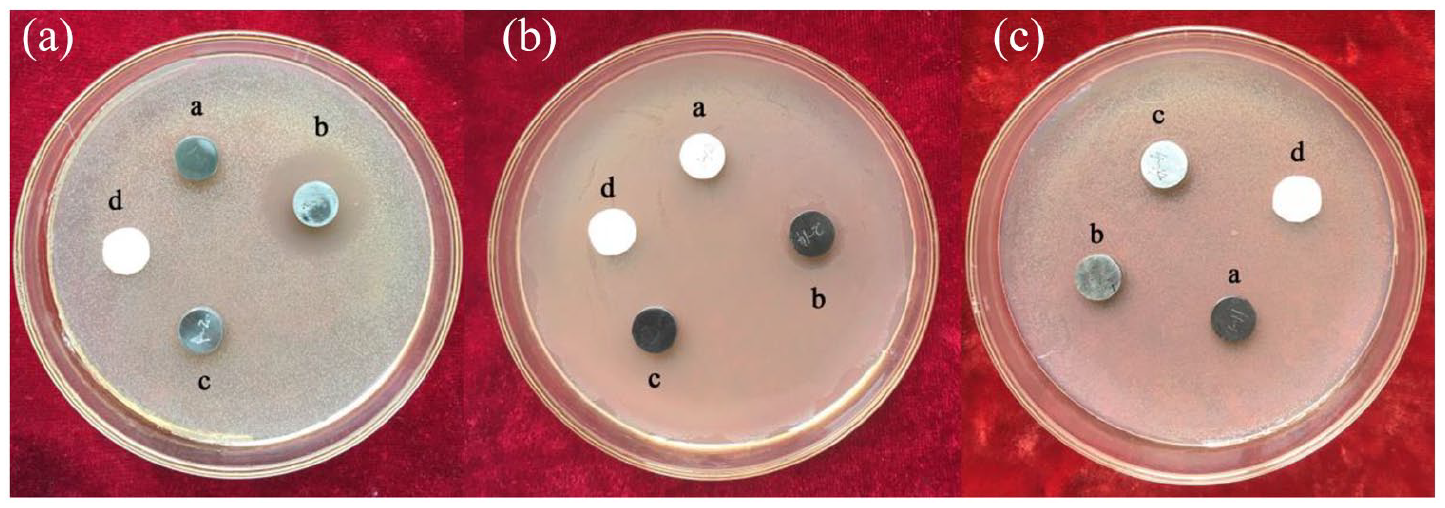
Inhibition zone of 304-2.5Cu SS (a), 304-4.5Cu SS (b), 304 SS (c), blank control group filter paper sheet (d) against S. mutans (A), S. sanguinis (B), and their mixture (C).
Cytotoxicity
As shown in Figure 6, the OD value decreased slightly with the Cu content, and those of both 304-2.5Cu SS (Figure 6 GroupA) and 304-4.5 Cu SS (Figure 6 GroupB) were lower than those of 304 SS (Figure 6 GroupC) and the blank control (Figure 6 GroupD), indicating that increase of the Cu content enhanced the cytotoxicity to a certain extent, however, the difference was not statistically significant (p > 0.05). In addition, with increase of the incubation time, the OD value showed an increased tendency, hinting that the cells grew well on all the sample surfaces. Detailed cytotoxicity grades (CGT) at each time point are shown in Table 2. CGT of groups A and B are grade 1, and the those of groups C and D are grade 0, indicating that Cu addition slightly enhanced the cytotoxicity of 304 SS to some degree.

Absorbance values (OD) of 3T3 cells in each group for 24, 72, and 120 h of co-culture time: Group A, 304-2.5Cu SS; Group B, 304-4.5Cu SS; Group C, 304 SS; Group D, negative control group.
Relative cell proliferation rate (RGR) and cytotoxicity grade (CTG) of different groups at different culture time.

Apoptosis rate
Figure 7 shows flow cytometry results of NIH3T3 cells suspension in each group cultured with metal extraction for 48 h. The apoptosis rates of the blank control (Figure 7(d)), 304 SS (Figure 7(c)), 304-2.5Cu SS (Figure 7(a)), and 304-4.5Cu SS (Figure 7(b)) were 12.1%, 15.5%, 16.7%, and 25.0%, respectively. Of note, compared with the blank control, there was no significant difference in the early apoptosis rate for 304-2.5Cu SS, 304-4.5Cu SS, and 304 SS. With prolonging the culture time, the apoptosis rates demonstrated an increased trend with increase of the Cu content in 304 SS.

Flow cytometry results of cell suspension in each group for 48 h: (a) 304-2.5Cu SS, (b) 304-4.5Cu SS, (c) control group 304 SS, and (d) blank control. Q1, dead cells; Q2, early apoptotic cells; Q3, viable cells; Q4, mid and late apoptotic cells.
Discussion
More than 700 microorganisms have been found to colonize in oral cavity. 22 Among these, S. sanguinis is one of the early colonized bacteria in oral cavity, which can interact with saliva on the surface of tooth and create an excellent growing environment, leading to the formation of dental plaque. Besides, it also acts as a habitat for other secondary colonization bacteria such as S. mutans.10,29,30 S. mutans is a causative agent of caries which has acid-producing and acid-tolerant properties, and can produce extracellular polysaccharides (EPS) in the biofilm.31,32 Therefore, it is meaningful that both S. mutans and S. sanguinis in this work were selected as the research objects to explore the antibacterial effect of Cu-bearing SS on the oral resident bacteria and pathogenic bacteria.
There are two antibacterial mechanisms of Cu2+ including contact killing and ion release killing, as shown in Figure 8. The former is mainly attributed to the strong reducibility of Cu2+, which can extract electrons from bacteria and thereby form reactive oxygen species (ROS). ROS can inhibit the respiratory chain and convert into hydroxyl radicals (HO.) which damages the bacterial cell walls, causing cytoplasm to run off and oxidize cell nucleus. The latter is because Cu2+ can easily enter the cell wall through osmosis, and firmly bind with the intracellular amino acids and proteases, resulting in the protein denaturation.33–37 In this study, by ways of plate counting, scanning electron microscopy, and DAPI staining excellent effect of 304-4.5Cu SS through contact killing was illustrated. Furthermore, the inhibition zone method could well differentiate the antibacterial mechanism of metal material. There were obvious inhibition zones around 304-4.5Cu SS samples on the plates with S. mutans and S. sanguinis, indicating that the Cu-bearing stainless steel can also inhibit the growth of surrounding bacteria through continuous release of Cu ions. However, for the mixed bacteria, the antibacterial effect of ions release was weaker than the contact killing, which might be due to the presence of water film between the Cu-bearing SS sample and the bacteria. While continuous contact of the bacteria with Cu-bearing SS would accelerate the entry of Cu ions into bacteria. 36

Schematic diagram of possible anti-bacterial mechanism of Cu ions released from Cu-bearing SS. (1) Cu ions on the surface of Cu-bearing SS extract electrons from bacteria, causing their cytoplasm to run off and oxidize their cell nucleus. (2) The released Cu ions accumulate in the bacterial cell membrane, which affects the permeability of the cell membrane, inhibits the respiratory chain, produces reactive oxygen species, and interferes with gene replication of bacteria.
In the past, most research on antibacterial SS ignored the influences of synergy and antagonism between different bacteria on the antibacterial properties of materials. 38 Dual-species biofilms were more resistant to antibacterial agents, because the interaction of bacteria would affect the formation, function, and structure of biofilms. 25 Unlike single-strain biofilm, dual-species biofilms produce more extracellular matrix, resulting in limited diffusion of antimicrobial substances into microbial cells. In this work, dual-species biofilm composed of S. mutans and S. sanguinis limited the diffusion of Cu2+ dissolving from the surface of 304-Cu SS, and even 304-4.5Cu SS also had no significant inhibition effect on the biofilm formation of dual-species, as shown in Figure 5. However, due to large contact area between the antibacterial SS and the dual-species biofilms, 304-4.5Cu SS in the direct contact experiment (shown in Figures 1–4) could play an effective antibacterial effect on the dual-species biofilms.
Antibacterial performance of Cu-bearing SS is closely related to Cu element and its content, so it is particularly important to optimize its performance by adjusting proper Cu content in the Cu-bearing SS. Xi et al. 39 reported that the antibacterial rates of 316L-2.5Cu SS and 316L-3.5Cu SS by solution treatment against Escherichia coli were over 90% by aging at 700°C for more than 3 h, and high Cu content in 316L SS possessed better antibacterial ability. Sun et al. 40 showed that the antibacterial rate of 317L-4.5Cu SS against Staphylococcus aureus reached 98.3%. Above results are consistent with the results of this work. For S. mutans, S. sanguinis and their mixture, the antibacterial activity of 304-4.5Cu SS was significantly stronger than that of 304-2.5Cu SS. Therefore, as the Cu content increases, the antibacterial SS can release more Cu2+ to significantly improve the antibacterial performance. Cu is a heavy metal element, a large amount of Cu may cause cytotoxicity and liver and nervous system diseases in body.14,41 Previous research showed that adding 5 wt.% Cu into pure titanium and 3.9 wt.% Cu into 304 SS did not affect the biological safety of the materials.11,42 Bai et al. 43 found that Ti-10Cu alloy had no obvious cytotoxicity in five simulated environments. In this study, the apoptosis experiment showed that the apoptosis rate of antibacterial SS with Cu content of 2.5 wt.% increased slightly compared with the control group, and when the Cu content rose to 4.5 wt.%, the apoptosis rate increased more significantly. Therefore, it can be speculated that when the Cu content is between 2.5 and 4.5 wt.%, there may exist an ideal Cu content the Cu-bearing SS, which has little effect on the apoptosis rate and can exert acceptable antibacterial properties.
In this study, although Cu-bearing SS with excellent antibacterial properties was prepared, its effect on the apoptosis still needs to be optimized. Moreover, long-term antibacterial effect of antibacterial SS still requires more deep study.
Conclusions
The 304-4.5Cu SS prepared in this study exhibits excellent antibacterial activity against S. mutans, S. sanguinis, and mixed bacteria, and has no obvious cytotoxicity to mouse NIH3T3 fibroblasts. Cu addition significantly improved its antibacterial properties, while it also slightly enhanced the cytotoxicity and apoptosis rate of 304 SS. The biological properties of 304-Cu SS still need to be further optimized, and its long-term antibacterial effect still still requires more deep study.
Footnotes
Author contributions
YL, CY, KY, and YL researched literature and conceived the study. YL, JY, and XL performed the experiments. HZ, XZ, and XY analyzed the data. YL wrote the first draft of the manuscript. All authors reviewed and edited the manuscript and approved the final version of the manuscript.
Declaration of conflicting interests
The author(s) declared no potential conflicts of interest with respect to the research, authorship, and/or publication of this article.
Funding
The author(s) disclosed receipt of the following financial support for the research, authorship, and/or publication of this article: This work was financially supported by the National Key Research and Development Program of China (Grant No. 2016YFB0300205), the National Natural Science Foundation of China (Grant Nos. 51771199 and 51631009), the Liaoning Province Key Research and Development Plan Project (2020JH2/10300038), the Foundation of Liaoning Educational Committee of China (No. L2019lkyjc-03), Youth innovation promotion association CAS (Grant No. 2018221), and Shi-changxu Innovation Center for Advanced Materials, Institute of Metal Research, Chinese Academy of Sciences.