
Abstract
Background:
The aim of this study was to compare the degradation resistance of nickel-chromium (Ni-Cr) and cobalt-chromium (Co-Cr) alloys used as a base material for partial dentures in contact with saliva.
Methods:
Wiron® 99 and Wironit Extra-Hard® were selected as representative casting alloys for Ni-Cr and Co-Cr alloys, respectively. The alloys were tested in contact with deionized water, artificial saliva and acidified artificial saliva. Material characterization was performed by X-ray diffractometry (XRD) and microhardness and nanohardness testing. The corrosion properties of the materials were then analyzed using open circuit potential analysis and potentiodynamic analysis. Alloy leaching in solution was assessed by inductively coupled plasma mass spectrometry techniques.
Results:
Co-Cr alloy was more stable than the Ni-Cr alloy in all solutions tested. Leaching of nickel and corrosion attack was higher in Ni-Cr alloy in artificial saliva compared with the acidified saliva. The corrosion resistance of the Co-Cr alloy was seen to be superior to that of the Ni-Cr alloy, with the former exhibiting a lower corrosion current in all test solutions. Microstructural topographical changes were observed for Ni-Cr alloy in contact with artificial saliva. The Ni-Cr alloy exhibited microstructural changes and lower corrosion resistance in artificial saliva. The acidic changes did not enhance the alloy degradation.
Conclusions:
Ni-Cr alloys are unstable in solution and leach nickel. Co-Cr alloys should be preferred for clinical use.
Introduction
Alloys such as cobalt- and nickel-based alloys are gaining popularity for the construction of both removable and fixed dental prostheses. This could be a result of the increase in prices of noble metals over the past 30 years, due to the expanding price of commodity raw materials (1). In many countries, nickel chromium (Ni-Cr) alloys have been replaced by cobalt-chromium (Co-Cr) alloys owing to growing concerns over the cytotoxic effects of leached nickel ions present when exposing the former alloy to the oral cavity (2). Nickel is found in very low concentrations in the human body; however, increased concentrations may cause it to become hazardous (3, 4). Lately nickel has begun to be considered a toxic element, and with the Nickel Directive introduced by the European Union in 1994, the position against nickel inclusion in materials was further strengthened. Around 1 in every 10 people has been found to be allergic to nickel (5), and it is possible that more people suffer from this allergy but that due to its nonspecific symptoms, the official rate is an underestimate. A number of systemic disorders have been linked to nickel exposure (6, 7).
Co-Cr alloys exhibit high strength (8) and are nonmagnetic. They are also resistant to creep, corrosion and wear (9). Apart from this, these alloys have been found to be cytocompatible (10-12). Thus they perform their desired function without inducing an unwanted local or systemic effect in the patient (13). These properties make them ideal alloys to be used in the oral cavity. Co-Cr alloys have also been found to be more resistant to corrosion than Ni-Cr alloys (10, 11), as indicated by in vitro potentiodynamic scans of both alloys immersed in several lactic acid and sodium chloride aqueous solutions. The corrosion of Ni-Cr alloys occurs by preferential dissolution of nickel-rich grains, which is very different from the mode of corrosion displayed by Co-Cr alloys, in which no preferential dissolution of Cr-rich grains is observed (7, 14). The casting procedure was reported to have a very minimal effect on the corrosion properties of either Ni-Cr or Co-Cr alloys (14). Temperature and pH both affect the corrosion resistance (15), with Ni-Cr alloy being more susceptible to acid attack.
The high corrosion susceptibility and leaching of nickel from Ni-Cr alloys results in decreased cell viability, increased oxidative and cellular toxicity levels and an increase in cytokine inflammatory expression (16). Some authors (17) have reported that the effect is enhanced by low pH conditions, which result in an increase of nickel ions leaching into the simulated oral environment, another study investigating the effect of gastro-oesophageal reflux disease (GERD) on corrosion susceptibility of metal alloy dentures found no difference between GERD patients and non-sufferers (7).
The objective of this study was to investigate corrosion resistance, surface topographical changes and physical and chemical changes of 2 base metal alloys in the presence of artificial saliva and acidified artificial saliva. The null hypothesis was that the mechanical properties and electrochemical responses of both Co-Cr and Ni-Cr alloys would not be affected by saliva or acidified saliva.
Materials and methods
Specimen preparation
Two base metal alloys were investigated: Co-Cr alloy (Co 63.0, Cr 30.0, Mo 5.0, Si 1.1, Mn 0.5, C 0.4; Wiron 99; Bego, Lincoln, RI, USA) and Ni-Cr alloy (Ni 65, Cr 22.5, Mo 9.5, Nb 1, Si 1, Fe 0.5, Ce 0.5, C max. 0.02; Wironit Extra-Hard, Bego).
Cylindrical specimens of 8-mm diameter and 1-mm height, and of 8-mm diameter and 15-mm height were cast for each material. The cylinders 8 mm in diameter and 1-mm high were used for leachate analysis, phase analysis, microscopy and assessment of microhardness and nanohardness, while cylinders of 8-mm diameter and 15-mm height were used for potentiodynamic testing.
Artificial saliva solution preparation
Two solutions were prepared: Fusayama-Meyer artificial saliva (18) and acidified artificial saliva. Fusayama-Meyer artificial saliva includes the right components to mimic the natural oral environment and has also been used for various potentiodynamic scans. The artificial saliva was acidified by adding 8 mL of lactic acid to 1,000 mL of artificial saliva solution. This mimics the oral environment after food intake and release of acidic media (19). The pH of the artificial saliva solution was assessed using a pH meter (Hanna HI 3221; Hanna Instruments, Sigma Aldrich, St. Louis, MO, USA) with a single-junction (Ag/AgCl) ceramic pH electrode (Hanna HI 1131). Temperature compensation was accomplished by simultaneously immersing a temperature probe (HI 7662) in the measurement solution. The pH meter was calibrated using 3 standard calibrating solutions (pH 4.01, 7.01 and 10.00). The pH of the artificial saliva was 6.7. Addition of lactic acid resulted in a drop in pH with a final pH value of 2.6 for the acidified Fusayama-Meyer solution.
Material characterization
The 8 × 1 mm cast cylinders were attached to aluminium sample holders and were ground with progressively finer grits of silicon carbide grinding discs (Struers, Ballerup, Denmark) with a manual grinding machine, followed by polishing with 3-µm polycrystalline diamond paste and finished with 1-µm diamond paste. These polished discs were then aged by immersing them in 5 mL of deionized water (control), Fusayama-Meyer artificial saliva or acidified artificial saliva for 30 days. The aged discs were then characterized by X-ray diffraction (XRD) and microhardness and nanohardness testing.
X-ray diffraction
The diffractometer (Bruker D8 Advance; Bruker, Billerica, MA, USA) used Cu Kα radiation at 40 mA and 45 kV, and the detector was rotated between 2θ of 35°-55° with a step of 0.02° and a step time of 0.6 second. The samples were spun at 15 revolutions per minute around the z-axis. Phase identification was accomplished using search-match software utilizing the International Centre for Diffraction Data (ICDD) database.
Hardness testing
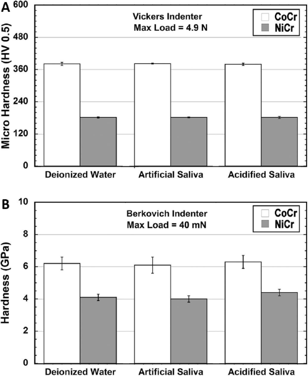
Microhardness testing was carried out using the Mitutoyo MVK-H2 (Mitutoyo, Tokyo, Japan) microhardness tester equipped with a Vickers indenter. Indentations were made on each sample’s surface using a load of 0.5 kg. Five hardness readings were taken for each sample, and the mean not median hardness was calculated from these readings.
Nanohardness of the alloys was assessed by indenting with a Berkovich indenter using a NanoTest nanoindentation system (Micro Materials Ltd., Wrexham, UK). Thirty indents per sample were made, in the shape of a grid with indents spaced 30 µm apart in both the x and y directions. The parameters were as follows: initial load 0.03 mN, loading and unloading rate 0.8 mN/s with a 1-second dwell time at a maximum load of 40 mN.
Immersion corrosion testing
Co-Cr and Ni-Cr cast discs 8 mm in diameter and 1 mm in height were immersion aged at room temperature in air (control), Fusayama-Meyer artificial saliva or acidified artificial saliva for 30 days. The solutions had a volume of 5 mL and were placed together with the coupons in a sealed container. The surfaces of these specimens postimmersion were characterized using a scanning electron microscope (SEM) coupled with an energy dispersive spectroscope (EDS). For SEM, the specimens were removed from the soaking solutions and dried in a vacuum desiccator. The specimens were then mounted on aluminium stubs and viewed under an SEM (Zeiss MERLIN Field Emission SEM; Carl Zeiss NTS, Oberkochen, Germany). Scanning electron micrographs of the different material microstructural components at different magnifications in secondary electron mode were captured.
Assessment of leaching
Co-Cr and Ni-Cr cast discs 8 mm in diameter and 1 mm in height were immersed in 5 mL of deionized water (control), Fusayama-Meyer artificial saliva or acidified artificial saliva for 30 days. At the end of the soaking period, the solutions were tested for traces of nickel, chromium, cobalt and molybdenum, using inductively coupled plasma (ICP) spectroscopy.
Potentiodynamic testing
The 8 × 15-mm cast cylinders had a blind hole measuring 2 mm in diameter and 5-mm deep prepared on one end. These cylinders in multiples of 4 were then immersion aged at room temperature in air (control), Fusayama-Meyer artificial saliva or acidified artificial saliva for 30 days. Following aging, a brass cylinder 2 mm in diameter and 4.5-mm deep with a threaded blind hole was press fitted in the cast Co-Cr and Ni-Cr cylinders. This was done to have an electrical circuit connection for the working electrode. The brass cylinder was totally isolated from contact with the solution by the O-rings shown in Figure 1.
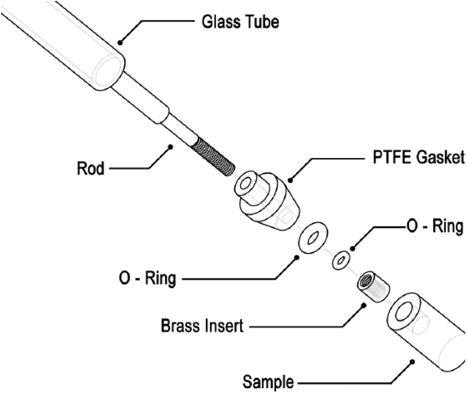
Working electrode assembly. PTFE = polytetrafluoroethylene.
The air-aged cast cylinders were potentiodynamic tested in a 9 g/L sodium chloride testing solution (control). The cast cylinders aged in the Fusayama-Meyer artificial saliva or acidified artificial saliva were potentiodynamic tested in a testing solution with an identical composition to that used for aging. The experiments were conducted following ISO 16428:2005 (20) and BS EN ISO 17475:2008 (21). Potentiodynamic testing was performed using a potentiostat (Gamry Interface 1000; Gamry, Warminster, PA, USA). The setup shown in Figure 1 shows a EuroCell™ electrochemical cell kit which was filled with 150 mL of testing solution and kept at 37°C ± 1°C via a heating jacket. The solution was deaerated by bubbling with nitrogen gas for an hour at a flow rate of 1 L/hour. Following the termination of bubbling, the specimen (working electrode; WE) assembly was inserted in the solution via the central 24/40 port through the 2 ace thread ports. The cell also contained a glass frit isolated platinum wire (counter electrode; CE) and a potassium chloride (3 g/L) fitted Luggin capillary, saturated calomel electrode (SCE) assembly (reference electrode; RE).
The time to set up the cell was kept constant, and testing commenced by measuring the open circuit potential (OCP) for an hour. At termination of the OCP test, a potentiodynamic test was performed at a voltage of between -100 mV vs. OCP and 1,000 mV vs. reference at a scan rate of 0.17 mV/s. Each solution was tested with 4 individual samples of each alloy. Graphs of current density (A/cm2) against linear voltage (V) were plotted. Current density was calculated by dividing the current recorded from the potentiostat by the surface area of the area specimen in contact with the solution.
To calculate the corrosion current density (icorr) for each of the samples, a number of steps were undertaken: (i) The graph of log(i) against potential was plotted for each of the representative potentiodynamic plots. (ii) The gradient of the tafel slopes was then calculated by differentiating each of them at a region around the OCP voltage. The polarization resistance of each sample, along with the gradient of the tafel slopes was then used to determine the actual corrosion current (icorr) according to Equation [1] (22).
Where ba and bc are the gradients of the anodic and cathodic regions, respectively.
Statistical analyses
The data were evaluated using PASW software (PASW Statistics 18; SPSS Inc., Chicago, IL, USA). Parametric tests were performed, as Kolmogorov-Smirnov tests on the results indicated that the data were normally distributed. Analysis of variance (ANOVA) with a p value of 0.05 and Tukey post hoc test were used to perform multiple comparison tests.
Results
Phase analysis
The XRD plots for both alloys in contact with the different soaking solutions are shown in Figure 2. The Co-Cr alloy shown in Figure 2B is a dual phase alloy, which contains face centered cubic (FCC) α phase (largest quantity) and an ε hexagonal closed packed (HCP) phase. The Ni-Cr alloy (Fig. 2B) has a FCC structure. Both alloys did not exhibit any phase changes in contact with the soaking solutions.
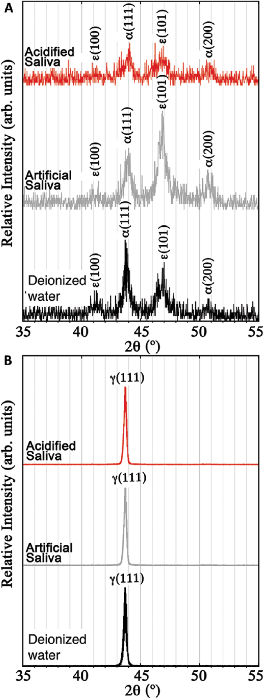
X-ray diffraction patterns for the Co-Cr (
Hardness testing
The mean microhardness and nanohardness of each alloy after a 30-day immersion in different solutions are shown in Figure 3A and B, respectively. It can be clearly seen that the Co-Cr alloy had a higher value of hardness than the Ni-Cr alloy. Furthermore, there was no difference in hardness when comparing the same alloy in different media (p>0.05) for either microhardness or nanoindentation. This means the medium had no effect on the surface hardness of the alloys.
(
Immersion corrosion testing
The SEM micrographs of Co-Cr and Ni-Cr alloys exposed to different solutions are shown in Figure 4. The corrosion attack on the Co-Cr in all solutions and on the Ni-Cr in the acidified saliva was very minimal. On the other hand, the Ni-Cr alloy exposed to artificial saliva had a greater tendency for dissolution, with more evident surface depressions when compared with the other sample electrolyte combinations.
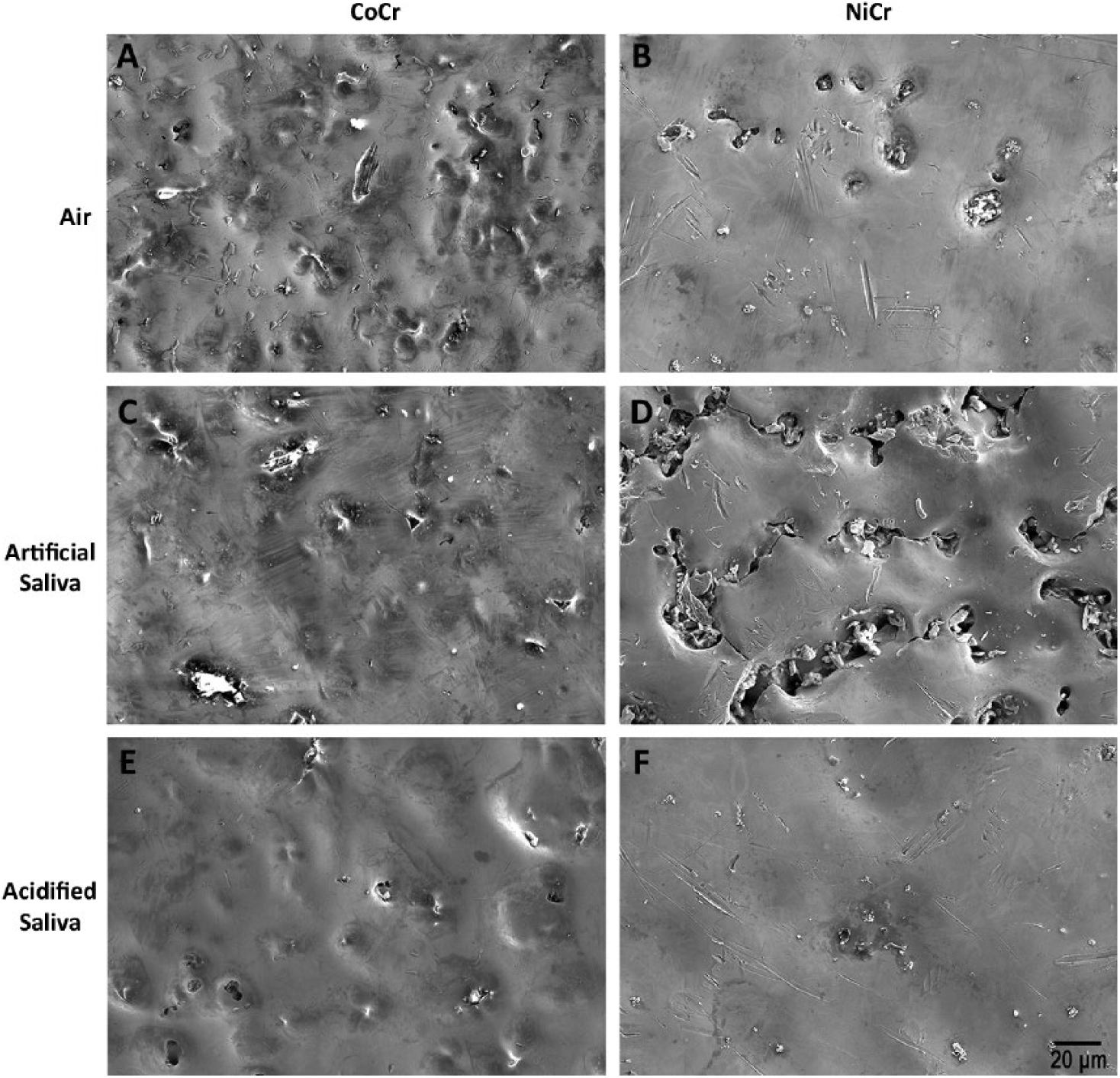
Surface scanning electron microscopy image after exposure of the Co-Cr or Ni-Cr alloys to air, artificial saliva or acidified saliva.
Assessment of leaching
The leaching of trace metal ions in the various solutions is shown in Figure 5. The release of nickel was particularly high in the Ni-Cr alloy immersed in artificial saliva, followed by acidified saliva, while the least amount of leaching was obtained from the control sample. Some leaching of Cr and Co were demonstrated in the Co-Cr alloy.
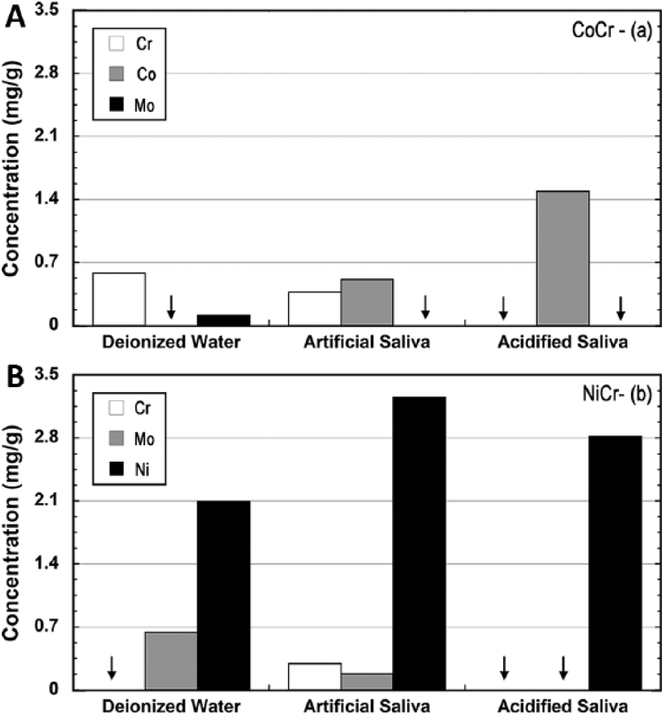
Concentration of Cr, Co and Mo ions leached from the Co-Cr (
Potentiodynamic testing
The results of potentiodynamic assessments for both Co-Cr and Ni-Cr alloys in different electrolyte solutions are shown in Figure 6A and B, respectively. A comparison between the materials can be found in Figure 7. The scan can be split into 4 regions – namely, cathodic, OCP, passive and transpassive. The OCP is the point at which the current density dips to virtually zero. The cathodic region lies at potentials lower than that of the OCP, while the passive part (anodic) lies at potentials higher than the OCP. The passive region is a region of a stable low current, while the transpassive region occurs after an approximate voltage of 700 mV.
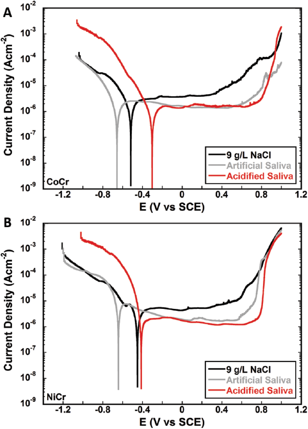
Representative potentiodynamic plots for the (
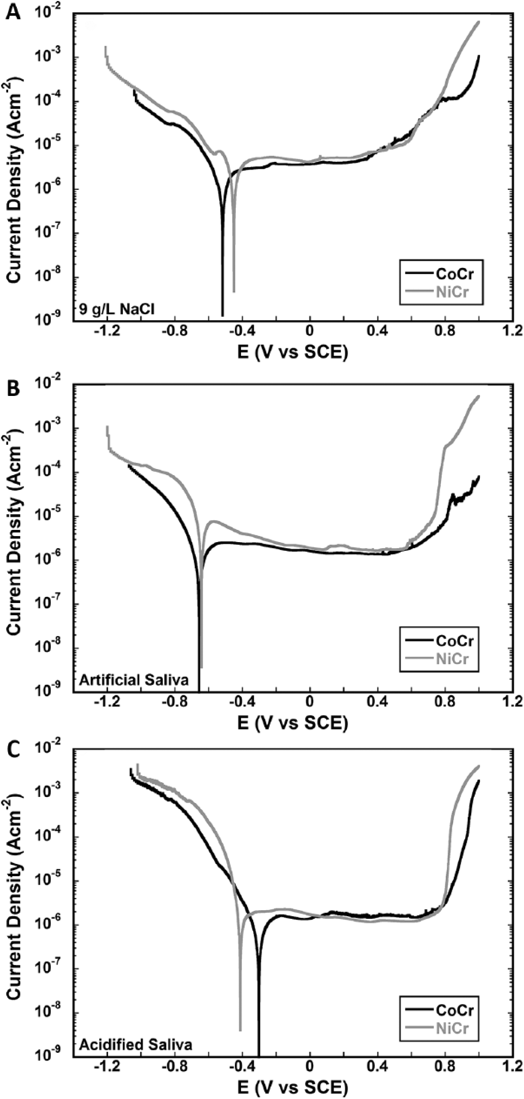
Representative comparative potentiodynamic plots for the Co-Cr alloy and Ni-Cr alloy exposed to 9 g/L NaCl (
As shown in Figure 8A, both alloys subjected to artificial saliva reached OCP at a lower voltage than the control (specimens tested in 9 g/L NaCl) and the alloy tested in acidified artificial saliva. When exposed to the control solution, both alloys were observed to have higher passive current densities compared with the same alloys in both test media. As shown in Figure 8B, the highest corrosion current density was produced when the Ni-Cr alloy was exposed to artificial saliva. A lower value was then obtained when the same alloy was exposed to acidified artificial saliva and the control solution. The Co-Cr alloy generally showed a very low corrosion current density in all solutions tested.
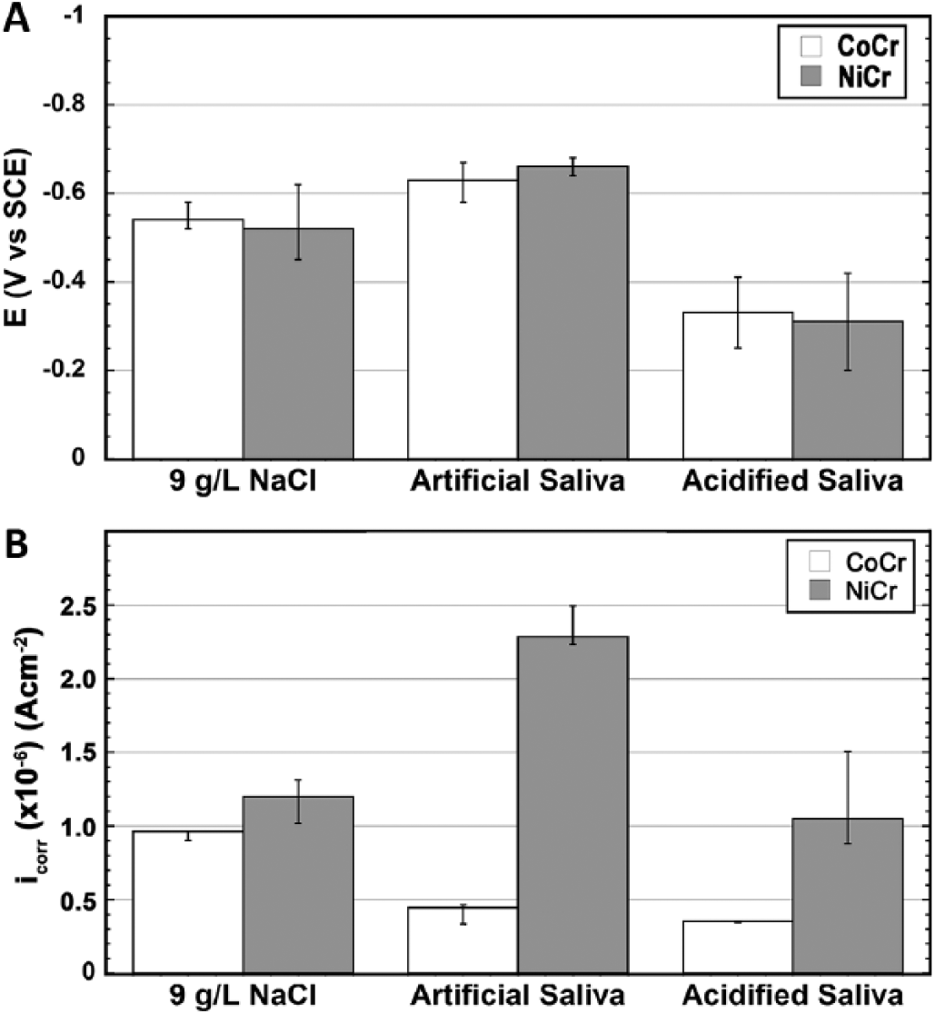
(
Discussion
Potentiodynamic scans (Figs. 6 and 7) provided information on the corrosion rate, pitting susceptibility and passivity. Both alloys exhibited transpassive dissolution in all media, as indicated by a sudden increase in corrosion current at similar potentials in the region of 700 mV/SCE. Oxidation of chromium from Cr3+ to Cr6+ has already been reported at these potentials in other works (23). However, no evidence of pitting could be found from the same potentiodynamic scans.
The corrosion resistance of the Co-Cr alloy was seen to be superior to that of the Ni-Cr alloy in all solutions tested as shown by the lower OCP corrosion current densities plotted in Figure 8B. The most pronounced difference could be observed in artificial saliva, in which the Ni-Cr alloy had a corrosion current that was approximately 350% greater than that of Co-Cr in the same solution. This was reflected in the quantity of ions released into solution as shown in Figure 5, where the Ni-Cr alloy can be seen to have released a far greater flux of ions compared with the Co-Cr alloy in each solution. This is mainly due to the amount of Ni released into solution.
When comparing results from the acidified artificial saliva with those of the nonacidified artificial saliva, Figure 8B shows that the difference in corrosion current was negligible when considering Co-Cr alloys. This is in accordance with previous findings (7) showing no differences in Co and Cr concentrations in saliva from patients with metal dentures suffering from GERD compared with those who do not suffer from GERD. In contrast, however, a major difference could be found when comparing the corrosion current and OCP potential of the Ni-Cr alloy when exposed to acidified and nonacidified artificial saliva. This partially contradicts the work presented by Borg et al (7) who also stated that no difference could be found in the amount of nickel ions released from Ni-Cr alloy dentures into saliva when comparing patients suffering from GERD versus control. This may be due to the difference in exposure time to the lower pH environment, which, as stated by the same authors, requires monitoring in a clinical trial.
The surface irregularities observed in Figure 4D on the Ni-Cr alloy samples after a 30-day immersion in artificial saliva are indicative of preferential corrosion from Ni-rich zones, due to preferential segregation of nickel-rich phase during solidification of the alloy (24). These nickel-rich regions appear to suffer localized selective dissolution as also reflected in clinical trials conducted by Borg et al (7) on Ni-Cr alloy dentures, and in in vitro experiments (14) on a similar alloy. This is once again confirmed in results from ion leaching experiments (Fig. 5), which show that in all media, leaching of nickel was high in comparison with other metal ions. On the other hand, when considering the Co-Cr alloy, no evidence of preferential corrosion could be observed. All of this leads the authors to the rejection of the null hypothesis.
On the other hand, Co and Cr ions were still released into solution during the in vitro testing, as can be observed in Figure 5. This is also demonstrated in the in vivo environment, as suggested by a recent clinical study in which patients wearing metal prostheses had higher levels of Co and Cr than non-denture wearers (7). The similar results obtained also show the robustness of the in vitro methods used in the current study.
Conclusions
Both alloys exhibited adequate corrosion resistance, with Co-Cr having a higher corrosion resistance when compared with Ni-Cr alloy across all solutions tested. The largest difference in corrosion resistance was found to be produced in artificial saliva, where the Ni-Cr alloy gave a corrosion current 350% greater that that given by the Co-Cr alloy. The electrolytic solutions were found not to modify the alloy’s mechanical properties. Furthermore, the Ni-Cr alloy suffered from a higher corrosion attack in artificial saliva when compared with an acidified artificial saliva. Surface topographical changes were observed in Ni-Cr alloy in contact with artificial saliva due to larger Ni dissolution. The null hypothesis was thus rejected.
Footnotes
Acknowledgements
The Department of Manufacturing Engineering of the University of Malta prepared the samples. The authors also thank Mr. Daniel Dimech and Ing. James Camilleri from the Department of Metallurgy and Materials Engineering of the University of Malta for their assistance.
Disclosures
Financial support: The Biomaterials Research fund financed this work, and Cherubino Limited, Malta, also gave their support. ERDF (Malta) financed the testing equipment throughout the project titled Developing an Interdisciplinary Material Testing and Rapid Prototyping R&D Facility (Ref. no. 012).
Conflict of interest: The authors declare they have no conflicts of interest.