
Abstract
The clinical application of anastomotic instruments improves the efficiency of the digestive tract surgery. However, the stapler with titanium nails implanted is still controversial in terms of anastomotic complications, and further improvement and optimization are needed. The purpose of this study was to explore the optimal microtextured parameters that could enhance the bioactivity of titanium implants in vitro. Laser microtexturing technology was used to construct the groove-type microstructural surfaces with different parameters, and human gastric mucosal epithelial cells (GES-1 cells) and mouse fibroblasts (3T3 cells) were cultured on the surface of the titanium plates in vitro. The data of cell adhesion, cell proliferation and cell activity were obtained and statistically analyzed. The textured titanium plates meet the expected design. GES-1 and 3T3 cell adhesion were better in the surface of titanium plates in microstructural group than that in the polished group. GES-1 and 3T3 cells also showed higher proliferative activity in the microstructural group compared with the polished group. The laser textured titanium plates have good groove-type microstructure, which increase the surface roughness, change the surface wettability, promote the adhesion, proliferating and orderly growth of GES-1 and 3T3 cells, and show good biological properties.
Introduction
The digestive tract is one of the most common sites of solid tumor. According to the data of the International Agency for Research on Cancer (IARC) in 2020, colorectal cancer, gastric cancer, and esophageal cancer are the third, fifth, and seventh most common cancer cases worldwide, and the second, fourth, and sixth common causes of cancer-related deaths, respectively. 1 The patients with above cancers can benefit from the homologous radical operation, especially with the early-stage cancer. But the occurrence of the anastomotic complications in postoperative period is an unpredictable event that can extend the length of hospital stay, reduce the quality of life and may lead to worse prognosis.2,3 The technique of instrument anastomosis has been widely used to improve the efficiency of digestive tract surgery, but the use of the staplers does not solve the problems of anastomotic bleeding and leakage, and can’t ensure the patency of the intestinal cavity at the site of anastomosis.4,5 More studies to achieve the improvement and optimization of these apparatus are still needed.
Titanium and titanium alloy materials have the characteristics of high specific strength, superior fatigue resistance, excellent corrosion resistance and good biocompatibility, so they are more suitable to be processed into biomedical materials and have been widely used in the preparation of surgical stapling mechanism. 6 The surface property of the implanted materials directly determines the biocompatibility and biological activity, and affect the tissue healing, tissue regeneration and the formation of organic interface between the surface of the implants and the cells.7,8 It has been shown that the proper adjustment of the surface characteristics can promote the proliferation of the cells, inhibit the growth of bacteria and promote tissue healing at the implantation site.8–10 The general specifications for stapler (YY/T 0245-2008 standard) from China stipulates that the surface roughness of the anastomotic nails should be less than 0.8 µm, and the surface of the anastomotic nail currently used for the gastrointestinal surgery from Johnson & Johnson, Medtronic, etc. is smooth. Therefore, this study assumed that the construction of microstructure on the staplers could improve their performance.
Surface laser microtexturing technology is a mature method to construct the surface microstructure, and the research and application of surface modification are mostly focused on dental and orthopedic implants. The optimal surface microtexturing treatment can enhance the antibacterial activity and the biocompatibility of these materials, promote the adhesion, proliferation, differentiation, and maturation of osteoblasts, and contribute to the good integration of the implant materials and the bone tissue.11–15 However, there are few studies on the surface modification of titanium materials in digestive tract tissue healing. In this study, surface laser microtexturing technology was used to find a superior titanium implant material with specific groove-type microstructure that may promote the tissue healing of the anastomotic site in gastrointestinal surgery and reduce the incidence of anastomotic complications especially the anastomotic leakage. The aim of this study is to reveal the relationship between the surface microstructure and the tissue healing.
Materials and methods
Materials and the surface pre-treatment
TA2 pure titanium plates (Grade 2 unalloyed titanium referring to ASTM B265-15 Specification) (diameter 15 mm, thickness 0.1 mm) (Baoji Yingnaite Nonferrous Metals, China) were used in this study due to their good biocompatibility. The round titanium plates with a base area of 2 cm2 were polished step by step with SiC sandpaper with different mesh, and then the polished titanium plates were cleaned by ultrasonic wave in acetone solution. Measure the surface roughness (Ra) of each polished titanium at five different locations by the Form Talysurf i-Series (Taylor Hobson, UK), and the polished titanium plates with the mean of roughness less than 0.8 μm were included. The titanium plates were etched to construct the groove-type microtextured surface with different parameters by laser marking machine (YLP-F20, GBOS LASER INC, China) with a maximum output power of 5 W, wavelength of 1064 nm, scanning speed of 100 mm/s, laser frequency of 20 kHz, and pulse width of 20–50 ns. The microtextured titanium plates were grinded by metallographic sandpaper to remove the burr, cleaned by ultrasonic in acetone solution and ethanol successively and sterilized by autoclaving. Three different groups of titanium plates were established for the following experiment and three identical titanium plates were set up for each group in this study.
Determination of the microtextured parameters
The central composite design (CCD) was used to determine the applicable parameters. A two-level two-factor (22) full factorial design was used to observe the effect of the variables influencing the Ra and contact angle. Recent research has shown that Ra values ranging from 20 nm to 10 μm have a significant influence on the biocompatibility of many materials because micro Ra affects the initial adhesion of cells. 16 And the most favorable contact angle range for cell adhesion is considered to be 40°–60°.17,18 Groove width (10, 20, or 30 μm) and spacing (50, 100, or 150 μm) have been considered as the variables and groove depth is always 10 μm, which can make sure the Ra and contact angle are within the above range. Analysis of variance (ANOVA) was used to build the model of Ra and the contact angle, and response optimizer of Minitab 18 was used with the goal of minimum contact angle and maximum Ra to determine the optimal microtextured parameters (width of 10.0 μm and spacing of 64.1935 μm) (Figure 1 ). We found that the titanium plates with width of 30 μm and spacing of 150 μm also had sufficient roughness and small contact Angle in the process of building the model (Figure 2). Finally, the polished plates (polished group), the plates with width of 10 μm and spacing of 60 μm (10–60 group) and the plates with width of 30 μm and spacing of 150 μm (30–150 group) were used in this study.

Optimal results of the width and the spacing performed by the response optimizer of Minitab. The optimal combination of the parameters is found with the goal of minimum contact angle and maximum Ra. The results showed that the better combinations were the parameter with groove width of 10 μm and spacing of 64.1935 μm.

Results of surface roughness (Ra) and contact angle of titanium plates after different treatments.
Analysis of surface characteristic
Scanning electron microscope (SEM) (Carl Zeiss AG, Germany) was used to observe the surface morphology of titanium plates in each group with working voltage 5 kV at different magnification (100×, 1.00K×, and 10.00K×). The profilometer (Talysurfi-Series, Taylor Hobson, UK) was used to obtain the profiles of titanium plates, and the contour arithmetic mean deviation (Ra) is obtained from the computer to evaluate the surface roughness. Five measurements were acquired for each titanium plate. The surface contact angle of the plates was measured by surface contact angle meter (PCA-11, HED Group of Beijing, China), and three measurements were collected for each plate to evaluate the surface wettability.
Cell culture
GES-1 cells (normal human gastric mucosal epithelial cells, purchased from Procell Life Science & Technology Co., Ltd, China) and 3T3 cells (normal mouse fibroblasts, purchased from National Biomedical Laboratory Cell Resource Bank, Beijing, China) were selected for this study. Sterile titanium plates were placed inside 24-well plates. GES-1 and 3T3 cells were sub-cultured, harvested and seeded in the sterile titanium plates at a density of 1 × 10⁵/mL with 1 mL medium per well. GES-1 and 3T3 cells were cultured in Dulbecco’s modified eagle medium (DMEM) with 10% fetal calf serum and 1% penicillin-streptomycin and maintained in the incubator (SANYO MCO-18AIC, Panasonic, Japan) at 37°C in 5% CO2 atmosphere. Both cells were maintained for 6 h for cell adhesion assay and 24 h for the observation of cytoskeleton, and cell proliferation experiment and detection of cell activity were performed on day 1, 3, and 7 after cell culture.
Observation of cell adhesion and cytoskeleton
DAPI fluorescent dye (Chongqing Pulikebio Co., Ltd., China) was used to stain GES-1 and 3T3 cells in dark for 5 min after 6 h of cell culture on the surface of titanium plates to observe the cell adhesion. When the cells were cultured for 24 h, GES-1 and 3T3 cells were fixed in 4% paraformaldehyde for 10 min and permeabilized in 0.1% Triton X-100 for 10 min. TRITC Phalloidin (Shanghai Fushen Biotechnology Co., Ltd., China) was used to stain GES-1 and 3T3 cells in dark for 30 min to evaluate the cytoskeleton. Confocal microscope (Olympus FV1000, Japan) was used to get the images of cells staining in five different random areas of each plate, and Image J software was used to count the number of the cell adhesion.
Cell proliferation experiment
Resazurin is a redox indicator, which is the main ingredient of Alamar blue (Shanghai Fushen Biotechnology Co., Ltd., China). Resazurin can be reduced in all processes of the cellular respiration metabolic reactions, and then a change in the fluorescent signal is produced. This change which can indicate Alamar blue reduction rate is caused by the cell’s consumption of oxygen molecules and can be detected by measuring absorbance to reflect the activity of cell proliferation. 19 Alamar blue was added in the corresponding culture medium of GES-1 and 3T3 cells on day 1, 3, and 7 after cell culture, and then the cells were further cultured in dark for 4 h. Multifunctional enzyme marker (TECAN, infinite F500, Austria) was used to measure the absorbance at the wavelength at 570 and 600 nm, and the Alamar blue reduction rate was calculated according to manufacturer’s instructions.
Detection of cell activity
GES-1 and 3T3 cells were trypsinized, harvested, lysed by the cell lysis buffer (per 1 mL lysis buffer with 10 μL PMSF, 10 μL phosphatase inhibitor, and 10μL protease inhibitor) and centrifuged at 4℃ at 14,000g for 30 min on day 1, 3, and 7 after cell culture. The BCR method was used to determine protein concentration. Colorimetry was used to detected the nitric oxide synthase (NOS) activity of GES-1 cells and alkaline phosphatase (ALP) activity of 3T3 cells. The absorbance was then measured at the wavelength of 520 nm for 3T3 cells and 530 nm for GES-1 cells using the spectrophotometer (NanoDrop 1000, Shanghai, China). ALP kit (NanJing JianCheng Bioengineering Institute, China) and NOS kit (NanJing JianCheng Bioengineering Institute, China) was used in this study. The formulas are as follow:

OD1 stands for the absorbance of sample hole; ODb stands for the absorbance of blank hole; OD0 stands for the absorbance of phenol reference material hole; Cph stands for the concentration of phenol reference material; Cp stands for the protein concentration of sample.

OD1 stands for the absorbance of sample hole; ODb stands for the absorbance of blank hole; ε stands for the molar extinction coefficient of the sample; Va stands for the total volume of the reaction solution; Vs stands for the sampling amount; Φ stands for the optical path of the cuvette; T stands for the reaction time; Cp stands for the protein centration of sample.
Statistical analysis
SAS 9.4 software was used for the data analysis. The data was presented as the mean ± standard deviation. one-way ANOVA was used to perform the multiple comparisons. The comparisons between two groups were performed by student’s t test or nonparametric test (U test). The p value less than 0.05 indicated that the result had significant difference.
Results
Surface topography
The SEM images of the three groups of samples at different magnification (100, 1.00K, and 10.00K×) showed microscopic differences in the surface characteristics. The polished surface was smooth and flat, and showed some relatively uniform small scratches under a microscope with a magnification of 10.00k. In the 10–60 group, the surface showed the groove-type microtexture which was smooth with width about 10 μm and spacing about 60 μm. In the 30–150 group, regularly distributed microgrooves can be observed and the interior of the grooves was rough with width about 30 μm and spacing about 150 μm. Polished and laser microtexturing treatment were in accordance with the expected design (Figure 3).
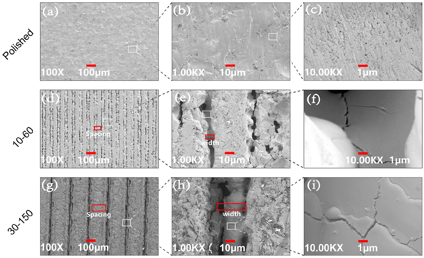
Scanning electron microscope images of titanium surface after different treatment. The laser microtexturing technology was used to construct different microtopography on the polished titanium plates, and the parameters of microtexture were observed and measured at different magnifications. (a–c) The images in the polished group at different magnification (100, 1.00K, and 10.00K×), (d–f) the images in the 10–60 group at different magnification (100, 1.00K, and 10.00K×), and (g–i) the images in the 30–150 group at different magnification (100, 1.00K, and 10.00K×).
Surface roughness and surface contact angle
The surface roughness of titanium plates in the polished group, the 10–60 group and the 30–150 group were 0.158, 2.645, and 1.825 μm (p < 0.001), respectively (Figure 2). The surface of titanium plates in the 10–60 group was roughest among the three groups (p < 0.05). The titanium plates in the 30–150 group had the strongest hydrophilicity (47.53° vs 69.70°, p < 0.05; 47.53° vs 55.60°, p < 0.05, respectively), which indicated that these plates might provide a more suitable environment for cell growth. We found that the contact angle in 10–60 group was not the smallest even though these plates had the optimal parameters. Similarly, although the plates in 30–150 group had the smallest contact angle, the Ra was not large enough. Since the optimal results of CCD experiment were obtained on the condition that the weight of roughness and contact angle are 50% each, further cell experiments are needed to verify the most suitable parameters.
Cell adhesion
The measurements and data analyses of cell adhesion are shown in Figure 4 and the representative images of each group are shown in Figure 5. There were only a few GES-1 cells to adhere to the surface of the polished titanium plates, but microtextured titanium plates seemed to be more suitable for cell adhesion. 3T3 cells obviously preferred to adhere to the surface of the titanium plates in the 30–150 group. However, 3T3 cells in 10–60 group only showed a tendency to promote cell adhesion compared with the cells in polished group (p > 0.05). The polished titanium plates presented the worst property in the cell adhesion for these two kinds of cells. The two cells tended to adhere to the inner surface of the micro-textured grooves, which may be related to the fact that the grooves can effectively increase the contact area between the titanium plates and the cells.

The number of the cells that attached to the surface of titanium plates after 6 h of cell culture in three groups. The cells were counted in one random field of view when the magnification of confocal microscope was 100 times.

Cell adhesion on the surface of the titanium plates in the polishing group, the 10–60 group and the 30–150 group. The images on the first line show the GES-1 cells adhesion in (a) the polished group, (b) the 10–60 group, and (c) the 30–150 group; the images on the second line show the 3T3 cells adhesion in (d) the polished group, (e) the 10–60 group, and (f) the 30–150 group.
Observation of cytoskeleton
The images of the cells stained by TRITC Phalloidin also show that more GES-1 and 3T3 cells adhered on the microtextured surface of titanium plates (Figures 6 and 7). The spreading and pseudopodia of the cells were observed in all groups for 3T3 cells but only in the 10–60 and the 30–150 groups for GES-1 cells. In the 30–150 group, the GES-1 and 3T3 cells spread well, and the pseudopodia were clear and obvious. The GES-1 cells in the grooves were fusiform, while GES-1 cells on the surface of polished titanium plates were round, oval or polygonal and gathered into clusters. The shape of 3T3 cells in the grooves was fusiform or irregular triangle. GES-1 and 3T3 cells tended to grow and extend in microtextured grooves.

Cytoskeleton morphology of GES-1 cells on the surface of titanium plates. The images on the first column show GES-1 cells nuclei in three groups; the images on the second column show the GES-1 cells actin in three groups; the images on the third column show the merged results of the nuclei and the actin in three groups; the images on the fourth column are the partially enlarged images of that on the third column.

Cytoskeleton morphology of 3T3 cells on the surface of titanium plates. The images on the first column show 3T3 cells nuclei in three groups; the images on the second column show the 3T3 cells actin in three groups; the images on the third column show the merged results of the nuclei and the actin in three groups; the images on the fourth column are the partially enlarged images of that on the third column.
Cell proliferation
The results of GES-1 and 3T3 cells proliferation are shown in Figure 8. On day 3 and day 7 after cell culture, both GES-1 and 3T3 cells cultured on the surface of the plates in the 30–150 group presented the optimal activity of cell proliferation. The activity of 3T3 cells proliferation in the 10–60 group was superior to that in the polished group on day 7 (43.06% vs 23.02%, p < 0.05), but the titanium plates in the 10–60 group didn’t show other more advantage for cell proliferation. However, the 30–150 group did not directly show a better effect on cell proliferation at all time than the 10–60 group in both cell lines. The reduction rate of GES-1 cells in polished group showed a downward trend from day 1 to day 7, and similar trend can be discovered for 3T3 cells from day 3 to day 7. The cell proliferation activity of both cells in 10–60 group and 30–150 group maintained a high level from day1 to day 7 (Figure 9).

Alamar blue Reduction of GES-1 and 3T3 cells on the surface of titanium plates in the three groups.

The trend of Alamar blue Reduction in GES-1 and 3T3 cells between three groups on day 1, day 3, and day 7 after cell culture.
Detection of NOS activity and ALP activity
The level of NOS activity and ALP activity can reflect the activity and function of GES-1 and 3T3 cells, respectively (Figure 10). GES-1 cells in the 30–150 group maintained a higher level of NOS activity during the whole process of cell culture than those in the polished group. On day 1 and 3, the NOS activity in the 30–150 group was significantly higher compared with the 10–60 group (82.21 vs 54.30 U/mgprot, p < 0.05; 73.47 vs 44.6 U/mgprot, p < 0.05, respectively). The NOS activity in the 10–60 group was superior to that in the polished group only on day 7 (6.51 vs 4.42 U/mgprot, p < 0.05). 3T3 cells in the 30–150 group showed the highest ALP activity on day 3 and day 7 among the three groups. But the titanium plates in the 10–60 group didn’t present better property in the ALP activity compared with the polished group.
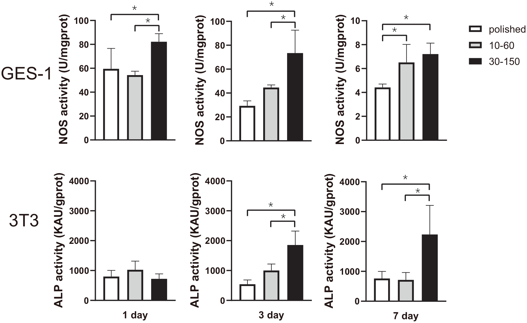
ALP activity of 3T3 cells and NOS activity of GES-1 cells on the surface of titanium plates in the three groups.
Discussion
The invention of anastomosis stapler realizes the anastomosis of the lumen and the closure of the stump in the state of digestive tract lumen closure, which reduces the incidence of abdominal cavity pollution in the process of digestive tract reconstruction, and this is a significant breakthrough in digestive tract anastomosis technology. Gastrointestinal wound healing is achieved through inflammation, proliferating, and remodeling. The anastomotic mucosa can be repaired by the migration and proliferation of epithelial cells, thus sealing the coloboma and forming a barrier to the lumen contents, and the appearance of granulation tissue marks the beginning of the proliferative phase, that is, fibroblasts reach the wound site. Wound collagen is dissolved and then re-synthesized at the initial stage, resulting in scar formation. The intensity of anastomosis depends on the newly synthesized collagen of fibroblasts and smooth muscle cells.20,21 However, many surgical factors, local factors and systemic factors can affect the tissue healing of digestive tract, and Instrument anastomosis in anastomotic complications is still controversial. 5 Surface modification has been applied to the studies on the properties of implant materials. In this study, groove-type microtextured surface with different parameters were fabricated by laser texturing technology, and the biological properties of titanium plates were estimated by the experiment of GES-1 and 3T3 cells culture in vitro.
It has been shown that the surface roughness of the implanted materials will affect the differentiation, proliferation, and matrix production of osteoblasts, as well as the production of local growth factors and cytokines. 22 In the field of biomedical engineering, the increase of surface energy and hydrophilicity of implant materials can promote bone tissue repair and accelerate the integration between implant materials and bone tissue.23–27 The lower contact angle between the implant materials and water means that the surface of the material has higher polar surface energy, better surface hydrophilicity and higher surface wettability.23,24 In this study, we use the central composite design (CCD) and analysis of variance to create the roughness and contact Angle model, and the final parameters were obtained by the response optimizer of Minitab, and these three kinds of titanium plates can be used for this study.
Cell adhesion is the prerequisite for cell spreading, growth, migration, proliferating, and differentiation, which will directly affect the integration of implant materials and tissues. GES-1 cells adhered better on the surface of the microtextured titanium plates, but there was no difference between the two groups with different microtexturing treatment. That indicated that both microtextured titanium plates can better promote the adhesion of this cell, and may accelerate the healing of tissue at the anastomotic site in vivo. In addition, these positive results also proved that laser microtexturing treatment was a reliable method to obtain new surface topography. The 3T3 cells adhesion was significantly better in the 30–150 group, and there was no significant difference between the 10 and 60 group and the polishing group. We suspect that different cells may have specific optimal parameters, so the implant material with specific surface treatment should to be selected for the surgery on a specific part of the body. Further experiments are needed to confirm whether there are better parameters for cell growth.
The proliferative activity of the cells on the surface of the implants is an important index of tissue healing. A small amount of NO can maintain the blood supply of gastric mucosa, promote the secretion of mucus and bicarbonate in gastric mucosa, inhibit platelet adhesion and aggregation, reduce the infiltration of inflammatory cells, and promote the repair of injured gastric mucosa. 28 As a key enzyme in the synthesis of NO, the change of NOS activity will directly affect the yield and biological effect of NO. GES-1 cells in the 30–150 group presented the optimal activity of cell proliferation compared with the polished group on day 3 and day 7. GES-1 cells with higher levels of NO in groups 30–150 might accelerate tissue healing at the anastomotic site.
ALP is involved in tissue formation, metabolism and regeneration in wound healing and chronic inflammation. 29 The increase of ALP activity is one of the phenotypic characteristics of fibroblasts, so it can indirectly reflect the activity and function of fibroblasts. 30 3T3 cells in the 30–150 group showed better proliferation activity and ALP activity, and we can infer that more fibroblasts participate in tissue healing and more collagen protein is secreted to increase the strength of tissue at the anastomotic site when this kind of titanium plate is applied into the digestive tract surgery. We can also discover that the proliferative activity of both GES-1 and 3T3 cells in the polished group showed a downward trend from day 3 to day 7. This’s probably because that the microtextured treatment increased the surface area of plates, which provided enough space for the adhesion and proliferation of both cells, while the appearance of contact inhibition on the surface of polished titanium plate suppressed the cell activity.
The results of this study were ultimately inclined to select the titanium plates in 30–150 groups for gastrointestinal anastomosis implants, which seemed to be contrary to the optimal results of the CCD experiment. This may be because the optimal results are obtained under the assumption that roughness and contact angle have the same effect on cell growth. We suspected that one of the two factors, Ra and contact angle, might be the main factor, so the results finally were biased. The 30–150 group had a smaller contact angle compared with the 10–60 group, which might more offset the adverse effect of insufficient roughness on cell growth. This might indicate that contact angle played a major role in promoting cell growth. Therefore, it is necessary to further optimize the model so that it can predict the optimal parameters better.
The limitation of this study is that we did not accurately measure the magnitude of the effects of roughness or contact angle on cell growth. It is reasonable to believe that there are better titanium plates in terms of biological properties than that in 30–150 groups. In future studies, we will quantify the effects of roughness and contact angle on cell growth. Then, accurate prediction models can be built according to the magnitude of the two factors to build the best implant materials.
Conclusions
In this study, the microstructure and relevant biological functions of titanium plates under laser texturing treatment were revealed. The surface of microtextured titanium plates had good groove-type microstructure, which increased the surface roughness and changed the surface wettability. The microtextured titanium plates promoted the adhesion, proliferating and orderly growth of GES-1 and 3T3 cells, and enhance the cells activity and function. The grooved microtextured titanium plates with a width of 30 μm, a depth of 10 μm, and a spacing of 150 μm showed better biological properties. The application of this technique in the digestive tract stapler with implantation of titanium nails maybe promote tissue healing and reduce the incidence of postoperative anastomotic leakage.
Footnotes
Acknowledgements
The authors would like to thank Tianjin Medical University General Hospital for support.
Declaration of conflicting interests
The author(s) declared no potential conflicts of interest with respect to the research, authorship, and/or publication of this article.
Funding
The author(s) disclosed receipt of the following financial support for the research, authorship, and/or publication of this article: This study was funded by the Natural Science Foundation of Tianjin City (15JCZDJC3280).