
Abstract
Nowadays, dental caries is one of the most common oral health problems, affecting most individuals. It has been found that, by remineralizing enamel at an early stage in the formation of enamel caries, teeth can be effectively protected from dental caries. In this work, a peptide with eight repetitive sequences of aspartate-serine-serine (8DSS) is applied as the bio-mineralizer in an in-vivo rat enamel caries model. Nondestructive quantitative light-induced fluorescence-digital (QLF-D) imaging and micro-computed tomography (micro-CT) are used to evaluate the remineralization of enamel carious lesions by measuring the total fluorescence radiance loss of the molar area (ΔQTotal), acquired using QLF-D imaging, and the mineral density and residual molar enamel volume, acquired using micro-CT. Correlations are explored between ΔQTotal and mineral density (strong correlation, r = 0.8000, p < 0.001) and ΔQTotal and residual molar enamel volume (moderate correlation, r = 0.6375, p < 0.001). Our results demonstrate that 8DSS is a promising in-vivo remineralization agent that exhibits comparable effects to NaF (p < 0.05), which has been verified using the classical Keyes method. Moreover, the nondestructive QLF-D and micro-CT methods can be combined to quantify the remineralization of enamel carious lesions three-dimensionally in vivo, making them broadly applicable in quantifying hard tissues.
Keywords
Introduction
Dental caries is a multifactorial universal infectious disease that has an underlying effect on almost every individual because of the high consumption of sucrose. The outermost layer of the teeth, the enamel, bears various kinds of physical and chemical erosion, such as bite, friction, wear, and erosion from acid-producing bacteria and fermentable carbohydrates, namely demineralization. 1 In recent years, numerous efforts to address this problem have focused on noninvasive approaches in the early stage before enamel lesions form, including efforts to inhibit enamel demineralization and to boost the remineralization of early enamel carious lesions. Over the past 30 years, the local application of fluoride-containing products has substantially reduced caries prevalence. 2 However, extensive use of fluoride has contributed to a rising incidence of dental fluorosis and skeletal fluorosis. 3 In addition, fluoride-containing products are still inadequate to overcome the high caries challenge in some individuals. 4 Thus, there is an urgent demand to explore alternative ways to regenerate hydroxyapatite crystals and repair lesions.
Biomimetic mineralization has appeared on the research horizon in recent years. This strategy involves taking organics as a template, regulating the growth and deposition of inorganic mineral ions, and forming organized apatite crystals with chemical structure and mechanical properties resembling those of enamel or dentine. In recent decades, various biomimetic systems have been investigated to mimic enamel-like microstructures, including calcium phosphate nanoparticles, peptides, amelogenin-inspired polymers, and other organic additives.5, 6 Inspired by the functions of proteins working in tooth formation, various peptides, and their derivatives, such as β-sheet-forming peptides (P11-4), self-assembly oligopeptide, and enamel matrix derivative have been synthesized to repair enamel defects.7–9 In a previous study, our group designed an amelogenin-derived peptide (QP5) and demonstrated its capabilities to nucleate hydroxyapatite and promote mineralization in vitro.9, 10 All these promising studies indicate that finding natural macromolecular polymers that can act as analogs of natural proteins involved in biomineralization may be an effective strategy for enamel remineralization.
Another inspiration derives from dentin phosphoprotein, which is secreted by odontoblasts and lies in the dentin mineralization front. It is the most abundant noncollagenous protein existing in the dentin extracellular matrix and has been employed as a nucleation template in forming hydroxyapatite to initiate dentin mineralization.11–14 There is an aspartate- and serine-rich region within the dentin phosphoprotein sequence,12, 13, 15 consisting primarily of a large number of repeats of the sequence aspartate-serine-serine (DSS). 16 This region is known to be highly phosphorylated 13 and highly flexible 17 and is thought to bind with high affinity to calcium phosphate compounds. 18 Moreover, these small peptides can act as initiator and modulator in generating hydroxyapatite minerals by providing an optimal alignment of calcium ion binding sites. 19 Of these multi-DSS peptides, the one containing eight DSS repeats (8DSS peptide) is the most active in the mediation of biologically directed mineral deposition, and it possesses the ability to promote mineral deposition. 18 Our group has shown that the peptide 8DSS can promote remineralization of initial enamel carious lesions induced in vitro by pH cycling. 20 We further illustrate the potential of 8DSS for inhibiting enamel demineralization and for enhancing the anti-caries effect of NaF. 21 However, all these studies have limitations because they were carried out in vitro. This raises the question of whether the peptide would exert similar effects in the complex oral environment in vivo, since saliva composition, flow rate, and pH, as well as the presence of biofilms, can influence the outcomes of anti-caries treatments. 22
Quantitative light-induced fluorescence-digital (QLF-D) imaging and micro-computed tomography (micro-CT) are two newly developing methods with working principles suitable for determining the mineral content of the teeth. Compared with other conventional assessment methods, the proposed techniques are nondestructive, thus allowing imaging tracking of the same site at different disposition stages. Additionally, in previous in vitro studies, QLF-D and micro-CT showed statistical correlation with transverse microradiography, the current gold standard method for evaluating two-dimensional measurement of remineralization in vitro.23, 24 As is well-known, space can be regarded as the superposition of numerous planes. So, if we can verify the correlation between QLF-D and micro-CT in vivo, we can further justify the effectiveness of the two methods and enlarge their application scope to determination of three-dimensional remineralization, in agreement with results from classical Keyes scoring.
The objective of this study was (1) to investigate the effects of 8DSS peptide on remineralization in a rat model of enamel caries in vivo; (2) to explore the feasibility of the QLF-D technique and micro-CT to evaluate three-dimensional remineralization effects in vivo; and (3) to analyze the correlation of the QLF-D technique and micro-CT for evaluating remineralization in vivo. To the best of our knowledge, this work is the first to observe remineralization effects of 8DSS peptide in a rat model of enamel caries in vivo, and the first to analyze whether the QLF-D technique and micro-CT give consistent results when evaluating remineralization.
Materials and methods
Peptide synthesis
The 8DSS peptide was synthesized by Ontores Biotechnologies (Hangzhou, Zhejiang, PR China). The peptide purity was determined to be >95.0%, using reverse-phase high performance liquid chromatography. The 8DSS was dissolved in distilled deionized water to a final concentration of 25 µM.
Incubation of bacterial strain
Streptococcus mutans UA159 was acquired from the State Key Laboratory of Oral Diseases at Sichuan University (Chengdu, China) and was cultured for 24 h in brain heart infusion medium (Oxoid, UK) anaerobically (80% N2, 10% H2, and 10% CO2). An aliquot (100.0 µl) of this overnight culture was subcultured in 10.0 ml of brain heart infusion medium broth overnight, until it achieved the stationary phase. For subsequent inoculation, the strain was modulated to 7 × 10⁸ CFU/ml based on turbidity measurements in an optical spectrophotometer (Spectronic 200, Thermo Fisher Scientific, Waltham, MA, USA).
Rat caries model
Animal experiments complied with the ARRIVE Guidelines (Animal Research: Reporting of In Vivo Experiments) and conformed to the US National Institutes of Health Guide for the Care and Use of Laboratory Animals (NIH Publications no. 8023, revised 1978). All procedures performed in studies involving animals were conducted in accordance with the ethical standards of West China Hospital of Stomatology, Sichuan University (ethical approval number, WCCSIRB-D-2016-002). The experimental Animal Center of Sichuan University supplied 30 male specific-pathogen-free, 12-day-old Sprague-Dawley rats and their dams. To avoid endogenous S. mutans infection, oral swabs immersed in their mouths were incubated in fresh Mitis salivarius agar medium (Difco) and cultured anaerobically at 37 °C for 48 h. Rats were weaned at the age of 20 days, and their mass recorded. On the following day, the pups were infected with S. mutans UA159 by soaking swabs in 1 ml of bacterial inoculum and applying them to the rats’ teeth and oral mucosa for three consecutive days. On day 25, the rat saliva samples were used to confirm the infection, again by anaerobic culture on Mitis salivarius agar medium. To induce caries, during the following 2 weeks, we fed the animals a 28% sucrose and 28% glucose diet, termed Diet 2000, and drinking water containing 10 wt % sucrose aqueous solution, which was available at will.
Treatments
From day 40 to day 63, animals were randomly and averagely divided into three groups with 10 rats each (five companions per cage), and their molars were treated with 25 µM 8DSS peptide solution, 1 g/l NaF (positive control) or distilled deionized water (negative control) according to the treatment protocol. Test solutions were applied orally twice daily in the laboratory, at 08:00 and 20:00, with saturated cotton swabs containing treatment solutions wiped on the molars during a uniform operating time of approximately 1 min. To assure the duration of treatment, food and water were not available within 2 h. On day 64, the rats were killed and their maxillaries were obtained. During the treatment period, the rats were weighed weekly. All animals received humane care in compliance with the Ethical Principles of Animal Experimentation.
QLF-D evaluation
QLF-D measurements were conducted as described previously. 10 Images were taken of the occlusal, buccal and lingual aspects of molars with the quantitative light-induced fluorescence (QLF) handpiece connected to a control box (Inspektor Research Systems BV, Amsterdam, Netherlands); all digital images were stored using proprietary software (C3 version 1.25, Inspektor Research Systems BV). Next, fluorescence images were analyzed using QLF-D software (QA2 v. 1.25, Inspektor Research Systems) to determine three parameters related to mineral loss: fluorescence radiance loss percentage ΔF (%), lesion area (mm2), and the product of the two, integrated fluorescence radiance loss ΔQ (%·mm2). The carious lesion areas of the teeth demonstrated by clinical examination manifested as dark areas on the QLF-D images, indicating a decrease in fluorescence radiance and mineral content.
Micro-CT analysis
Mandibles were scanned at a 7 µm isotropic voxel resolution using the Scanco Medical µCT 50 (Scanco Medical AG, Brüttisellen, Switzerland). Sample data were obtained at an operating voltage of 70 kV and a current of 200 µA. By scanning five hydroxyapatite disks (0, 100, 200, 400, and 800 mgHA/cm3) as reference phantoms, a mineral density calibration curve could be acquired. The mineral density of enamel was calculated using the curve-fitting equation.
After scanning, three-dimensional images were reconstructed using Scanco Evaluation software v. 1.1.11.0 (Scanco Medical AG); since enamel mineral density differs between rats and human beings, a fixed threshold of 550‰ was used, as this was capable of differentiating enamel and dentin in rats. 25 Three-dimensional images were viewed using Visualizer software (Scanco). Mineral density and residual molar enamel volume of molar areas were measured using Evaluation software (Scanco).
Correlation between QLF-D and micro-CT
The total fluorescence radiance loss of all three tooth surfaces over an entire molar area was measured using the QLF-D technique as

The value of ΔQTotal was compared with the mineral density and residual molar enamel volume acquired by micro-CT, which reflected the mineral content of the same molar area. The data acquired using the two methods were displayed as scatter plots to compare the ability of the two methods of measuring remineralization of early carious lesions.
Caries scoring
After micro-CT scanning, jaws were dyed by 0.4% murexide solution for 12 h to stain carious lesions and then hemisectioned in the mesiodistal direction with a cutter (0.1 mm, Struers Minitom, Copenhagen, Denmark). Smooth surface lesions and sulcal lesions and their severities were evaluated under a stereoscopic microscope (Nikon SMZ1000, Tokyo, Japan) based on the Keyes method for diagnosing and scoring caries 26 by a single examiner blinded to the study.
Statistical analysis
Statistical analysis and depiction were performed using the software GraphPad Prism 5 (GraphPad Software, CA, USA). Analysis of variance (ANOVA), followed by Tukey’s multiple-comparison test for pairs were carried out to analyze the difference in ΔF, ΔQ, caries scores, and micro-CT analysis between the three treatment groups. Pearson’s correlation coefficients (with 95% confidence intervals) were calculated for mineral density against ΔQTotal and residual molar enamel volume against ΔQTotal. Linear regression was also executed to provide a best-fit line relating ΔQTotal to mineral density and residual molar enamel volume. The level of statistical significance was set at 0.05. Samples were measured in triplicate in each experiment, and each experiment was performed at least three times.
Results
Body mass
A flowchart of the rodent caries study is displayed in Figure 1(a). Animals remained in good health throughout the experimental period (Figure 1(b)). No mice died in the experiment period, nor did swelling and other oral mucosal disease emerge, which verified the relative safeness of the 8DSS peptide, as there were no noticeable side effects.
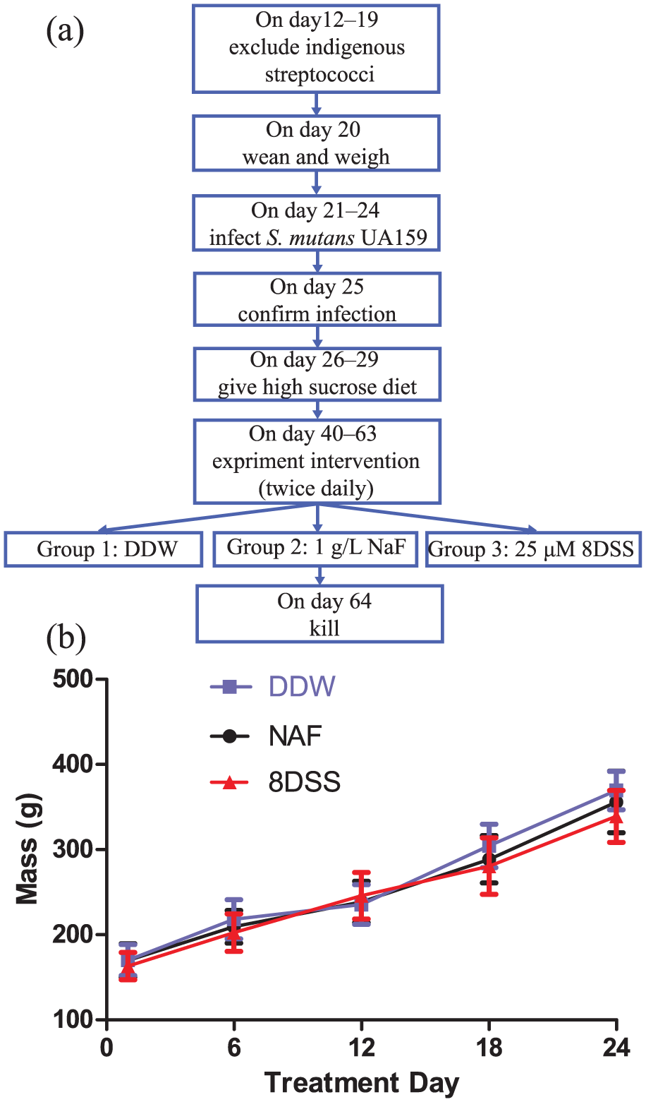
(a) Flowchart of the rodent caries study. (b) Mass changes in Sprague-Dawley rats infected with Streptococcus mutans, after which the molars were treated with distilled deionized water, NaF, or 8DSS peptide. Data shown are mean ± SD for 10 animals per treatment. Mass was similar for all groups throughout the treatment period.
QLF-D evaluation
Figure 2(a) shows post-treatment pictures taken by white-light digital (upper row) and QLF-D (lower row) imaging. In QLF-D images, areas of lesions are readily identifiable as darkened spots and red fluorescence. The quantity ΔF, defined as the percentage change in fluorescence radiance in each image point, represented the mean mineral loss of the selected area. The quantity ΔQ, defined as the total volumetric fluorescence change, calculated as the product of ΔF and lesion area, represented the integrated mineral loss from one tooth surface. According to the definition of ΔQ, we can sum the ΔQ values from the occlusal, buccal, and lingual surfaces to obtain a new index involving all three surfaces, denoted ΔQTotal. It can be seen from Figure 2(b) and (c) that there was a statistically significant reduction in ΔF and ΔQ on rat molar occlusal, buccal, and lingual surfaces in the NaF and peptide group, with poor results in the distilled deionized water group (p < 0.05). These results suggest that the 8DSS peptide can enhance the regression of carious lesions and promote remineralization under our experimental conditions.
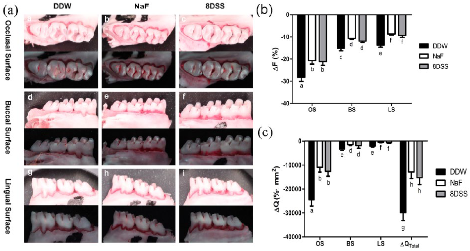
Determination of fluorescence radiance loss in molars in rats by nondestructive Quantitative light-induced fluorescence-digital (QLF-D) imaging across experimental groups. (a) Post-treatment pictures taken after the applying of distilled deionized water, NaF, or 8DSS peptide. The upper row shows white-light images of the occlusal, buccal, and lingual surfaces, while the lower row shows the corresponding QLF-D images. (b, c) QLF-D parameters (b) ΔF and (c) ΔQ on occlusal, buccal, and lingual surfaces, as well as on all three surfaces of rat molars with demineralization that had been subsequently treated with distilled deionized water, NaF, or 8DSS peptide. Bars labeled with different letters denote significant statistical difference.
Micro-CT analysis
Micro-CT performs well in qualitative and quantitative evaluation of dental hard tissue. Figure 3(a) shows three-dimensional images of the molar areas. The mineral density and residual molar enamel volume of the molar areas was measured using micro-CT. Calculation of mean mineral density profiles (Figure 3(b)) and morphometric volume analysis (Figure 3(c)), respectively, revealed that the mineral density of molar area and the residual molar enamel volume of the molar enamel treated with peptide were greater than those of the enamel treated with distilled deionized water (p < 0.05) and approximately equal to those of the enamel treated with NaF.
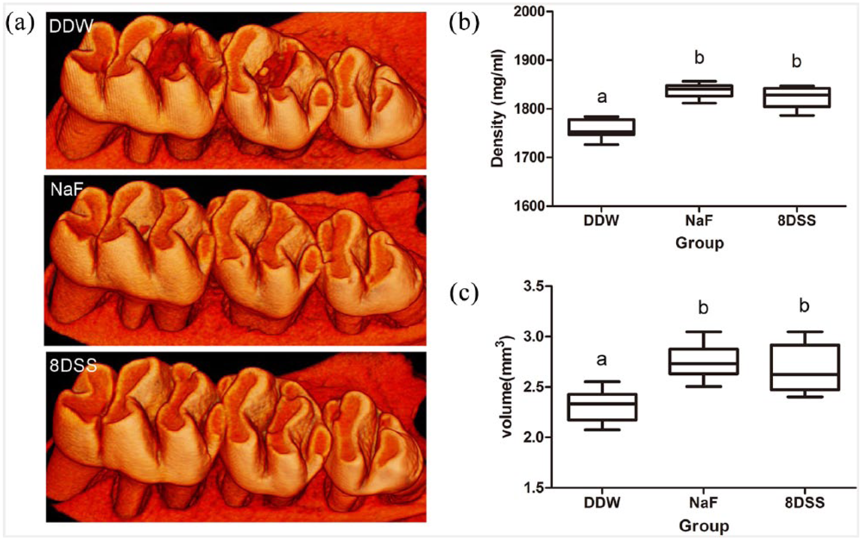
Determination of mineral density and residual molar enamel volume of molars in rats by nondestructive micro-computed tomography (micro-CT) across experimental groups. (a) Three-dimensional image of molar area. (b) Mineral density and (c) residual volume of the molar area in the presence of NaF, 8DSS and distilled deionized water (p < 0.05). Graphs labeled with different letters show significant statistical difference.
Correlation of QLF-D and micro-CT
The scatter plots in Figure 4(a) show the relationship between ΔQTotal determined using the QLF-D technique and mineral density determined by micro-CT, while Figure 4(b) showed the relationship between ΔQTotal and residual molar enamel volume. The linear regression lines with their respective 95% confidence intervals indicate a positive association between both sets of measurements. Linear regression analysis demonstrates a strong correlation between ΔQTotal and mineral density (r = 0.80, 95% confidence interval (CI) = 0.6177–0.9007). Pearson correlation coefficients indicated moderate but statistically significant positive associations between ΔQTotal and residual molar enamel volume (r = 0.6375, 95% CI = 0.3598–0.8114; p < 0.001).
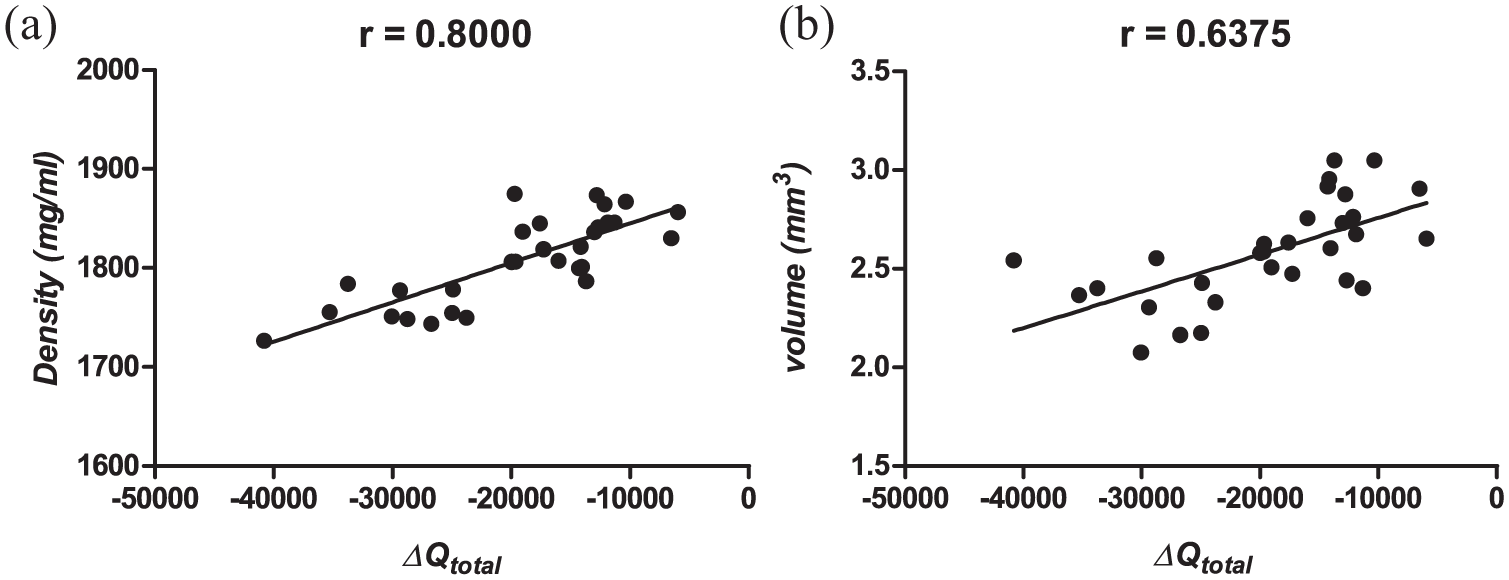
Correlation analysis of the QLF-D technique and micro-CT for evaluating remineralization in vivo. (a) Correlation between ΔQTotal (determined by QLF-D imaging) and mineral density (determined by micro-CT). (b) Correlation between ΔQTotal and residual enamel volume (determined by micro-CT). The black line shows a linear regression fit to the data.
Caries scoring
We can observe two types of pit and fissure lesion in the occlusal surface based on lesion severities: enamel only and slight dentinal. The lesions in the labial and buccal surfaces only affected the enamel level. The Keyes scores of fissure lesions on occlusal and smooth surfaces were determined and recorded (Table 1). The remineralization layers were less distinct in the group treated with distilled deionized water than the layers in the other two groups, in both the occlusal surfaces and the smooth surfaces. The enamel-only and slight dentinal scores indicated less extensive fissure in the samples treated with peptide and NaF than in the control samples treated with distilled deionized water. Similar results could also be achieved for the smooth surfaces. In other words, for all lesion types and locations, the values of Keyes scores for the peptide and NaF groups displayed similar reductions relative to the distilled deionized water group.
Evaluation of Keyes caries scoring method on occlusal and smooth surfaces of molars in rats compared across experimental groups.
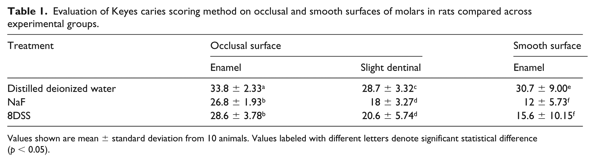
Values shown are mean ± standard deviation from 10 animals. Values labeled with different letters denote significant statistical difference (p < 0.05).
Discussion
Both QLF-D imaging and micro-CT are nondestructive techniques for measuring mineral content; here we use both to show that our 8DSS peptide, derived from dentin phosphoprotein, may help remineralize enamel in a Sprague-Dawley rat model of early enamel lesions induced by S. mutans. Furthermore, these findings were corroborated using a classical caries scoring method for lesion depth and severity.
How 8DSS peptides promote remineralization remains only partly understood. Earlier studies emphasized the electrostatic force between negatively charged amino acids and positively charged calcium and phosphate ions,18, 19, 27 leading DSS peptides to bind tightly with cationic calcium and phosphate ions. Computer-constructed models of the DSS repeat domain show that the most possible secondary structure of 8DSS peptide is a ribbon-like, twisting, trans-extended chain structure with repetitive arrays of carboxylate and phosphate groups on both edges of the chain. 15 Thus, it is not surprising that DSS can function effectively for interaction with Ca2+ and can increase the occurrence of Ca2+-mediated bridging between parallel chains or between chains and a hydroxyapatite surface. At the same time, these peptides possess strong affinity for the hydroxyapatite surface. 19 In other words, 8DSS peptides prevent calcium and phosphate ions from leaching out of enamel, while simultaneously promoting the capture of calcium and phosphate ions from the surrounding medium into new mineral deposits on the enamel surface.
The QLF-D technique exhibits much superiority over visual examination for detecting early carious lesions.28–30 Lesions cause light scattering and fluorescence loss that appear as dark spots on a bright background. Furthermore, QLF has the capability to detect red fluorescence generated by carious lesions that are metabolic by-products of intraoral bacteria. Changes in fluorescence luminance resulting from enamel and dentine alterations can be used as a surrogate measure of mineral density. 31 Indeed, successful cases of applying this technique to gauge mineral content alternations and lesion size have been achieved not only in vitro,29, 31 but also in animal protocols to quantify dental fluorosis. 32 In this study, ΔF and ΔQ indicated that distilled deionized water was less effective than peptide or NaF in decreasing mineral loss, suggesting that remineralization occurs with the peptide group to a similar extent as with NaF under our experimental conditions. The results are in accord with an earlier study of our group, in which we verified the feasibility of the QLF-D technique for use in a rat model. 10
Micro-CT is a promising technology for dental hard tissue. Not only can it be used to visualize the morphological characteristics of teeth in a detailed and accurate manner, but it can also be used to map the mineral-related parameters precisely. What makes it superior to other detection methods is its nondestructiveness and high resolution. Moreover, the technique has been considered a reliable substitute for the evaluation of dental hard tissue volume and mineral density. 33 Many trials have adopted the technique for analyzing bone34–36 and quantitatively assessing loss of tooth mineral density in vitro.37–41 Recently, Hamba et al. 24 described correlations between enamel lesion parameters determined by micro-CT and by transverse microradiography; the latter is considered the gold standard method for two-dimensional measurement of mineral density in vitro. In the present study, we calculated the residual molar enamel volume and mineral density of the enamel of the mandibular molar. The larger residual enamel volume indicated stronger anti-carious and regression effects, and higher mineral density values provided direct evidence of an enhanced remineralization effect. These parameters proved to be more accurate than visual examination under a stereoscopic microscope and showed that 8DSS can prominently induce remineralization compared with control samples treated with distilled deionized water. To our knowledge, the present study is the first report of micro-CT to examine the remineralizing effects of 8DSS peptide in a rat model of enamel caries in vivo.
Since both micro-CT and the QLF-D technique measure parameters related to mineral content in a stereoscopic way, we wished to investigate whether the mineral loss measured using QLF-D correlates with the mean mineral density and volume of the molar enamel area measured using micro-CT. A plot of ΔQTotal as a function mineral density shows an apparently linear relationship between the two properties. Linear regression analysis demonstrated a strong correlation between ΔQTotal and mineral density (r = 0.8, 95% CI = 0.6177–0.9007) and a moderate correlation between ΔQTotal and residual molar enamel volume (r = 0.6375, 95% CI = 0.3598–0.8114). Agreement between these methods is reasonable, since both methods largely assess surface mineral content changes. Remarkably, our work demonstrates the feasibility and validity of both the QLF-D technique and micro-CT for evaluating remineralization effects in vivo.
Conclusions
Taken together, our results suggest that 8DSS peptide enables the regression of enamel demineralization and boosts enamel remineralization in a rat model, which will lay the foundation for further work under a wider range of cariogenic conditions and is one of the primary procedures for future clinical therapy in human beings. The correlation shown here between the QLF-D technique and micro-CT in rats may provide more technical inspirations and present steps toward the development of new study methods of biomimetic anti-caries. Finally, the two nondestructive techniques, if explored positively, can be applied in long-time repeat imaging tracking of the same individual, which can not only improve the comparability of data, avoiding the impact of individual differences on the results of the experiment, but also removes the need to kill the model animals every time and save on scientific cost.
Footnotes
Acknowledgements
The study was completed at the State Key Laboratory of Oral Diseases, West China Hospital of Stomatology, Sichuan University.
Ethical standards
All procedures performed in studies involving animals were conducted in accordance with the ethical standards of West China Hospital of Stomatology, Sichuan University (ethical approval number, WCCSIRB-D-2016-002).
Declaration of conflicting interests
The authors declared no potential conflicts of interest with respect to the research, authorship, and/or publication of this article.
Funding
The authors disclosed receipt of the following financial support for the research, authorship, and/or publication of this article: This work was supported by the National Natural Science Foundation of China (grant numbers 81470734 and 81771062).