
Abstract
This work discusses the biomechanical testing of 3 elbow bones, namely the humerus, ulna, and radius. There is a need to identify the mechanical properties of the bones at the organ level. The following tests were performed: 3-point bending, fracture toughness, and axial compression. Six sets of whole-bone samples of human male cadaveric humerus, ulna, and radius (age of donor: 35 to 56 years) were tested. The results were analyzed for statistical significance by 2-stage, repeated-measure analysis of variance (ANOVA). The difference between the bending strength of the humerus, ulna, and radius was statistically significant (P = .001) when compared to one another. However, the fracture toughness and compressive strength were observed to be similar for the 3 bones. The knowledge of mechanical properties of elbow bones can aid in the design of elbow implants and upper limb protection systems, and also allow us to identify criteria for injury. Further, knowledge of the mechanical properties of the elbow bones can aid in calibrating simulations through finite elements analysis.
Introduction
The human elbow joint is a hinge joint that allows flexion and extension of the arm. The elbow joint is the junction of 3 bones: the humerus, ulna, and radius. In the junction of the elbow joint the distal end of the humerus and proximal ends of the ulna and radius participate. During the normal daily life of a human being performing routine tasks, the elbow bones undergo various types of loadings such as tensile, compression, bending, and torsion. Understanding the mechanical properties and mechanism of failure of the elbow bones under different loading conditions can help designers and researchers develop successful orthopedic implants that can work long term without sacrificing function. The fundamental bone properties of greater importance are maximum force absorbed at failure, ultimate strength, elastic modulus, fracture toughness, and hardness. Appropriate selection of implant materials as well as design can avoid problems such as aseptic loosening of the implant, stress shielding, or fracturing of bone by the implant.1–3
Literature data on quantifying the mechanical properties of bones in the human elbow are scarce. Of the few research publications, Jurist and Foltz studied the prediction of maximum bending strength at failure of the human ulna (whole bone) by measuring bending stiffness, mineral content, and geometry. The team reported correlation coefficients relating to: (i) breaking load and bending stiffness as 0.958, (ii) breaking load and mineral content as 0.947, and (iii) breaking load and cross-section as 0.754. From the correlation coefficients above, Jurist and colleagues concluded that in the case of the ulna, bending strength can be estimated from bending stiffness or mineral contents as compared to cross-section of the bone. 4 Fracture toughness is the ability of material to resist fracture. In other words, toughness is the amount of energy needed to fracture a sample or is the energy to create 2 surfaces. Higher fracture toughness value indicates higher resistant for crack propagation and material separation. Kc is the measure of fracture toughness for initiating the crack. Koester et al analyzed the effect of aging on the fracture toughness of humerus cortical bone in the transverse orientation. 5 Values for fracture toughness are given in Table 1, which lists the few reported works on mechanical properties of human elbow bones.
Literature on Mechanical Properties of Human Elbow Bones as Compared to Femur Bone.
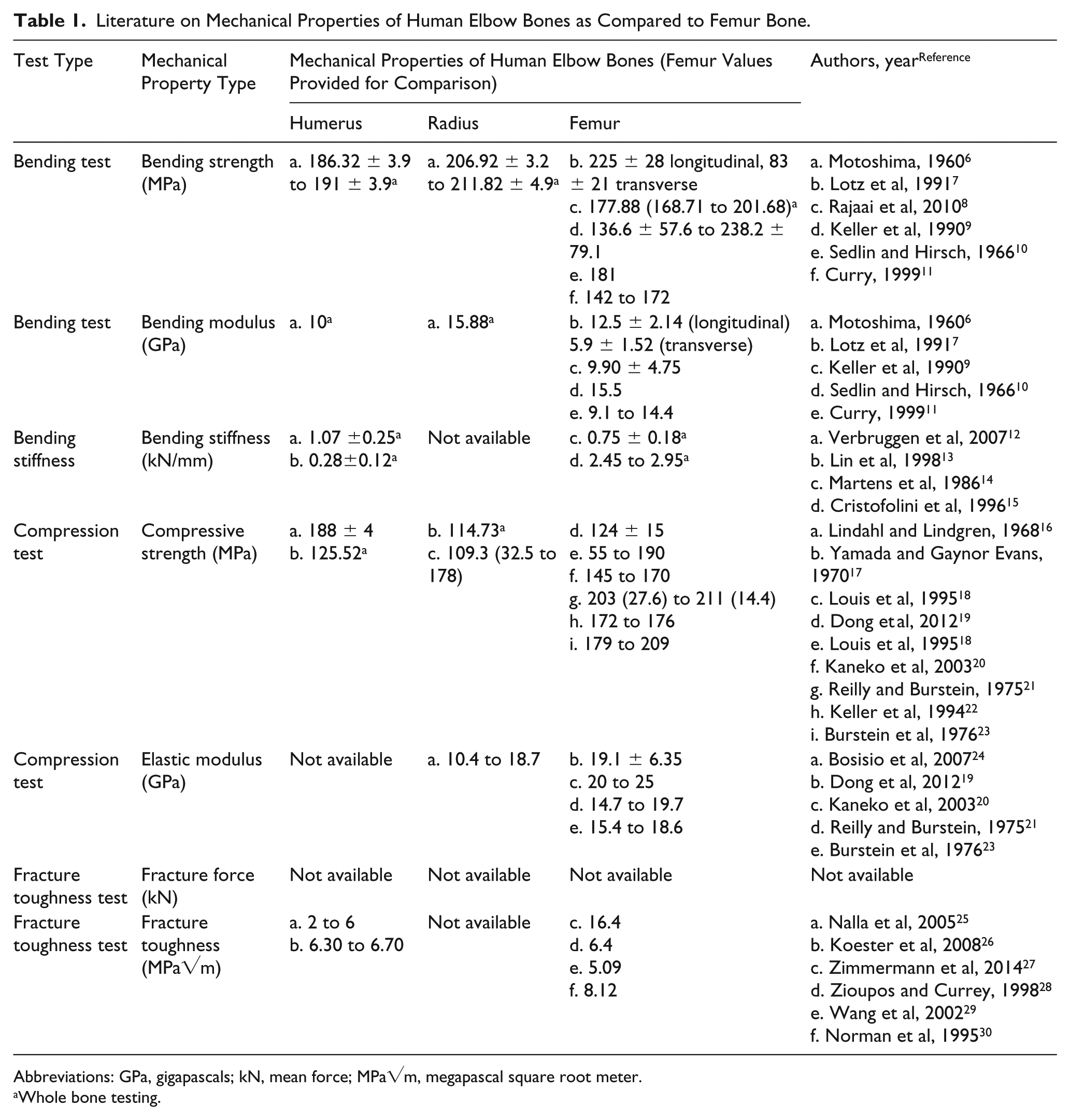
Abbreviations: GPa, gigapascals; kN, mean force; MPa√m, megapascal square root meter.
Whole bone testing.
Knowledge of the mechanical properties of the elbow bones aids in analytical as well as numerical simulations such as in evaluating stresses generated in the elbow joint at various flexion and extension angles 31 and postimplantation of prostheses. 32 Burkhart et al developed a finite element (FE) model to simulate the impact of forward fall on the distal radius bone. 33 The authors adopted an elastic modulus of 25.1 GPa, adopted from Burstein et al, 23 from experiments conducted on frozen femur cortical bone. 33 Johnson and Troy 34 used high-resolution peripheral computed tomography (CT) to develop a multiscale FE model for the prediction of the stresses of elbow bones under physiological loading. 34 The research group used an elastic modulus of 15 GPa from the results of nano-indentation studies carried out by Hoffler et al on frozen radius bone samples. 35 Sakai and colleagues developed an FE model of humerus bone to analyze stress distribution and fracture conditions during throwing actions in javelin and baseball. 36 Sakai et al used an elastic modulus of 17 GPa from the experimental results of sectioned frozen femur cortical bones reported by Reilly et al. 37 In a study carried out by Greybe et al, the authors developed an FE model to study the effect of ulnar shortening (a treatment for ulnocarpal impaction syndrome) on distal radioulnar joint mechanics. 38 Greybe et al used an elastic modulus of 18 GPa of sectioned frozen cortical bone samples of femur and tibia by Rho and colleagues. 39 There is no uniformity of reference values because of the unavailability of the data. Further, the adoption of properties of sectioned as well as whole-bone samples of femur and tibia for the FE analysis of the ulna and radius may not yield accurate results for the analysis of elbow bones. In short, the material properties of whole-bone test results of the elbow bones are required for better estimation.
In the literature, descriptions of elbow bones’ properties are scarce. Owing to the geometric stability of the femur, researchers have worked on femur bones. To the best of our knowledge, to date the properties of elbow bones have not been studied comprehensively in any research article. Therefore, in this article, a comprehensive study is conducted to identify mechanical properties of the elbow bones at the macro level such as the 3-point bending test, fracture toughness test, and compression test. The following mechanical properties of the humerus, ulna, and radius of human male cadavers in the age group of 35 to 56 years were experimented on and their statistical significance analyzed: (i) bending strength in the 3-point bending tests, (ii) bending modulus, (iii) bending stiffness, (iv) fracture toughness from the 3-point bending tests, (v) compression force and strength, (vi) elastic modulus under compression, and (vii) compression stiffness.
Sample Preparation
Testing Procedure for 3-Point Bending Test and Fracture Toughness Test
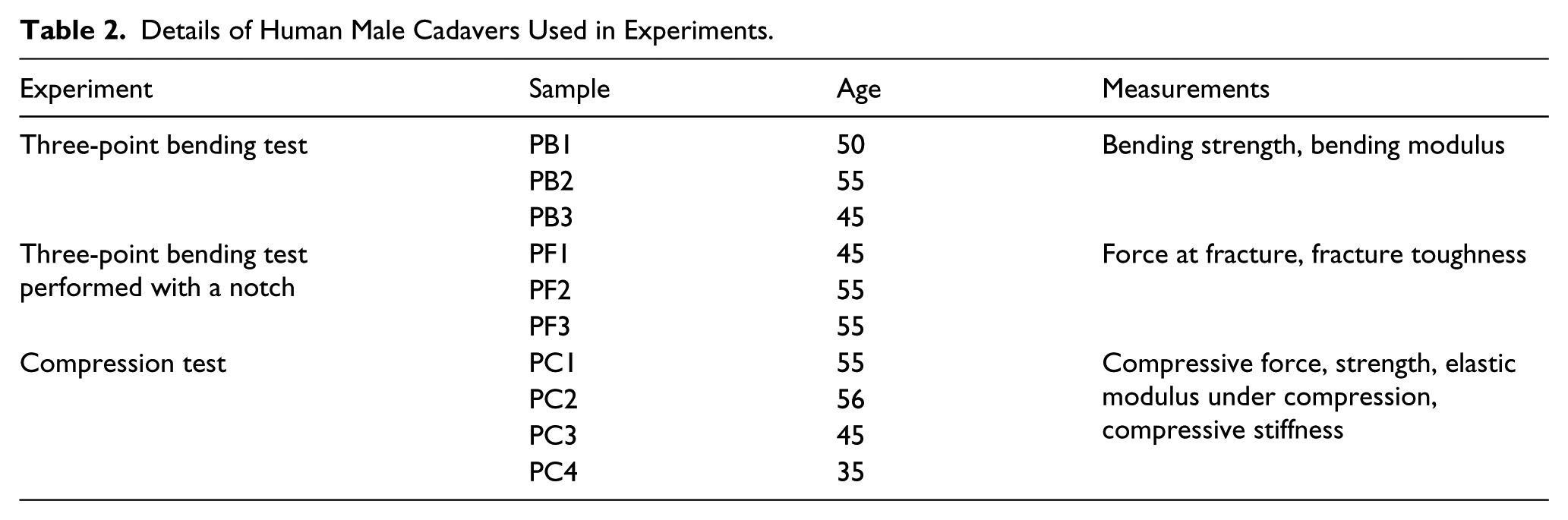
The humerus, ulna, and radius of 3 cadavers (3 left and 3 right elbows) were sectioned to a length of 120 mm from the distal side of the humerus, and from the proximal side of the ulna and radius. Human male cadaveric samples were obtained (age 45 to 55 years) from the All India Institute of Medical Sciences (AIIMS) for testing. Table 2 shows the complete list of samples used in the study. The humerus, ulna, and radius samples were dissected from deep-frozen (–20°C) cadavers. All the muscle fibers and cartilage were removed from the bones before experimentation. Twenty-four hours before experimentation, the samples were stored in saline water (0.9% NaCl). All length and diameter measurements were performed using a Vernier caliper (Insize, India). The samples were irrigated with saline water at all times during the test. 40 The 3-point bending test was performed on a universal testing machine (H5KS, Tinius Olsen, UK) with 2 bottom roller-type supports and 1 moving plunger/pin from the top coinciding with the midpoint of the length between the 2 bottom rollers. The metallic fixture for the bending test was specifically designed and fabricated in house. The material used for manufacturing the fixtures was EN 31 steel, a high-carbon (~1.3%) alloy steel with a hardness of 63 Rockwell C scale (HRC) (approximately) and elastic modulus of 215 GPa. 41 The roller supports were machined of D2 die steel material having a hardness of 62 HRC and elastic modulus of 210 GPa. 41 The distance between the 2 bottom supports was varied to achieve an aspect ratio of 6. The aspect ratio was calculated by dividing the length of the bone by the average diameters at the diaphysis portion of the bone. Loads were gradually applied at a test speed of 1 mm per minute.
Details of Human Male Cadavers Used in Experiments.
In the 3-point bending test, the topmost fiber or the osteons above the neutral axis undergoes compressive loading, and the osteons below the neutral axis undergo tensile loading. Local mineralization and microcracks at the site of the loading pin play an important role in the failure mechanism of the bone. 11 To simulate real-life conditions for fracture, the posterior part of the bone is placed on the support pins, while the anterior portion faces the loading pin as reported in Leppänen et al. 42 The bending strength was determined using classical beam theory by assuming the bone as a hollow beam.
Fracture toughness test samples were prepared from the diaphysis portions of the humerus, ulna, and radius for another set of 3 cadaveric (identification code: PF1, PF2, PF3) subjects with age ranges from 45 to 55 years. The earlier-used 3-point bending test protocol was followed for sample preparation and testing of bones in the fracture toughness test. In addition, a notch was created using a mini-hand hacksaw under a continuous stream of saline water (NaCl 0.9%) on the posterior portion of the bones. Notch provides the concentration of the stresses at a point. A precrack was formed at the tip of the notch using a razor blade. The current experiment demonstrates the 3-point bending test on the sample with a circumferential notch/crack. During the test, Mode I fracture occurs. The critical fracture toughness Kc is the minimal energy required for a crack to propagate into the sample material. Fracture toughness Kc, assuming the sample material to be a cylindrical tube, is Equation 1 (Eq). 43
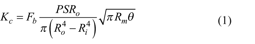
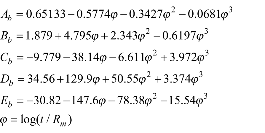
where Fb is a geometric factor in KI solution for a circumferential cracked cylindrical tube. P is the maximum load at fracture, S is the length between the 2 supports in the 3-point bending test, Ro is the outer radius of the bone, and Ri is the inner radius of the bone. Rm is the mean radius of the bone and θ is the half crack angle.
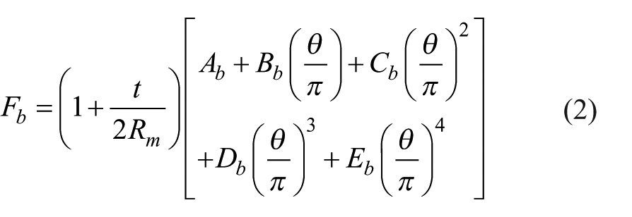
where t is the thickness of the cortex of the bone sample, θ is the half crack angle, and Ab, Bb, Cb, Db, and Eb are the dimensionless terms given by
Compression Test, Measurement of Compressive Force and Compressive Strength
Eight elbow joint bone samples for the humerus, ulna, and radius bones from 4 male cadavers (identification code: PC1, PC2, PC3, PC4) were used for compression tests. The samples were sectioned from the mid-diaphysis of the bones; a height-to-diameter ratio (H/D ratio) of 2 was maintained for all samples. Keaveny et al suggested an H/D ratio of ~2 provides optimal value for strength without buckling, shearing, and double barreling of the sample. 44 Samples were sectioned using a mini-hand hacksaw to maintain slow-speed cutting under a continuous stream of saline water. Twenty-four hours before the start of the experiments, the samples were stored in saline water (0.9% NaCl). The testing speed was maintained at 1 mm per minute. The testing was performed using a Zwick ProLine Universal testing machine of capacity of 100 kN and with a servo test system control. The compressive strength was determined by assuming the bone as a hollow rod. Owing to its irregular cross-section, the average diameter was considered for the calculations of strength and elastic modulus.
Statistical evaluation
Statistical analysis was performed among the 3 groups of bones: the humerus, ulna, and radius. Two types of analysis were performed: (a) comparison among the group (humerus, ulna, and radius), and (b) multiple comparisons between any 2 groups (humerus vs ulna, humerus vs radius, ulna vs radius) at a time. IBM SPSS software 20.0 (IBM Corp, Armonk, NY, USA) was used for the analysis. First, to check the difference between the right and left elbow bones of the same subject individually, the paired t test was performed.
The normality of the data was tested using the Shapiro–Wilk test. Thereafter, in the case of normally distributed data, repeated-measure ANOVA was used to evaluate the variation of the mean among the 3 groups of bones. An overall P value less than .05 was considered significant. For multiple comparisons between 2 groups (humerus vs ulna or humerus vs radius or ulna vs radius), repeated-measure (two-way) ANOVA followed by post-hoc comparison using Bonferroni test (with P < .05 considered significant) was executed.
In addition, linear regression was performed to establish a relationship between stiffness vs age and strength vs age for the bending and compression tests. P values < .05 were identified as significant.
Results and Discussions
Postexperimentation, the following mechanical properties were normally distributed: (i) strength, modulus, and stiffness (in 3-point bending tests), compressive force, compressive strength, modulus, and stiffness (in compression tests), fracture force (in 3-point bending test with notch), and fracture toughness. The summary of the results is provided in Table 3, and the results are discussed in the corresponding sections.
Results of Bending, Compression and Fracture Toughness Tests Along with Statistical Significance.
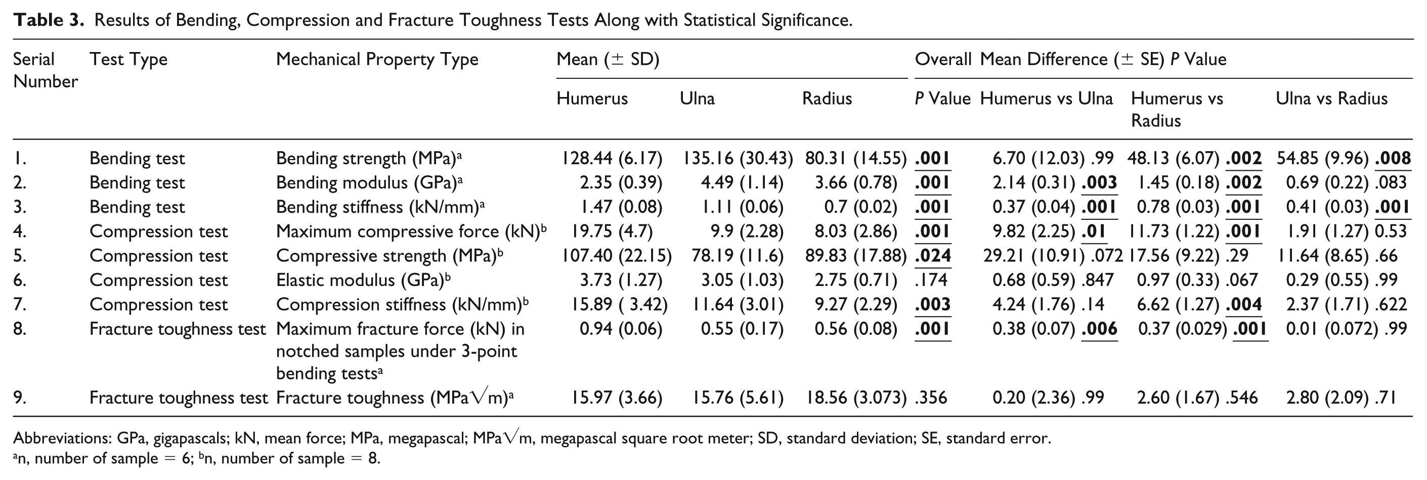
Abbreviations: GPa, gigapascals; kN, mean force; MPa, megapascal; MPa√m, megapascal square root meter; SD, standard deviation; SE, standard error.
n, number of sample = 6; bn, number of sample = 8.
Bending Strength from 3-Point Bending Tests
The average bending strength of the humerus, ulna, and radius were 128.43 ± 6.17 MPa, 135.16 ± 30.43 MPa and 80.31 ± 14.55 MPa, respectively. Figure 1A shows samples with typical brittle material fracture behavior. Repeated-measure ANOVA depicts the difference in strength for humerus vs radius and ulna vs radius as statistically significant with a P value of .01; Figure 1B depicts the same. The overall difference in average bending strength for the humerus, ulna, and radius was significant with a P value of .001. The difference in the strength between the elbow bones can be attributed to several factors, including bone mineral density 45 and the mineral-matrix ratio of the bone. 46 Further investigation is required, however, for identification of the cause of variation in strength. The biochemical and microscopic investigation are beyond the scope of the current work and can be explored in the future. Loading during in vivo conditions is complex. As per the accepted literature, the 3-point bend test is an important evaluation criterion.
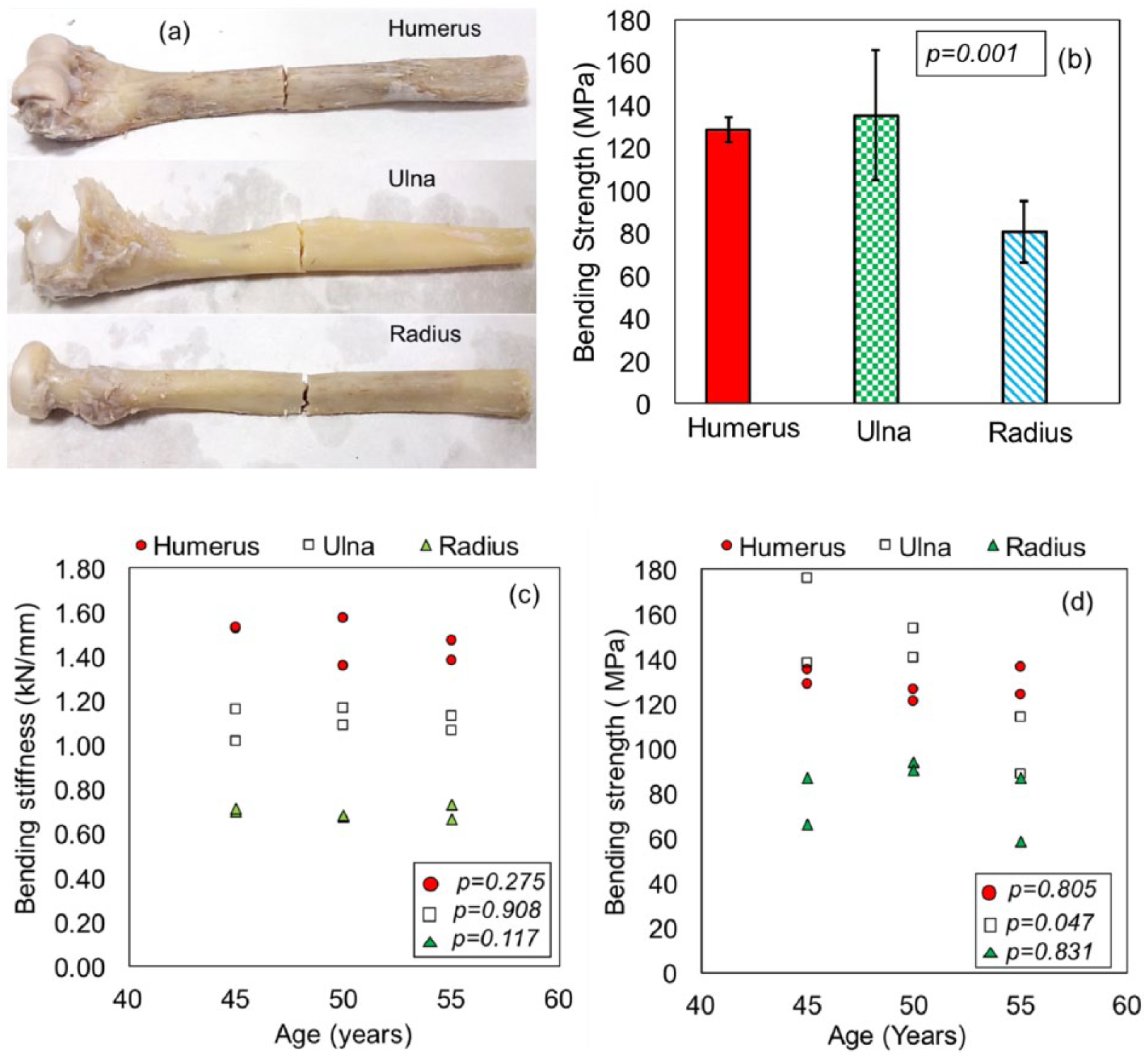
Three-point bending test: (a) humerus, ulna and radius samples after testing, (b) average bending strength, (c) relationship between bending stiffness and age of bones, (d) relationship between the bending strength and age of bones.
The average bending strength of whole-bone samples of humerus, ulna, and radius reported by Motoshima 6 for ages ranging between 50 and 59 years were 191.22 ± 3.9 MPa, 206.92 ± 3.9 MPa, and 205.9 ± 2.94 MPa, respectively. The sample storage method and experimented strain rates are unreported. To the best of our knowledge, no other research article is available on whole-bone testing of the elbow. A few research groups, including Burkhart et al, 33 Johnson and Troy, 34 Sakai et al, 36 and Greybe et al 38 have used femur bone values for modeling and design of elbow-related mechanics. However, the bending strength of whole-bone femur samples for the same age group is 195 ± 3.9 MPa. 6 Rajai and colleagues reported a femur bending strength of 177.88 MPa. 8 The bending strength of the sectioned cortical portion of femur bone (~ less than 10 mm × 10 mm) varies between 117 and 238 MPa as reported in the literature.7,9,10 So, the use of femur bone properties is not appropriate for the analysis of elbow bones.
Bending Modulus from 3-Point Bending Tests
Subsequently, the bending modulus for bone samples was determined. The average values were 2.35 ± 0.39 GPa, 4.49 ± 1.14 GPa, and 3.66 ± 0.78 GPa for the humerus, ulna, and radius, respectively. The difference in mean bending modulus is statistically significant for the humerus, ulna, and radius with an overall P value of .001. The difference between the bending modulus of humerus vs ulna and humerus vs radius is statistically significant with a P value of .01. Using the classical beam theory and with the assumption of bone as a tubular structure, bending strength and modulus was determined. However, this assumption has a disadvantage. The area moment of inertia for tubular structures is higher and hence can result in the prediction of lower bending strength. 47 Nevertheless, this method is good for a first approximation. As reported by Motoshima, the bending modulus of humerus, ulna, and radius bones was 10, 15.39, and 15.88 GPa, respectively. 6 In the current work, the mean elastic modulus of the humerus, ulna, and radius was 2.35, 4.50, and 3.67 GPa, respectively. For the sake of comparison, femur bone properties are cited here. The elastic modulus of whole femur bones were observed in the range of 3 to 18.33 GPa6,14,15,48 while, in the case of sectioned femur bones, the values were found in the range of 9.9 and 19.7 GPa.7,9,10 The above-mentioned values demonstrate that the elbow bones have a lower modulus than femur bones. For the purpose of design and analysis of prostheses or elbow bones, it is appropriate to use the currently reported values.
Bending Stiffness from 3-Point Bending Test
The bending stiffness was identified from 3-point bending test experiments. The bending stiffness for the 3 bones was 1.47 ± 0.08 kN/mm, 1.1 ± 0.06 kN/mm, and 0.7 ± 0.02 kN/mm for the humerus, ulna, and radius, respectively. The difference between the mean bending stiffness for 3-point bending is statistically significant for the humerus, ulna, and radius with an overall P value of .001. The difference between the bending stiffness of humerus vs ulna, humerus vs radius, and ulna vs radius is statistically significant. According to Verbruggen et al, 12 the bending stiffness of frozen humerus bone was 1.03 ± 0.22 kN/mm, while the bending stiffness of embalmed ulna bone was 0.73 ± 0.30 kN/mm as reported by Jurist and Foltz. 4 The reported values in the literature are consistent with the values obtained in the present work. However, in the case of saline-preserved cadaveric humerus reported by Jin and colleagues, 13 the bending stiffness was 0.28 ± 0.12 kN/mm, which is lower than the reported values in the current work as well as that in the literature. 13 The humerus bones were experimented on in the posterior-anterior direction, whereas in the current work the bones were tested in the anterior-posterior direction. For the sake of comparison, a few examples of the literature on the stiffness of femur bones are provided here. Cristofolini et al 15 reported a bending stiffness of 2.45 kN/mm and 2.95 kN/mm for rehydrated femur samples and frozen femur samples, while Martens et al 14 reported a bending stiffness of 0.75 ± 0.18 kN/mm for frozen femur specimens. Cristofolini and colleagues in a subsequent manuscript reported a bending stiffness of 1.9 kN/mm for formalin-preserved tibia bone specimens. 48 The above literature observes that the stiffness of elbow bones is in the range of 0.7 to 1.5 kN/mm. Nevertheless, the stiffness of the femur and tibia is higher than that of the elbow bone.
Relationship Between the Bending Stiffness and Age
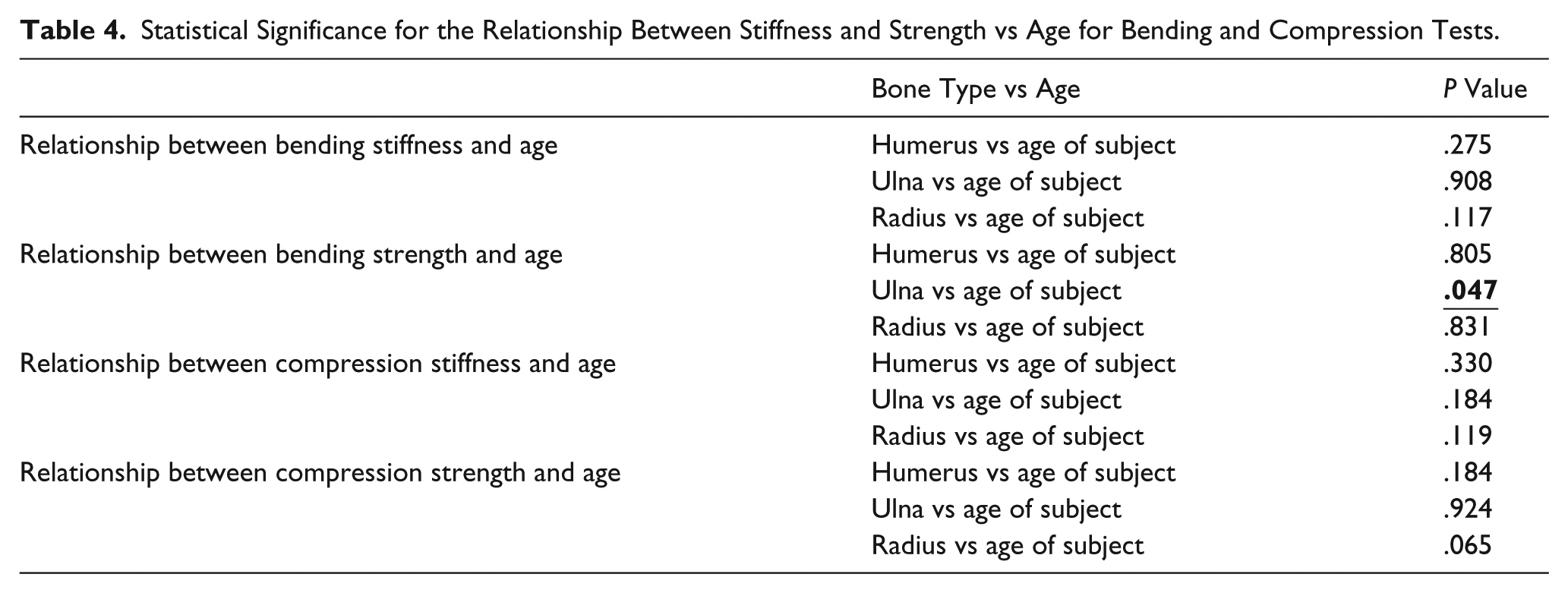
Figure 1C depicts the bending stiffness in the elastic regimen as a function of age for all 3 bones. Bending stiffness was found to decrease for the humerus, ulna, and radius with the increase in age. The P value for the linear regression analysis between the bending stiffness and age is shown in Table 4. Though the P value suggests nonsignificance in the variation of bending stiffness as a function of age for the humerus and radius, the stiffness values seem to decrease. The relationship of bending stiffness of the ulna with age was observed to be significant with a P value of .047. Out of very few studies conducted on the relationship of bone properties with age, our results are in line with the observations of Jurist and Foltz 4 and Zioupos and Currey. 28 The decrease in stiffness with increase in age could be attributed to low mineralization of the bone in aging. 49 The alteration in the dynamics of the bone cells results in a change in the natural process of bone formation and resorption because aged bone offers less resistance to deformation in the elastic zone.
Statistical Significance for the Relationship Between Stiffness and Strength vs Age for Bending and Compression Tests.
Relationship Between the Bending Strength and Age
Figure 1D depicts the maximum bending strength as a function of age. The maximum bending strength was found to decrease with increase in age for the humerus, ulna, and radius. The P value for the linear regression analysis between age and bending strength is shown in Table 4.
Fracture Force and Fracture Toughness from 3-Point Bending Tests on Notched Samples
The average fracture force (kN) for 6 samples each of humerus, ulna, and radius with a notch was 0.94 ± 0.06 kN, 0.55 ± 0.17 kN, and 0.56 ± 0.08 kN, respectively, with an overall P value of .001. P values of the multiple comparisons also yielded results less than .05 and hence were observed to be significant (Table 3). Figures 2A-C represent force vs deformation plots for fracture toughness-tested samples. Figures 2D and 2E show the notched samples before and after mechanical testing. The fracture toughness was determined using Eqs. (1) and (2). Figure 2F represents the correlation of crack length with fracture toughness for the humerus, ulna, and radius for all samples. The average fracture toughness for the humerus, ulna, and radius is 15.97 ± 3.66 MPa√m, 15.76 ± 5.6 MPa√m, and 18.56 ± 3.07 MPa√m as shown in Figure 2G. Statistically, there is no difference between the fracture toughness of all 3 bones. The values obtained for fracture toughness of whole-bone samples are on the higher side compared to the fracture toughness of sectioned bone samples reported in the literature.5,50 This could be attributed to quasi-static loading by which the travel head speed was kept at 1 mm per minute. Zimmermann et al pointed out that higher fracture toughness values are observed at lower strain rates. 27 In line with the observations of Zimmermann et al, we also observed a similar trend for fracture toughness.
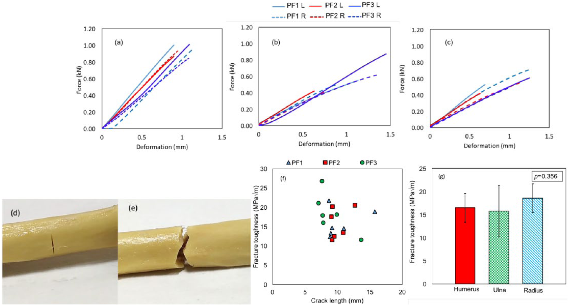
Three-point bending fracture toughness test: Force versus deformation graph for (a) humerus (b) ulna and (c) radius (d) bone sample before fracture (e) bone sample after fracture, (f) crack length versus fracture toughness graph, (g) average fracture toughness for humerus ulna and radius.
There are no established standards available in the literature for testing mechanical properties of bones compared to engineering materials such as metals, ceramics, and polymer composites.51,52 Research groups have experimented on the fracture toughness both on sectioned-bone samples25,26,53,54 as well as on whole-bone samples.43,47,55 In the case of the characterization of human elbows for fracture toughness, whole-bone samples have not been reported yet. Research groups have used various ASTM standards for mechanical testing of bones such as D-790 for bending, 56 C-469 and D-1621 for compression testing,44,57 and E-399 and E-182018,30,53,58 for fracture toughness. In the current experiments, a method developed by Ritche et al was adopted for evaluating bone fracture toughness. 43
Maximum Compression Force and Compressive Strength from Compression Tests
Compression tests were performed on the third set of 8 elbows. Eight samples of humerus, ulna, and radius bones were prepared and tested. Compressive force vs deformation plots of humerus, ulna, and radius are shown in Figures 3A-3C. Figure 3D shows the micro-CT 3-dimensional reconstructed image for a whole-bone sample. A typical fractured sample postcompression test is shown in Figure 3E. Out of a total of 8 samples each of humerus, ulna, and radius, 70% fractured along the longitudinal direction (loading direction) and 30% fractured both in the longitudinal and oblique direction. The average maximum compressive failure force of the humerus, ulna, and radius was 19.75 ± 4.7 kN, 9.90 ± 2.8 kN, and 8.03 ± 2.86 kN, respectively; Figure 4A depicts the same. The difference between the average breaking force for humerus, ulna, and radius was found to be statistically significant with an overall P value of .001. Among multiple comparisons, the compressive force of the humerus is statistically more significant than the ulna and radius (Table 3).
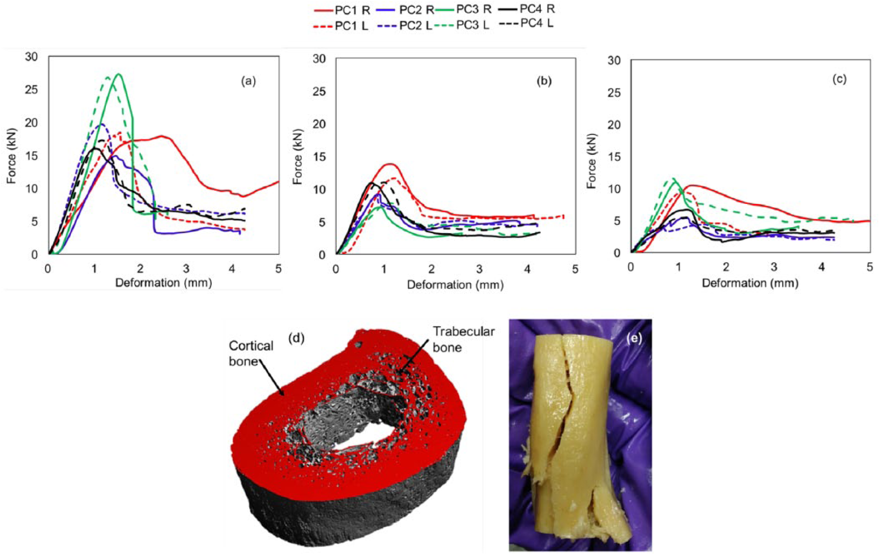
Force versus deformation graph for compression loading of (a) humerus, (b) ulna, (c) radius, (d) 3D reconstructed micro-CT image for bone sample exhibiting cortical and trabecular bone, (e) compression tested bone sample after fracture.
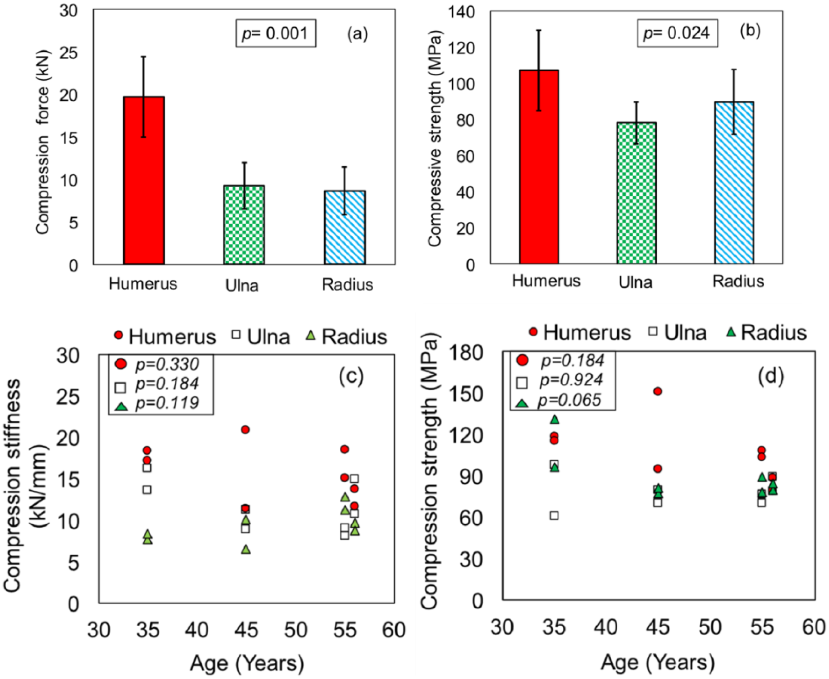
(a) Maximum compressive failure force for humerus, ulna and radius, (b) compressive strength, (c) relationship between compression stiffness and age, (d) relationship between the compression strength and age.
Maximum compressive strength was determined as a ratio of maximum compressive force to the effective cross-sectional area (ie, difference of the outer to the inner cross-sectional area). The average compressive strength for the humerus, ulna, and radius was 107.40 ± 22.15 MPa, 78.19 ± 11.60 MPa, and 89.83 ± 17.88 MPa, respectively, as shown in Figure 4B. These strength values are in close agreement with those reported in the literature for elbow bones. 18 The variation between the compressive strength of the 3 bone types was found to be significant. The compression strength of femur bone reported in the literature ranges from 124 to 211 MPa.19–21,23,59The above-cited values correspond to sectioned samples of the cortical portion of femur bone.
In addition, elastic modulus under compression loading was determined. The values obtained are 3.75 ± 1.27 GPa for humerus, 3.05 ± 1.03 GPa for ulna, and 2.75 ± 0.71 GPa for radius. The difference between the elastic modulus was found to be nonsignificant. Lindahl and Lindgren 16 reported the elastic modulus of bone to be 10.79 ± 0.49 GPa. This difference could be attributed to the sample size selection and the anatomic plane selection of the tested sample. No information about the anatomic plane of the tested sample was provided by the authors. Lindahl and Lindgren used the rectangular sample size of less than 4 mm length at a testing speed of 0.05 mm per minute. 16 To the best of our knowledge, there was no research paper found quantifying the elastic modulus of ulna in compression loading. The elastic modulus (in compression loading) of the sectioned cortical bone sample of femur ranged between 14.7 and 25 GPa.19–21,23,59 The elastic modulus of femur is higher than that of elbow bones.
The compressive stiffness was 15.89 ± 3.42 kN/mm, 11.64 ± 3.01 kN/mm, and 9.27 ± 2.29 kN/mm for the humerus, ulna, and radius, respectively. The compressive stiffness is the measure of how well the load can be dispersed under compression loading in the elastic region.
Compressive stiffness was statistically significant among the 3 bones with a P value of .003.
Relationship Between Compressive Stiffness and Age
The compressive stiffness in the elastic region as a function of age for all 3 bones is depicted in Figure 4C. The compressive stiffness of the humerus, ulna, and radius were found to decrease with the increase in age. The corresponding P value for the linear regression analysis between the compression stiffness and age is shown in Table 4. Though the P value suggests nonsignificance in the variation of compressive stiffness as a function of age, the mean stiffness values display a decreasing trend. To the best of our knowledge, there are no specific studies reported in the literature relating compressive stiffness and age. A decrease of bending stiffness with increase in age, however, has been reported in the literature.4,28,49
Relationship between Compressive Strength and Age
Figure 4D depicts the maximum compressive strength as a function of age. The maximum compressive strength was found to decrease with increase in age for the humerus, ulna, and radius. The P value for the linear regression analysis between the age and the bending strength is shown in Table 4.
Conclusion
This study was conducted to evaluate various mechanical properties of whole/bulk bone samples of human male elbow bones: humerus, ulna, and radius. The following properties can greatly aid both in experimental and computational design as well as for analysis (using finite element or finite difference methods) of implants, modeling of aseptic loosening in the implant-bone interface, and/or modeling of collision/impact on elbows. The following key observations are summarized herewith.
Bending tests: Brittle-type fracture was observed in 3-point bending tests. The bending strength for the humerus was the highest with an average of 128 MPa, ulna at 135 MPa, and radius at 80 MPa. The modulus of the humerus, ulna, and radius was 2.35, 4.50, and 3.67 GPa, respectively. The bending stiffness was observed to be 1.5, 1.1, and 0.7 kN/mm.
Fracture toughness tests: The average force for notched samples in 3-point bending tests for the humerus, ulna, and radius was 0.94 kN, 0.55 kN, and 0.56 kN. The average fracture toughness for humerus, ulna, and radius was 16 MPa√m, 16 MPa√m, and 19 MPa√m, respectively.
Compression tests: The average compressive force for the humerus, ulna, and radius was 20 kN, 10 kN, and 8 kN. The average compressive strength for the humerus, ulna, and radius was 107 MPa, 78 MPa, and 90 MPa. The compression modulus of the 3 bones is approximately 3 GPa. Consequently, the compressive stiffness was observed to be 16, 12, and 9 kN/mm.
There was a significant difference between humerus vs ulna and humerus vs radius with a P value of .001 for bending modulus, bending stiffness, maximum compressive force, and maximum fracture force for notched samples (in 3-point bend tests). However, in the case of compressive strength and modulus for bending, no significant difference was observed among the elbow bones. The elbow bones’ properties such as bending strength, bending modulus, compression strength, compression modulus, fracture toughness, and elastic modulus are observed to be different from the femur bone properties available in the literature.
Footnotes
Acknowledgements
We would like to thank Prof Puneet Mahajan of the Department of Applied Mechanics, IIT Delhi, for providing us access to the characterization equipment. We would like to thank Prof R.M. Pandey, Department of Biostatistics, AIIMS, New Delhi, for his guidance in statistical analysis. We would also like to acknowledge Dr Khursheed Raza, Dr Sanjeev Lalwani and Dr Nazim for their help.
The ethical committee of AIIMS, New Delhi, approved the request for study on cadaveric bone samples according to letters IEC/NP-400/14.11.2014, RP-14/2015, and RP-24/2015, dated 6 February 2015.
Declaration of conflicting interests
The author(s) declared no potential conflicts of interest with respect to the research, authorship, and/or publication of this article.
Funding
The author(s) disclosed receipt of the following financial support for the research, authorship, and/or publication of this article: This work was supported by the Naval Research Board (NRB/4003/PG/359), Department of Science & Technology – Science and Engineering Research Board (SERB) (YSS/2014/000880), Department of Science & Technology – Instrumentation Development Programme (IDP) (IDP/MED/O5/2014), and Ministry of Human Resource Development, Government of India (Design Innovation Center, IIT Delhi).