
Abstract
Background:
Great interest has recently been focused on tooth and tooth derivatives as suitable substrates for the treatment of alveolar bone defects. Here, we propose the use of demineralized baby teeth (BT) as potential grafting materials for bone augmentation procedures.
Methods:
Particles of human BT (Ø < 1 mm) were demineralized by means of a chemical/thermal treatment. Demineralized BT particles were thoroughly characterized by scanning electron microscopy/energy dispersive X-ray analyses to evaluate the effects of the demineralization on BT topography and mineral phase composition, and by enzyme-linked immunosorbent assays (ELISA) to quantify collagen and bone morphogenetic protein-2 (BMP-2) protein contents. The response of SAOS-2 cells to exogenous BMP-2 stimulation was evaluated to identify the minimum BMP-2 concentration able to induce osteodifferentiation in vitro (alkaline phosphatase (ALP) activity).
Results:
The demineralization treatment led to a dramatic decrease in relative Ca and P content (%) of ≈75% with respect to the native BT particles, while preserving native protein conformation and activity. Interestingly, the demineralization process led to a rise in the bioavailability of BMP-2 in BT particles, as compared to the untreated counterparts. The BMP-2 content found in demineralized BT was also proved to be very effective in enhancing ALP activity, thus in the osteodifferentiation of SAOS-2 cells in vitro, as confirmed by cell experiments performed upon exogenously added BMP-2.
Conclusions:
In this study we demonstrate that the BMP-2 content found in demineralized BT is very effective in inducing cell osteodifferentiation, and strengthens the idea that BTs are very attractive bioactive materials for bone-grafting procedures.
Introduction
Over recent years, the field of biomaterials has made significant progress in the search for functional and bioactive materials for bone repair procedures and regeneration. Current clinical approaches for bone defect treatments involve the grafting of autologous (autograft), homologous (allograft), and heterologous (xenograft) bone. 1 Among them, autografts are considered the gold standard because they are biocompatible and non-immunogenic, and they contain every essential component to trigger osteoinduction, osteogenesis, and osteoconduction. As an autologous bone transplant involves the harvesting of bone from the patient’s body, however, it relies on a relatively limited availability of tissue, and it is sometimes associated with a significant donor site injury and morbidity. In this context, allografts may represent a viable alternative to autografts in bone-grafting procedures. Unfortunately, allogenic grafts are associated with the risks of immunoreactions and transmission of infections, and they come with substantial cost issues. As an alternative, a wide range of biomaterials has been envisioned as scaffolds to foster and support tissue formation in vitro and in vivo.2,3 They include metals, 4 ceramics, 5 as well as synthetic,6,7 and natural polymers such as collagen and some composites.8–10
In recent years, tooth11–13 and tooth-derived materials14,15 have come to the fore as very promising substrates for bone regeneration because they closely resemble bone tissue, in terms of matrix composition and topography. It is of note that, unlike bone autografts/allografts, which are rarely available at need, teeth are extracted every day and customarily discarded as waste.
Dentin has recently gained growing interest as a potential osteoinductive material for bone regeneration. 16 Indeed, it is known that bone morphogenetic protein-2 (BMP-2), one of the most potentially effective osteoinductive proteins able to boost bone formation and repair in vitro,17–20 is inherently present in, and released from, native dentin. Interestingly, since demineralization of highly mineralized tissues was found to increase BMP-2 bioavailability, 15 particular interest has been devoted to dentin in its demineralized form.21,22
In this work we have attempted to take a step forward towards the use of tooth derivatives. We herein propose deciduous teeth as potential grafting materials in bone augmentation procedures. In particular, we examined the effects of the well-defined demineralizion protocol recently published; 15 thus we analyzed the morphology and composition of demineralized baby teeth and compared physico-chemical parameters with respect to those of their native counterparts. We also shed light on the effects of demineralization on deciduous teeth organic matter, evaluating whether or not collagen and BMP-2 protein contents were preserved after the chemical treatment. Moreover, we evaluated in vitro the response of osteoblastic cells to exogenous BMP-2 stimulation (at different protein concentrations) with the aim of identifying the minimum BMP-2 concentration able to induce the expression of alkaline phosphatase, the early marker of the osteoblastic phenotype.
Materials and methods
Materials and reagents
Demineralization and sterilization reagents were provided by TT Tooth Transformer S.r.l. (Milan, Italy). Enzyme-linked immunosorbent assay (ELISA) kits (human type I collagen: SEA571Hu; human BMP-2: SEA013Hu) were purchased from Cloud-Clone Corp. (Katy, TX, USA). BCA protein assay kit was supplied by ThermoFisher (Monza, Italy). SAOS-2 (human osteogenic sarcoma cell line) cells were purchased from the American Type Culture Collection (ATCC, Manassas, VA, USA). AlamarBlue® cell viability assay was purchased from Life Technologies Italia (Monza, Italy), while an alkaline phosphatase activity (ALP) colorimetric assay kit was purchased from BioVision Inc. (Milpitas, CA, USA). All other chemicals were from Sigma-Aldrich (Milan, Italy) if not otherwise specified.
Sample preparation
Deciduous (baby) teeth (n = 3; obtained from one child undergoing deciduous tooth extraction) were provided by TT Transformer S.r.l. Human teeth were minced by means of a Tooth Transformer TT machine (TT Tooth Transformer S.r.l.) to give particles (hereafter referred to as BT) of Ø < 1 mm. Following mincing, BT particles were subjected to a demineralization and sterilization process according to a protocol previously described. 15 Briefly, particles were: i) demineralized using a demineralization reagent, at 70° C under shaking by means of a thermomixer (1000 rev/min); ii) washed sequentially with two washing solutions at room temperature (RT) for 2 min; and iii) treated with a sterilization reagent at 70° C under shaking by means of a thermomixer (1000 rev/min). Particles were finally washed at R.T. for 2 min, twice. Demineralized BT particles (hereafter referred as to dBT) were next collected in sterile 1.5 mL polypropylene tubes and stored at RT until use.
In control samples, BT particles were subjected to the same thermal process, but no chemical reagents were used, and deionized water (dH2O) was used, instead.
Evaluation of the effects of demineralization on BT morphology and composition
Analyses of the surface morphology of demineralized and control BT particles were performed with an environmental scanning electron microscope (ESEM Zeiss EVO50, Carl Zeiss, Milan, Italy) connected to a secondary electron detector for energy dispersive X-ray (EDS) analysis. Briefly, following 1.5% (v/v) glutaraldehyde fixation (2 h) and dehydration in gradual ethanol, samples were gold-sputtered, mounted onto scanning electron microscopy (SEM) stubs and examined using an acceleration voltage of 15 kV. SEM images were acquired at ×5000 magnification. EDS analysis was also performed to examine the surface composition (elemental analysis, atomic percentage of carbon, C; nitrogen, N; phosphorus, P; and calcium, Ca) of three different particles from each group. EDS spectra refer to whole SEM pictures.
Type I collagen and BMP-2 protein content quantification
Type I collagen (COL-I) and BMP-2 protein content in demineralized and control BT samples were quantified by ELISA. 15 Briefly, following the thermal/demineralization treatment, ≈50 mg of both BT and dBT particles were transferred into 1.5 mL polypropylene tubes containing 500 μL of extraction buffer (50 mM HEPES pH 7.4, 1 mM PMSF, 2 μg/mL leupeptin, 2 μg/mL aprotinin, 1 μg/mL pepstatin, and 1% (v/v) Triton X-100). After incubation at 4° C overnight (ON), three freezing/thawing cycles, and sonication, proteins were extracted. Total protein content was determined by BCA protein assay kit according to the manufacturer’s instructions. For ELISA assays, 15 μg of total protein extracts (n ⩾ 4 per condition) were loaded and tests were carried out according to the manufacturer’s instructions. Absorbance was then measured at λ = 450 nm using a GENios Plus reader (Tecan, Monza, Italy), and values were referred to an internal standard curve. Results from ELISAs were normalized to the weight of each sample (expressed as grams of sample particles).
Dose–response activity of exogenous BMP-2 on osteoblast cell lines
Mycoplasm-free SAOS-2 cells were cultured in McCoy culture medium containing 1 mM sodium pyruvate, 100 U/mL penicillin, 0.1 mg/mL streptomycin, and 2 mM glutamine, and supplemented with 10% (v/v) FBS, at 37° C in a humidified atmosphere under constant supply of 5% CO2.
Cells were seeded at a density of 3 × 10⁴ cells/cm2 in a 96-well plate in 120 μL/well of culture medium enriched with exogenous BMP-2 at different concentrations (two-fold serial dilution from 200 ng/mL to 0 ng/mL = unstimulated CTRLs) (n = 4 per condition). Cells were cultured for three days, then cell viability and osteodifferentiation were evaluated. Specifically, cell viability was assessed using the AlamarBlue® assay. Briefly, the medium was removed and every well was loaded with 100 μL of specific medium containing 10% (v/v) of resazurin dye. Cells were incubated in standard culture conditions for 2 h and the fluorescence of the medium was read by means of a GENios Plus reader (λex = 540 nm; λem = 595 nm). Viability of CTRL cells was designated as 100%. Osteodifferentiation was evaluated by means of the ALP activity colorimetric assay according to the manufacturer’s instructions. Briefly, at the end of the cell culture period, the supernatant from each well was recovered and assayed, evaluating the hydrolysis of nitrophenyl phosphate in the presence of secreted ALP enzyme. After 60 min incubation at 25° C, the absorbance was read at λ = 405 nm using a GENios Plus reader and values referred to an internal standard curve.
Statistical analysis
Statistical analysis was carried out by GraphPad version 6 (GraphPad software, La Jolla, CA, USA). All data were initially analyzed using the D’Agostino and Pearson omnibus normality test. Comparisons among groups were performed by t-test. Significance was retained when p < 0.05. Data are expressed as mean ± standard deviation (SD, n ⩾ 4).
Results and discussion
In recent decades, a great deal of effort has been devoted to the regeneration and de novo formation of bone tissue. Attempts included bone-grafting procedures, segmental bone transport, and the use of biomaterials for the augmentation of alveolar bone defects.
We have recently proposed the use of tooth derivatives, namely demineralized dentin and enamel, as suitable substrates for bone regeneration due to the inherent similarity of such materials to natural bone tissue.15,23 We have here focused our attention on deciduous baby teeth as a very promising, readily available source of bone-like tissue. Indeed, while most parts of the body are perennial from birth onwards, deciduous teeth are normally replaced and lost through the development and eruption of adult permanent teeth (a process that starts at about six years and stops after 12 years of age). It is worthy of note that every year four million baby teeth fall out and customarily discarded. In addition, although primary teeth are of a smaller size and they contain less dentin and have a thinner enamel layer, baby teeth have the same basic structure as permanent teeth, i.e. enamel, dentin, cementum, and pulpal (nerve) tissue. Therefore, they may represent a valuable biological alternative to the synthetic materials currently used in bone augmentations.
In this context, to the best of our knowledge, this is the first study focusing on the use of deciduous teeth to repair bone defects. In this work native and demineralized BT particles (Ø < 1 mm) were obtained by means of the Tooth Transformer TT machine, according to a protocol previously described. 15 It is known that demineralization improves the in vivo resorption rate of highly mineralized materials while favoring their osteoconductivity and osteoinduction.13,22,24,25 We have thus evaluated the physico-chemical features of demineralized deciduous teeth particles and compared such properties with those of native counterparts.
Demineralization effects on BT surface morphology and chemical composition
The tooth is, basically, a composite of organic constituents, such as collagen and other non-collagenous proteins (e.g. BMPs), and inorganic components, including five types of calcium phosphates (e.g. hydroxyapatite, HAp, and fluorapatite). 23 Highly mineralized materials cannot be easily resorbed, thus limiting or even hampering their osteointegration and bone formation in vivo.13,24,25 In this light, demineralization was found to be very promising for overcoming such drawbacks through the improvement of the in vivo performance of tooth derivatives. 26
In this study we evaluated the effects of the demineralization process on morphology and topography as well as on the chemical compositions of tooth particles by means of SEM/EDS analyses. As shown in Figure 1(a), the SEM image of native BT particles revealed the presence of mineral crystals studding the particle surface. The presence of an inorganic phase was confirmed by the analysis of the EDS spectrum reported in Figure 1(b), which displayed typical Ca and P peaks. As a result of a substantial removal of mineral components, SEM analyses revealed that demineralized BT samples (Figure 1(c)) had a smoother surface, with more and wider dentin tubules when compared to native BT particles. EDS analyses confirmed these observations (Figure 1(d)). Indeed, the demineralization treatment led to a dramatic decrease in relative Ca and P content (%) of ≈75% with respect to the native BT particles, as reported in Table 1.
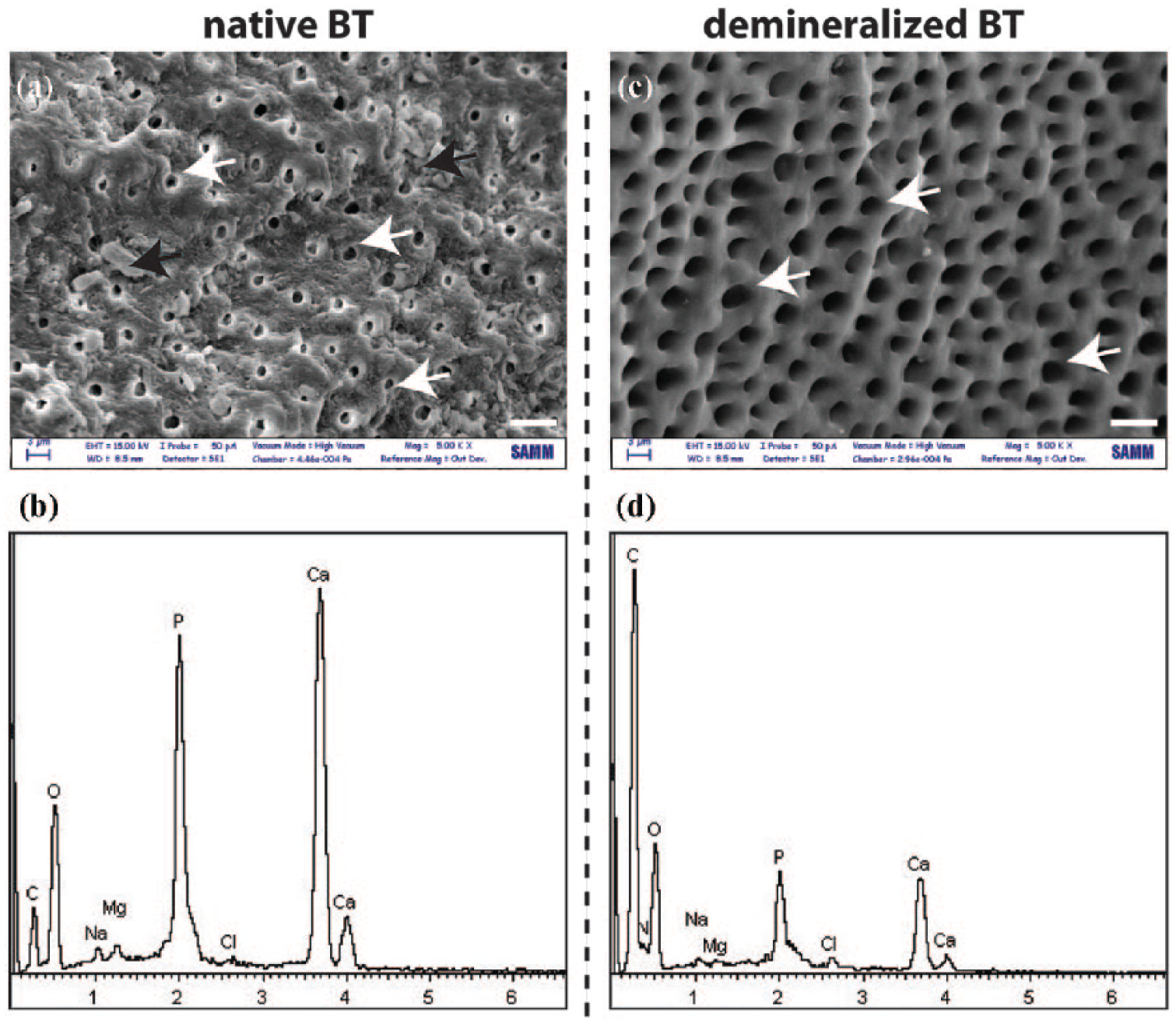
SEM micrographs of (a) native and (c) demineralized baby tooth particles; and (b,d) their corresponding EDS spectra. White arrows point to dentinal tubules ((a) and (c)), while black arrows indicate mineral deposits (a). White scale bar ((a) and (c)) = 6 µm. Magnification: ×5000. x axis = Energy (keV); y axis = intensity (a.u.).
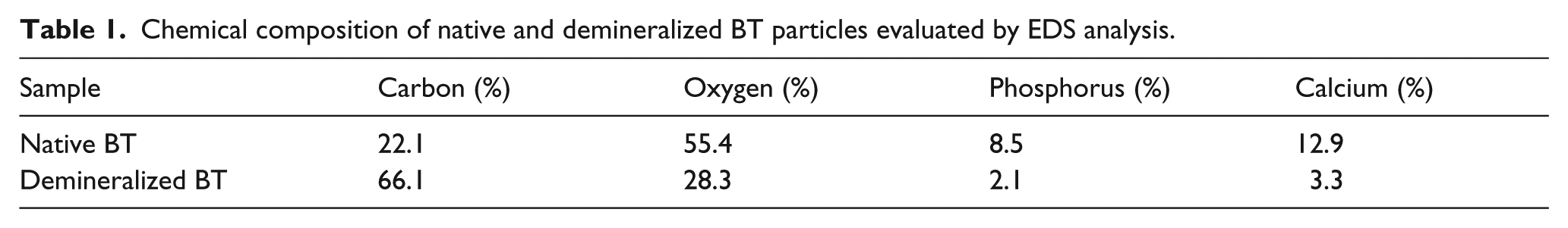
Chemical composition of native and demineralized BT particles evaluated by EDS analysis.
Overall, such findings are in agreement with those previously reported. 15 It is noteworthy that the demineralization process was very effective on the BT samples, and proved to be non-destructive on the protein components that were preserved after treatment.
Demineralization effect on type I COL-I and BMP-2 protein content in BT
Although teeth are one of the most mineralized materials in the human body, they also contain organic components that are mostly collagenous proteins, type I collagen (COL-I) being the most abundant, 27 and non-collagenous proteins, including bioactive growth factors (GFs) such as BMPs. It is well known that tooth collagen is more cross-linked than bone collagen. This implies that tooth collagen is more favorably resistant than any other to thermal and chemical denaturation. 28 On the other hand, the mineral matter has been shown to restrict the bioavailability of bioactive molecules, such as BMPs, 29 whereas the reduction of the mineral phase proved to favor their release. 30 Conversely, some reports have highlighted how some kinds of demineralization process may induce a dramatic damage to the tooth protein matrix.31,32
We thus shed light on the possible adverse effects of the demineralization process we applied to BT particles on the composition of the organic matrix in terms of COL-I and BMP-2 protein content. Besides, as proteins are only biologically functional if folded in a native-like fashion, we decided to quantify only the folded proteins. It is of note that the antibodies used in sandwich ELISAs targeted conformational rather than linear epitopes, which therefore allowed the detection of conformationally native COL-I and BMP-2 proteins and their quantification in BT particles.
The results from the ELISA tests are displayed in Figure 2(a). It is of note that, irrespective of the demineralization, COL-I was invariably present in the BT particles. Specifically, we found a COL-I content of ≈3 ng per gram of BT particles (corresponding to ≈45 ng of COL-I per mL of total particle lysate) in both native and demineralized particles. Such findings are in line with our previous data, 15 and support the idea that the demineralization process allows the preservation of the integrity of the tooth organic (protein) matrix.
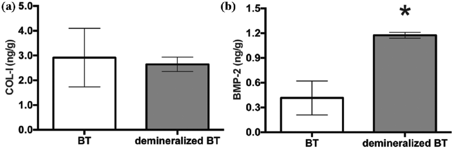
Protein quantification performed on native and demineralized baby tooth (BT) particles evaluated by ELISA assay. (a) COL-I; (b) BMP-2. Protein content was normalized with respect to the weight of each sample (expressed as grams of sample particles). Data are expressed as mean ±SD. * p < 0.05.
It has also been reported that extensive demineralization may induce moderate to severe BMP depletion in teeth and other bone-like tissues. 33 In line with this, Kim et al. 13 found undetectable levels of BMP-2 in tooth homogenates, probably because of the use of chemicals that had denatured and/or depleted some tooth proteins. Instead, the demineralization process we have adopted allowed preservation of the conformation and content of COL-I, one of the most important structural proteins of the extracellular matrix (ECM), and, by the same token, BMP-2, which is a bioactive molecule that plays a key role in osteoinduction. It is also worthy of note that significantly higher levels of BMP-2 were found in demineralized BT particles when compared with their native counterparts (Figure 2(b); p < 0.05). In more detail, demineralized BT contained 1.2 ± 0.3 ng of BMP-2 per g of particles (corresponding to 21.4 ± 6.2 ng of BMP-2 per mL of total particle lysate), which was roughly three-fold higher than the content found in native BT (0.42 ± 0.3 ng of BMP-2 per g of particles, corresponding to 8.7 ± 4.1 ng of BMP-2 per mL of total particle lysate). Again, our results support the idea that the demineralization process we used allowed an increase of the bioavailability of BMP-2 in tooth-derived materials; this in turn giving rise to more osteoinductive bone substitutes. 34 Interestingly, our data suggested that demineralized deciduous teeth contain much more BMP-2 with respect to native and demineralized dentin, 15 which are currently considered to be among the most suitable substitutes used for bone repair and regeneration.25,35–37
Such findings all strengthen the idea that BTs are very attractive bioactive materials for use in autologous bone-grafting procedures.
Effect of exogenous BMP-2 stimulation on osteodifferentiation
BMPs are members of the transforming grow factors beta (TGF-β) family, which play pivotal roles in the commitment and differentiation of progenitor cells in the osteoblastic lineage.38,39 Among BMPs, BMP-2 is a potent trigger in vitro and in vivo. 40 While osteodifferentiation is fundamental for normal bone development, it is important with respect to bone repair strategies as well. In this regard, the development of clinically relevant materials able to promote osteointegration and de novo bone tissue formation, such as those inherently containing a considerable amount of BMP-2, is of capital interest and utmost importance.
As we are aware, in this study we evaluated the dose–response effects of BMP-2 on the ostedifferentiation of osteoblastic cells in vitro. We thus challenged SAOS-2 cell cultures with exogenously added BMP-2. Upregulation of endogenous cellular ALP is a widely used marker for the assessment of cell differentiation into the osteoblastic phenotype. 41 As shown in Figure 3, the stimulation of SAOS-2 cells for three days with variable doses of BMP-2 protein induced a dose-dependent increase in ALP activity. The minimum concentration of BMP-2 able to induce a significant upregulation of ALP protein was 12.5 ng/mL (Figure 3(c); p < 0.05 with respect to unstimulated controls), corresponding to an increase of ≈1.5-fold over unstimulated controls (i.e. 0 ng/mL of BMP-2). As expected, the maximum ALP activity was found when cells were stimulated with 200 ng/mL of BMP-2, corresponding of a ≈1.75-fold increase in ALP levels with respect to controls. Viability of stimulated cells was also comparable with that of unstimulated cells. This is a very intriguing result because it points out that the differences in ALP activity mirrored the real osteodifferentiation of cultured cells (Figure 3(a) and (b)), instead of their mere overgrowth. Such data are in good agreement with previous observations in other cell lines. 41
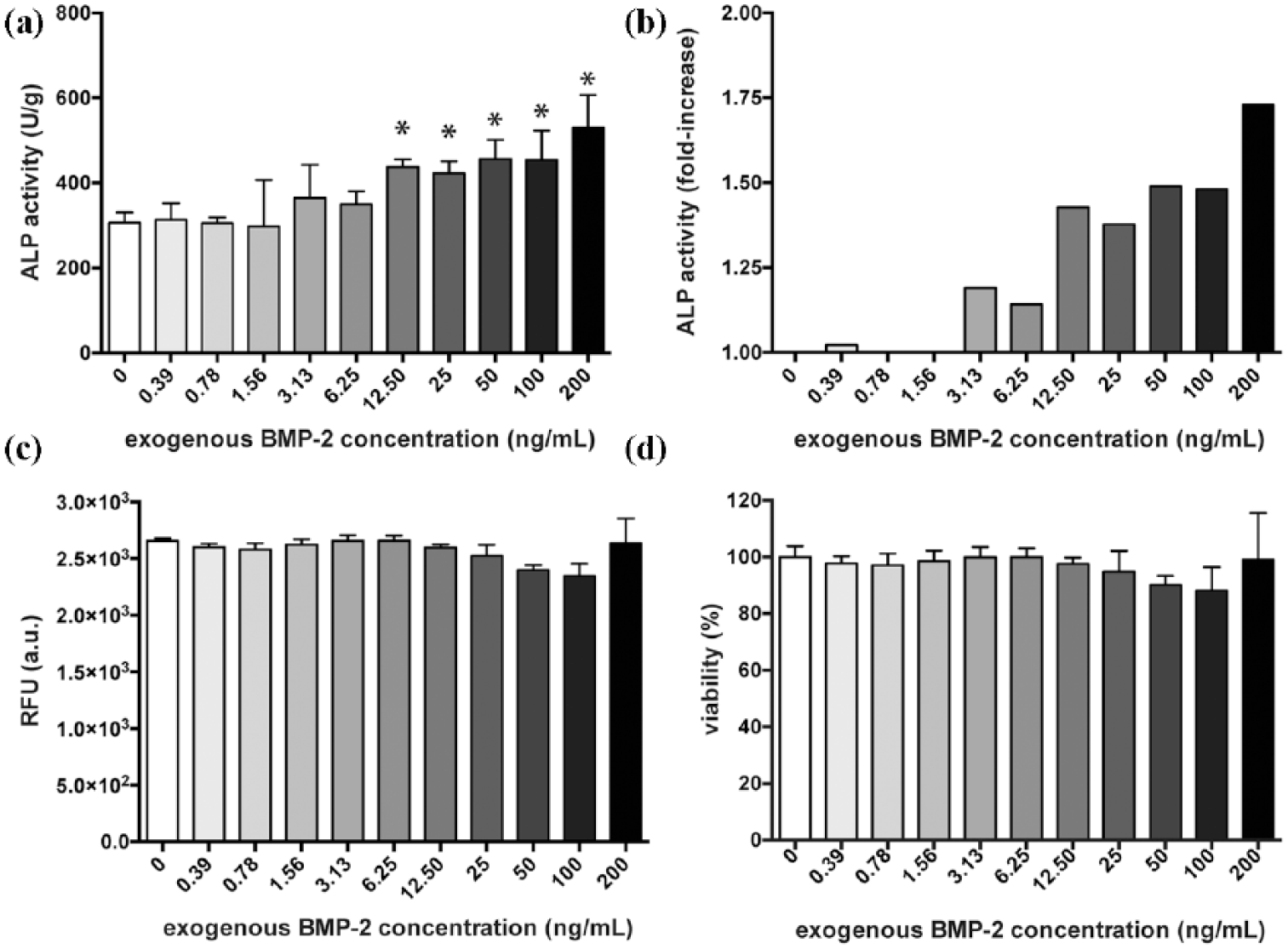
(a), (b) Evaluation of osteodifferentiation using alkaline phosphatase (ALP) activity assay of SAOS-2 cells after three days of culture upon exogenous BMP-2 stimulation. Data are expressed as means ±SEM in terms of: (a) arbitrary units of fluorescence (RFU) normalized to the total protein content in each sample; (b) fold-increase. * p < 0.05. (c), (d) Evaluation of SAOS-2 cells viability after three days of culture upon exogenous BMP-2 stimulation. Unstimulated SAOS-2 cells were used as internal reference (i.e. positive controls for cell growing, denominated as CTRL; 100% cell viability). Data are expressed as means ±SD in terms of: (a) arbitrary units of fluorescence (RFU); as well as (b) percentage of cell viability with respect to the CTRL.
On the basis of ALP activity as a function of the BMP-2 dose used to stimulate cells, we can speculate that the BMP-2 content found in demineralized BT, corresponding to a concentration of ≈22 ng of BMP-2 per mL of total particle lysate, is very effective in inducing cell osteodifferentiation.
Conclusions
In recent years there has been growing interest in teeth and tooth derivatives as valuable alternatives to synthetic biomaterials for bone-grafting and replacement procedures. In this context, due to their high similarity to bone tissue and their wide availability, deciduous baby teeth are very promising candidates for the augmentation or repair of alveolar bone defects. On the other hand, highly mineralized natural biomaterials, such as teeth, bone, and bone-derived materials, display unsuitably low resorption rates in vivo. In this study, we thus propose the use of demineralized BTs as osteoinductive substrates for bone regeneration and replacement. Demineralized BTs were thoroughly characterized by SEM/EDS analyses and compared to native teeth. It is of note that the demineralization process we used did not damage tooth microstructure. Indeed, the organic content was preserved in demineralized deciduous teeth, as demonstrated by ELISA assay used to quantify COL-I content. Interestingly, the demineralization process allowed an increase of the BMP-2 bioavailability in treated BTs, as compared to the untreated counterparts. The BMP-2 content found in demineralized BT also proved to be very effective in enhancing ALP activity, thus in osteodifferentiating SAOS-2 cells in vitro, as confirmed by cell experiments performed upon exogenously added BMP-2. With this perspective, deciduous teeth are very promising candidates as bone substitutes for bone repair and replacement. In this light, in vivo investigation is thus urged to strengthen our findings and to shed light on the performance of BTs for bone grafts in vivo.
Footnotes
Acknowledgements
The authors would like to thank Dr. E. Minetti for providing the deciduous teeth used in this study.
Declaration of Conflicting Interests
The author(s) declared no potential conflicts of interest with respect to the research, authorship, and/or publication of this article.
Funding
The author(s) disclosed receipt of the following financial support for the research, authorship, and/or publication of this article: This work was supported by TT Tooth Transformers S.r.l.