
Abstract
Optical radiation sources, and in particular lasers, find an ever-increasing number of applications in the medical field. It is essential that personnel who are in the presence of an optical radiation source, whether operator, patient or researcher, know precisely the risks inherent in the exposure of the human body to radiation. In order to reduce the risk of biological damage, beyond the provisions of the law on safety regulations, the precise information and accurate preparation of personnel are the main guarantee for the correct use of these sources. In all the application fields, the possibility of a biological damage cannot be completely eliminated, assuming the connotation of occupational risks. In order to understand the risks and operate their effective mitigation, the basic knowledge of the fundamental concepts at the basis of laser-matter interaction will be presented and discussed, with a focus on the physical parameters needed to efficiently estimate and mitigate the related occupational risks, in both a laboratory and clinical context.
Keywords
Significance statement
The use of artificial light sources and in particular lasers is growing rapidly in the healthcare environment for both diagnostic and therapeutic applications. Although light is considered a non-hazardous physical agent for organs other than the eye, it is very important to know how light interacts with biological tissues and to understand its undesirable effects. The precise information and accurate preparation of personnel are the main guarantee for the correct use of these sources.
Introduction
Management of safety issues 1 associated with the use of optical radiation in medicine has increasingly caught the attention of both the medical and physical counterparts, not to mention specific legislation in the field. In this respect, the scientific principles are to be found in the physics of light-matter interaction from two complementary sides: (i) how radiation exerts its therapeutic/diagnostic action; (ii) how the same radiation can contemporarily be a source of risk by interacting with both the operator and the patient. In this scheme, radiation interaction with both biological matter (the operator, the patient) and inanimate matter must be considered. In fact, it is clear that undesired laser reflection by the surgeon’s scalpel may constitute a risk for the eyes and the skin, and the same could be invoked for reflected/diffused light by other specular and/or opaque surfaces such as the flooring, the wall, the surgeon’s coat etc. The effects associated to undesired light-tissue interaction can relate for example, to excessive local heating, burns, temporary or permanent sight impairment, tissue ablation. Of course, light can interact with clothes, furnishings and furniture, and can constitute a possible fire hazard depending on the specific conditions.
Laser-matter interaction can be described by different approaches, ultimately relying on the interaction between photons and the atoms/molecules forming the specific encountered medium. Following a brief description of the relevant physical quantities and photon-matter interaction in the UV-visible and near infrared range, we will first consider the phenomena at the basis of laser-matter interaction (reflection, refraction, absorption, scattering, transmission), then illustrate the light-induced effects in terms of photo-thermal, photo-chemical and photo–mechanical effects. In both cases, the dependence of the considered phenomenon on both the light and matter properties will be considered.
In most phenomena related to laser-matter interaction in the biomedical field, extreme spectral purity (monochromaticity) is not a must-have property. This is associated with an increasing use of non-directional and/or non-coherent radiation whose spectrum can be relatively broad (e.g. FWHM of a few tens of nanometres). In a first instance and to a good approximation, those sources can be included in the following considerations by associating them with their peak wavelength.
Let us briefly illustrate some fundamental concepts at the basis of optical radiation interaction with the elementary constituents of organic matter. As is well known, the description of the structure of matter at the microscopic level is a very complex problem, which requires the concepts of quantum mechanics. It is possible to give a very qualitative picture of the physical phenomena affecting safety problems by using extremely simplified model situations which, however, maintain the salient elements of the characteristics of the processes in which we are concerned.
Primary constituents of matter are atoms and their aggregates, such as molecules, metals, semiconductors, etc. Quantum mechanics tells us that an isolated atom can only absorb precise discrete values of energy, differently from the classical mechanics interpretation where every system can take any possible amount of energy. These discrete energy levels define different atomic configurations, that is, different arrangement of electrons around the atomic nucleus. The separation between two consecutive energy levels is not constant but decreases as the energy of the atom increases. A similar description is also valid for molecules, while in the case of an atomic aggregate such as metals or semiconductors, the complex interaction among the various atoms compacts and groups the energy levels in a way to form bands. Inside each band, the energy levels are distributed continuously, but different bands can be spaced, thus defining an energy interval without allowed energy levels called ‘energy gap’.
To understand the light-induced effects, let us start by describing the fundamental optical properties needed to understand the propagation of optical radiation in tissues, as well as inanimate matter, such as absorption, transmission, reflection and diffusion.
Mechanisms of optical radiation-matter interaction
Biomedical optical applications rely on light sources that are generally based on lasers or on Light-Emitting Diodes (LEDs). Table 1 briefly resumes the definition of the most relevant physical quantities used to characterise the light sources and their interaction with matter, in the context of both clinical applications and for the management of the possible safety issues associated with their use.
Definition of the most relevant physical quantities used to characterise the light sources and their interaction with matter.

Each quantity is described and provided with its more widespread symbol and unit.
Light can behave as a wave (i.e. electromagnetic radiation) or as a particle, called photon. Although these two descriptions are very different, they are consistent with each other. The electromagnetic radiation is classified according to the wavelength. The most relevant regions for biomedical applications are: the ultraviolet (UV-A: 315–400 nm, UV-B: 280–315 nm, UV-C: 100–280 nm), the visible (approx. 400–780 nm) and the near-infrared (780–2500 nm). If we consider a laser beam characterised by wavelength
Let us consider a typical situation, in which a beam of light impinges on a slice of matter (biological tissue, plastic sample, fabric, etc. . .), as depicted in Figure 1. There are essentially four effects that govern the light transport: reflection and refraction, scattering and absorption. The first three take place whenever the propagating radiation encounters a discontinuity in the index of refraction (defined as the ratio between the speed of light in vacuum and the speed of light in the medium). In biomedical laser application, reflection and refraction play a significant role only when very transparent media are irradiated, such as the corneal tissue. In opaque media, they are subtle effects while absorption and scattering are the dominant ones.
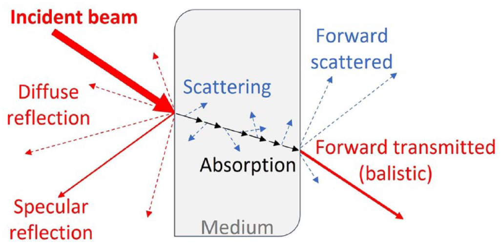
Scheme of the effects governing light transport in a medium.
Absorption
Photon absorption processes are ultimately responsible for the therapeutic effects of light. The study of light absorption by biological media is also very important in the framework of safety issues (for both the operator and the patient), together with the knowledge of the scattering processes. In general, absorption can be characterised by micro- and macroscopic parameters, depending on the properties of both the impinging light and the encountered tissue.
The absorption process is the phenomenon in which atoms and molecules, interacting with an electromagnetic radiation, absorb some energy from the field and convert it into internal energy, which can be described in terms of energy levels. If we consider a molecule in a well-defined energy level
Excited molecules tend to release the absorbed energy and return to the initial state, via a so-called relaxation process. The relaxation process may be accompanied with radiation emission (fluorescence or phosphorescence) or by non-radiative relaxation processes. These processes can occur with direct transition from the excited state towards a lower energy level, or with a series of intermediate transitions in which the released energy is transferred more gradually to the surrounding medium, generally in the form of heat. The excited molecule can also use its energy to rearrange its bond orbitals, resulting in chemical reactions that can end up in new stable molecular species. In this case we speak of a photochemical channel. A typical structure of energy levels for polyatomic molecules is indicated in Figure 2, where the most important relaxation processes are depicted.
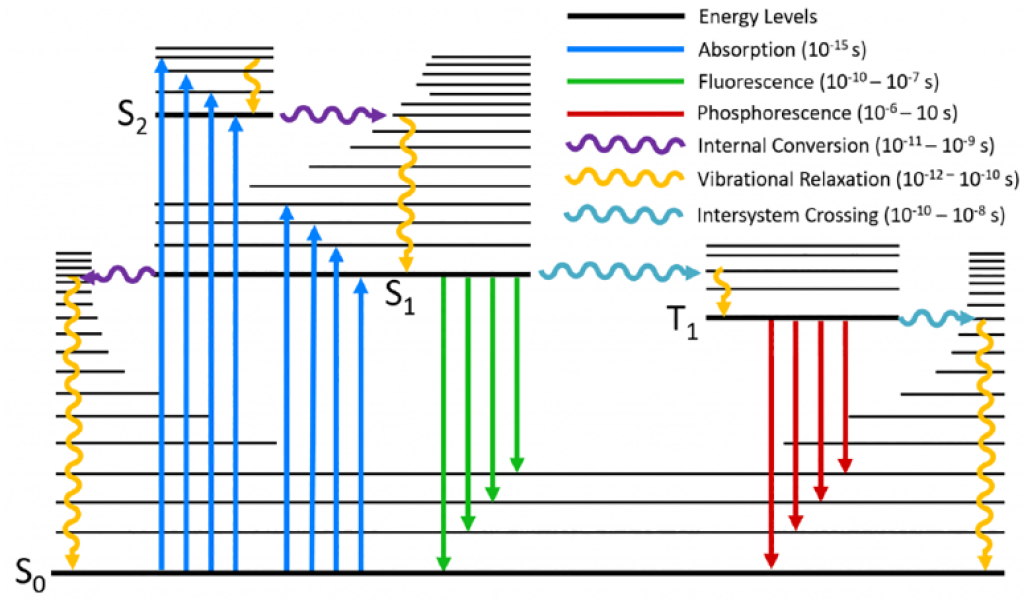
A typical Jablonski diagram showing the possible radiative and non-radiative transitions following a photo-excitation of a molecule (pointing-up arrows). Different energy levels of the molecules are depicted by the horizontal lines (electronic and vibrational states depicted with thick and thin lines respectively), while the arrows represent the various transitions that can happen between different layers: straight and wavy arrows indicate radiative and non-radiative transitions respectively. The label Sn and Tn stands for singlet and triplet state (referred to the basis of net spin quantum operator), while the subscript n indicates the n-th excited state (0 stands for ground state). For the different transitions, the typical timescale is also indicated. Source: Edinburgh Instruments www.edinst.com.
At the microscopic level, absorption is described by the absorption cross section

where
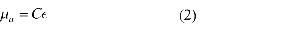
where

The absorption coefficient is a fundamental parameter to describe the optical properties of a material because it is wavelength dependent, thus very useful when comparing absorption from different lasers.
In reference to Figure 3, let us consider a collimated radiation field of intensity
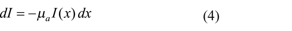

Illustration of light absorption by a purely absorption medium (in grey).
Integrating the previous equation from the surface of the material, assumed to be at

describing the exponential attenuation of a light beam propagating inside a purely absorbing medium. A very useful quantity used in the biomedical and chemical field is the absorbance
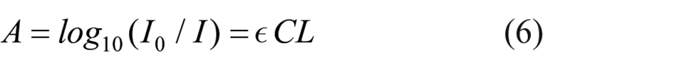
where

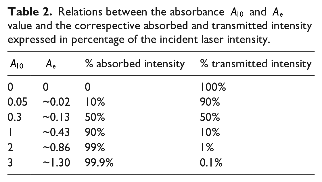
Relations between the absorbance
For the sake of completeness, when the number of photons per unit time and volume investing the medium exceeds a given threshold, nonlinear phenomena can arise, such as two-photon absorption or upconversion processes, that are not considered in equation (5). These multiphoton processes are only possible with the use of very powerful and short laser pulses, with increasing applications in the field of bioimaging and nanomedicine,6,7 especially associated with the use of externally-delivered nanoparticles.
Scattering
Scattering in tissues is the result of light interacting with random variations in refractive index (whose average in tissue lies in the range 1.36–1.40, and 1.46 in adipose tissues). Considering a biological sample, the membrane, organelles and the other intracellular structures are characterised by a larger index of refraction if compared to the intracellular liquid embedding them. A similar relation is also valid for the extracellular structures and proteins with respect to the extracellular fluids. The overall result of this heterogeneity ends up in the scattering effect.
From a physical point of view, scattering can be studied using a simple model where scattering particles have a characteristic dimension represented by a radius

A scattering event occurs when an incident photon, travelling in the direction
On encountering one scattering particle within a homogeneous medium, photons travelling in a direction
Laser penetration in tissues
The behaviour of a light beam propagating inside a tissue mainly depends on λ and on the specific tissue type, being described in terms of absorption and scattering. We now want to merge these two effects to define a single model describing light penetration.
Let us consider a collimated laser beam impinging on a tissue surface in the case of a pure absorbing medium. This hypothesis of absence of scattering may look like a brutal simplification for biological tissues, even if it will be possible to include the scattering effect by a proper modification of this simple model. This assumption allows us to start from the Beer-Lambert law derived in the previous section (see equation (5)), that describes the light intensity at various depths in the medium. If we introduce a new parameter called ‘penetration depth’ or ‘extinction length’

Equation (5) can be written as:

The penetration depth
It is important to note that the definition of
It is worth noticing that
Up to now we have considered purely absorbing media, but for a comprehensive description of the light-tissue interaction, we need to merge absorption with scattering. If we now add scattering properties to the medium, both absorption and scattering will contribute to deplete the beam of photons. We can calculate the amount of unscattered light intensity

where

Example of a simple setup used to measure the unscattered light intensity, also called coherent or ballistic component, that arise from the propagation of a collimated light beam through an absorbing and weakly scattering media. The usage of an aperture allows for the detection of only the unscattered light.
The situation becomes more complicated if we want to determine the overall intensity of light at a given depth inside a medium, considering both the unscattered and the scattered light, besides the absorbed one. Media where the scattered light component is non-negligible respect to the unscattered one are called turbid media. The complexity in turbid media is introduced by the presence of multiple scattering events. The medium can be considered as a group of absorbing and scattering centres whose random interactions with the photons are governed by quantitative coefficients. The Boltzmann transport model establishes the right physical approach to solve a similar problem. Without entering mathematical details, we consider only two biologically relevant limiting cases: dominant absorption and dominant scattering.
In the absorption-dominant limit
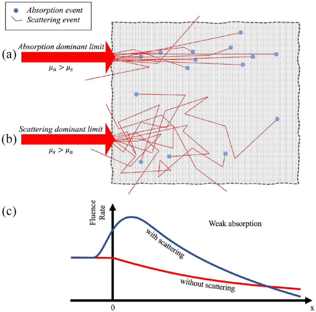
Schematic illustration of a laser-tissue interaction in the absorption (a) and scattering (b) dominant limit. The big arrow represents the laser that impinges on a turbid medium (dot-filled grey space). The thin lines represent the possible photon paths where the single scattering or absorption events are illustrated. (c) Represents the fluence rate, in case of weak absorption, showing the difference between scattering and not scattering medium.
In the scattering-dominant limit (also known as diffusion limit) photons undergo many scattering events before being absorbed, which results in a much greater path before being absorbed (see Figure 6(b)). In such a scenario it is convenient to introduce the reduced scattering coefficient µ' s defined as:

Its physical meaning is clear considering its inverse
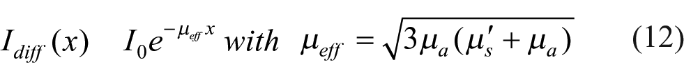
where


Optical penetration depth of light into human mucosa and skin over the wavelength range from 400 to 2000 nm. Data adapted from. 13
It is clear that an increase in µ
s
leads to an increase in µ
eff
and a decrease in
Even if it is clear that the penetration depth does not change with laser intensity, it is worth remembering that a higher impinging intensity results in a higher energy fluence at any depth in the tissue, once the irradiation time (i.e. the treatment) is fixed. Vice versa, a higher energy fluence can be obtained in deep tissue layers by increasing the interaction or treatment time, once the laser intensity (at the first tissue layer) is fixed. These are non-trivial considerations, as many light-matter interaction phenomena depend upon the absorbed energy, which in turn is a function of the energy fluence arriving at a given depth. For example, in the case of photo-chemically induced production of cyto-toxic species (like ROS, Reactive Oxygen Species), a threshold can be defined and associated with irreversible effects, leading to cell death like in the case of photodynamic therapy. This threshold can be expressed in terms of total number of produced ROS per cell; alternatively, it can be expressed in terms of number of absorbed photons and ultimately of light dose. Therefore, it is clear that any deep target can in principle be reached by an over-the-threshold dose. In this scheme many drawbacks can be present, related to possible tissue overheating in the more superficial layers or other undesired effects.
Photo-induced effects
When laser light interacts with biological tissues, a variety of mechanisms take place that depend on both the specific characteristics of the tissue and the parameters of the laser emission. In the following, we will focus on non-radiative processes, discarding radiative processes due to their negligible importance with regard to laser safety, being of interest mainly for diagnostic purposes.
Non-radiative processes can be classified according to a chart (Figure 8) which relates them to the laser irradiance

Map of photo-induced effects according to exposure time and light source irradiance.
Once
According to Figure 8, there are three main photo-induced effects: photochemical, photothermal and photomechanical, according to increasing values for the energy fluence.
Photochemical effects
In general, photochemical processes occur for much lower power and longer excitation time than those needed to obtain photothermal and photomechanical effects (see chart in Figure 8).
Photochemical effects can be modelled by chemical reactions where the photon is one of the reagents, leading to one or more products upon photon absorption. These reactions represent the first step in a series of complex processes, eventually leading to a biological response and accompanied by possible damage in the exposed tissue(s). In fact, the photoproducts formed upon photon absorption by the target chromophore(s) elicit reactions which can directly or indirectly alter a cellular or enzymatic or biochemical function, such as in the case associated with the photo-induced production of reactive oxygen species (ROS). For example, solar erythema (sunburns) is a common and well-known photo-induced effect in the case of solar UV exposure of the skin, based on photochemical processes. Drug-induced photosensitivity (photoallergy and phototoxicity) are other undesired effects associated with photochemical processes elicited by the presence of both light and light-absorbing drugs, especially if systemically administered. As reported in Kowalska et al., 17 ‘photoexcitation and photoconversion of drugs trigger multidirectional biological reactions, including oxidative stress, inflammation, and changes in melanin synthesis’.
Among the various photochemical processes (see Figure 9), the ones of greatest interest for safety issues are molecular photodissociation, photo isomerisation, inter- and intramolecular energy transfer. Examples of photochemical effects are UV-induced synthesis of vitamins or DNA and cellular damage. In the visible range, the eye (and in particular the retina) can be damaged starting from reasonably low laser irradiance, with a threshold which can be far lower than the one causing skin damage. A different damage induced by visible laser radiation can occur on the skin of a patient who is being treated with exogenous photosensitising products. In fact, a patient undergoing a photo-dermatological treatment for a skin pathology, may be administered drugs which increase the photosensitivity of the region to be treated prior to irradiation. This is the case of the Photodynamic Therapy (PDT) approach, 18 where exogenous light absorbers (called ‘photosensitisers’ – PS) are delivered in situ and irradiated with a laser whose emission matches the PS absorption spectrum. In these cases, undesired and possible PS distribution inside the healthy tissues can occur and its activation must be properly limited by avoiding irradiation outside the area under treatment.
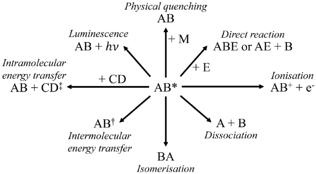
Chart of the photochemical reactions following photon absorption by the molecular species AB, being AB* the excited species.
The skin is the target organ for various visible and UV light phototherapy treatments having a strongly wavelength-dependent effect that generally causes inflammation.19,20 During this process, the DNA is one of the most important targets of UV light, having an absorption maximum at about 260 nm. Wavelengths in the 240–300 nm range interact mainly with nucleic acids, amino acids, urocanic acid and melanin, which is the chromophore acting as a shield for radiation between 300 and 400 nm. The amount and location of chromophores in the different skin layers, as well as variations in the thickness of the epidermis and stratum corneum, determine the degree to which UV radiation is absorbed.
Recently, partly as a result of the COVID 19 pandemic, there has been much increased use of UV radiation below 300 nm for sterilisation of airborne pathogens, with particular interest for
Photothermal interaction
The conversion of optical energy into thermal energy is at the basis of many laser applications, both in the biomedical and industrial fields. In this case, the photon energy is deposited into the target tissue via energy transfer towards rotational and vibration modes of the target molecules. This increases the molecular mean kinetic energy with the results of a local temperature increase.
The photothermal reaction responsible for temperature increase in a biological tissue can be described according to the following two-stage process:
A
inelastic collision of A* with a molecule M of the medium transfers the excess energy ∆E of A* towards M
To analyse the photothermal effects we must consider three steps: heat generation, heat transport and heat effects.
Heat generation is determined by the laser parameters (mainly irradiance and exposure time) and the tissue optical parameters, such as the absorption coefficient, the thermal conductivity, the mass density and the specific heat. The magnitude of the biological effects of heat are largely controlled by the target molecule(s) absorption coefficient as depicted in Figure 10: water, emo proteins, pigments (such as melanin, carotenoids, flavin, bilirubin etc.), other macromolecules (e.g. nucleic acids and aromatic molecules) and possibly nanoparticles, whose de-excitation path(s) may be optimised to perform localised photothermal therapy.

Absorption spectra of the main body pigments and water in the 100 nm – 10 μm range. Image from. 22
From the theory of heat diffusion,
23
in a time

where

corresponding to the time needed for heat to propagate along a distance equal to the light penetration length. Equivalently,

This corresponds to the heat confinement regime: heat remains confined into a volume

In this case, heat has the opportunity to diffuse in the tissue over lengths greater than the optical penetration. Thermal damage of the tissue adjacent to the volume affected by the radiation is therefore possible.
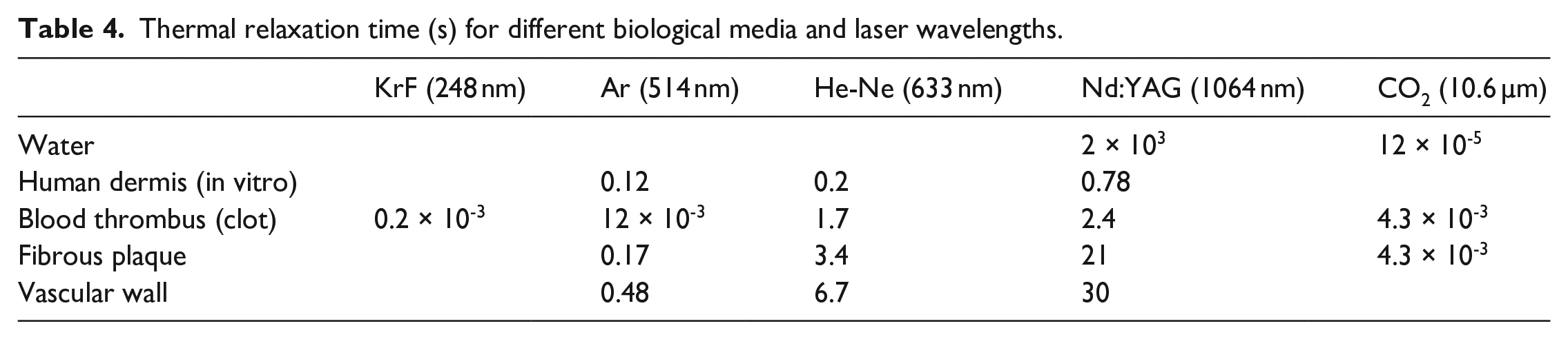
These parameters are very important for both therapeutic applications and safety issues, as they can give an estimate of the tissue volume damaged by a light-induced temperature increase. See Table 4 for a list of thermal relaxation times for some biological tissue relative to the more widespread biomedical-laser sources.
Thermal relaxation time (s) for different biological media and laser wavelengths.
The heating of the biological tissue in the range 37°C–42°C has no measurable effects. The hyperthermic regimen (42°C–50°C) is accompanied by changes in macromolecular conformation, destruction of molecular bonds and alterations of the cell membrane with consequent tissue retraction. Cell mortality via apoptosis is also observed, in particular for cells of oncological type. In the coagulation regimen (50°C–60°C), enzymatic activities are modified and reduced, while macromolecule denaturation begins; this is the basis of the coagulation process. Of particular interest is heat-induced denaturation of collagen. This transition is accompanied by a noticeable structural contraction of collagen fibres and by a visible change in the absorption and scattering properties, such as for thermal denaturation of the egg white. Vaporisation starts at around 100 °C, mainly due to the heating of the water contained in the tissues. The transformation of water into steam produces a great increase in volume, accompanied by cell wall explosive rupture and escape of steam. The high vaporisation heat of water (about 2300 J/g) is responsible for excess heat dissipation by vapour formation and escape, preventing further temperature increase. After complete water evaporation, the residual tissue fragments undergo a rapid increase in temperature until reaching 300–400 ° C, when the tissue blackens and carbonises producing gas and smoke. If the temperature exceeds 500 °C, the tissue burns and evaporates. All these thermic regimens are summarised in Table 5.
Photothermal effects of laser-tissue interaction as function of the tissue temperature.

Photomechanical interaction
When the duration of the laser pulse is less than microseconds, in general, photomechanical effects develop alongside processes of a purely thermal nature. These manifest themselves as pressure pulses which propagate both in the area in front of the irradiated surface and in the tissue itself. When the pulses are of short duration of the order of a nanosecond or even better than a picosecond, the mechanical shock waves that are generated can damage the very structure of the medium in which the wave propagates, especially if this is biological matter.24,25 This phenomenon can also be used for medical purposes, such as laser lithotripsy, 26 intravascular treatment 27 such as laser angioplasty and endovenous laser ablation, dermatological applications such as skin rejuvenation 28 or even cosmetic purposes such as the removal of tattoos.
In some of these applications, laser light is delivered intra-operatively while in others the laser beam is propagating in the air before reaching the target organ (e.g. the skin). This should be considered when analysing safety issues, being it clear that the basic safety principles in the use of optical radiation sources must be followed in any case.
Practical considerations
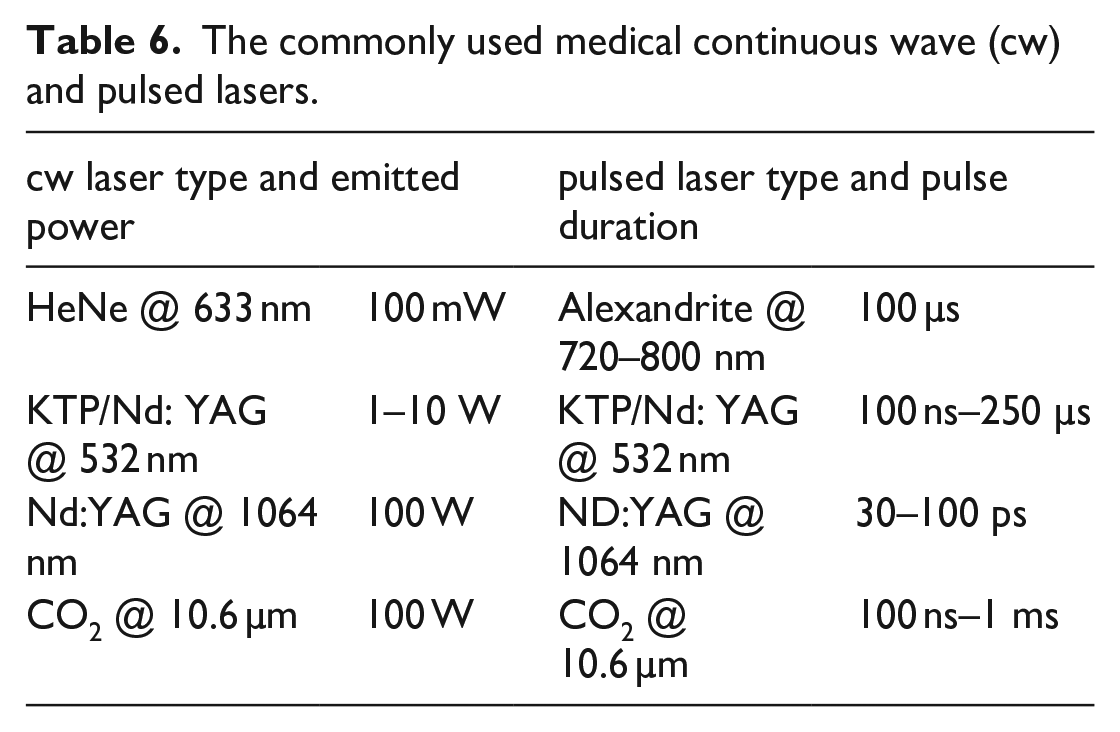
To further clarify the most relevant photo-induced effects and connect them with biomedicine applications, let us consider Table 6. This table reports some of the most employed continuous (cw) and pulsed biomedical lasers, together with some important characteristics such as the radiant power for the cw lasers and the pulse duration for pulsed ones. According to Figure 8, a proper combination of the irradiance and exposure-time allow to initiate a specific photo-induced effect on the target tissue. Once the laser operation regime has been chosen between cw or pulsed, as well as its power, the irradiance can be modified by a proper optical system (e.g. optical lenses) that modifies the laser beam diameter, further expanding or focalising it. We recall that the irradiance is increased by a factor 10 if the same power is delivered onto a circular surface with one-third smaller diameter. In this respect, an often underestimated risk consists in the potential production of hazardous airborne contaminants obtained during the laser-tissue interaction. Laser induced pyrolytic products of biological samples can contain toxic by-products among which known carcinogens. 29 The operating environment must be properly ventilated to reduce worker and patient exposition risk.
The commonly used medical continuous wave (cw) and pulsed lasers.
Conclusions
All the light-matter interaction phenomena are intrinsically dependent on both λ and the tissue optical properties. Therefore, very different physical and biological effects can be obtained by different lasers irradiating the same tissue type or, vice versa, by the same laser irradiating different tissue types.
Light interaction with biological matter can be described with a sequence of distinct events, which can be grouped into two main phases: damage induction and damage response. The first is completed in a time span of the order of seconds and includes the processes described above. In general, the ray propagation model can be invoked for a first understanding of the possible sources and/or conditions of danger while using laser beams in a biomedical environment. This can apply for example to the identification of reflective surfaces to be kept far from the laser beam, or estimate the beam dimensions at any distance from the source from its divergence. At the same time, the laws describing laser absorption and scattering inside a given tissue or medium drive the knowledge of the main effect(s) associated to their interaction (photochemical, photothermal, photomechanical). For example, in the absence of suitable chromophores able to selectively increase the extinction coefficient of the biological material (e.g. tattoo pigments, melanin), the effects of thermal increase have poor specificity, as in the case of infrared light absorption by water. On the contrary, processes based on photochemical reactions are highly specific and strongly dependent on the radiation wavelength.
Out of the knowledge of laser – biological matter interaction principles, a list of questions may arise and be applied to the specific working conditions, for analysis of the possible risks and the relative countermeasures: (i) which are the laser emission characteristics and the relevant tissue type(s)? (ii) which is the interaction geometry? are non-negligible reflections to be expected (which is the surface(s) type(s) and quality?) (iii) are there known absorbers? Which is their depth into the tissue? How is this related to the laser penetration depth and the expected photo-induced effects? (iv) Which emission and/or geometry parameters can be controlled/changed to maximise treatment efficacy and safety?
Together with the increasing therapeutic use of laser radiation, new and more complex issues arise regarding their safety of use, not only for the operator but also for the patient. For example, in intraoperative use of lasers30,31 internal organs become direct light targets, being the surrounding tissues/organs part of the possible indirect/undesired targets. In the last years, an increasing number of solutions have been developed to reach ‘difficult’ districts by light, such as the bladder, prostate, stomach, lungs, throat etc. mainly in the development of alternative and/or coadjuvant antitumoral therapies. At the same time, new ways to deliver light are being developed and commercialised, consisting in for example, diffusive optical fibres, modified gastroscopes or bronchoscopes, intracutaneous needles, ingestible or even inhalable sources. 32
Together with the research advancement in terms of light sources, treatment planning is rapidly evolving in the optical field, including sophisticated modelling of the interaction between light and the target organ(s) to predict light distribution in space and time, together with the analysis of the relevant photo-induced effects. This makes this field exciting but introduces new challenges from the safety point of view. For these reasons, the knowledge of light-matter interaction principles will be more and more necessary in the years to come.
Footnotes
Acknowledgements
The research activity reported in this manuscript has been performed in the framework of the regional project ‘Suppression of Airborne Viral Epidemic Spread by Ultraviolet light barriers (SAVES-US)’ and the project ‘Endoscopio luminoso per il trattamento dell’Helicobacter pylori (EndoLight)’, POR FESR 2014-2020.
Declaration of conflicting interests
The author(s) declared no potential conflicts of interest with respect to the research, authorship, and/or publication of this article.
Funding
The author(s) disclosed receipt of the following financial support for the research, authorship, and/or publication of this article: These research projects are funded by Tuscany Region.