
Abstract
MicroRNAs (miRNAs) are deregulated in human cancers. A large number of studies have shown that miRNAs play critical roles in tumorigenesis, either as oncogenes or tumor suppressors. This review provides an overview of current research on the roles of miRNA in cancer development. Furthermore, we also discuss the current progress and limitation of therapeutic application of miRNAs in preclinical studies.
Introduction
MicroRNAs (miRNAs) are a group of small, endogenous, noncoding RNAs that are evolutionarily conserved and widely distributed among species. These small RNAs can regulate the gene expression through messenger RNA (mRNA) degradation or translation inhibition, which generally forms imperfect base pairing to the 3′ untranslated region (3′UTR) of target mRNAs. 1 Like most mRNAs, miRNAs are initially transcribed as long primary precursors with a 5′ cap and 3′ poly-A tail, which are then processed by Drosha/DGCR complex into pre-miRNA and then spliced by Dicer into 18- to 22-nt duplex RNAs. 2 After that, one strand of miRNAs will be loaded into the RNA-induced silencing complex (RISC), in which another strand, referred to as passenger strand, is degraded. 3 Besides the above-mentioned canonical miRNA biogenesis pathway, alternative pathways, including Drosha/DGCR8-independent or Dicer-independent pathways, have also been proposed. 4
Unlike conventional protein modulators, miRNAs have several unique features, including tissue-specific or multiple-target regulation. For example, miR-1, miR-133, or miR-206 is highly expressed in muscle tissues. 5 Deletion of miR-1 in flies resulted in embryonic lethality, whereas targeted inhibition of miR-1-2, one copy of miR-1, in mice caused 50% embryonic lethality with cardiac effects,6,7 thus indicating that these muscle-specific RNAs play a key role in development and physiological functions. More interestingly, each miRNA may regulate hundreds of function-related target genes, thus fine-tuning the cell’s response. The human genome contains at least 1000 distinct miRNAs, which potentially regulate more than 30% of the transcriptome. It has been demonstrated that miRNAs are involved in varieties of basic biological processes, including cell cycle, proliferation, migration, apoptosis, and development.8–11 In this review, we mainly summarize the roles of miRNA in cancers and highlight promising advances for these small RNAs in cancer therapy.
miRNAs in Cancer
A great number of studies have shown that miRNAs are highly involved in cancer development. First, miRNAs are not randomly distributed in the human genome. 12 For example, chromosomes 17 and 19 have significantly more miRNAs that other chromosomes, among which chromosome 4 has fewer than average miRNAs. Indeed, more than half of miRNAs are located at chromosomal regions that are associated with cancers. It was reported that the miR-15 and miR-16 genomic locus is frequently deleted in chronic lymphocytic leukemia, promoting tumorigenesis due to the increase of BCL2, one of target genes regulated by miR-15 and miR-16.
Second, miRNAs have been proved to be globally deregulated in human tumors. When examining the expression of 200 miRNAs in more than 300 human samples, Lu and colleagues 13 found distinct expression profiles of miRNAs between six pairs of normal and tumor samples. Subsequently, the expression patterns of miRNAs have been intensively investigated in human samples, especially because of the development of high-throughput sequencing technologies. A large number of miRNAs have been found downregulated or overexpressed in different types of tumors. For example, miR-21 or the miR-17-92 cluster is upregulated in colorectal, liver, or other carcinomas. 14
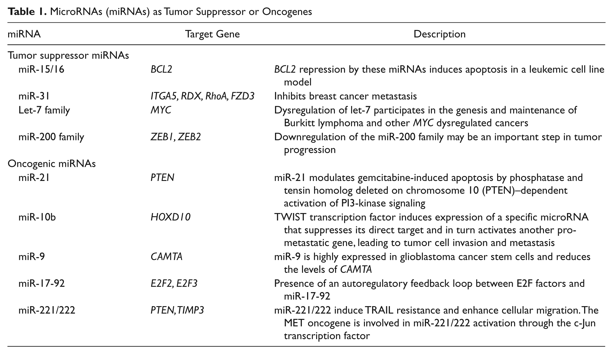
Third, many miRNAs have been shown to suppress or promote carcinogenesis, acting as either tumor suppressors or oncogenes ( Table 1 ). The decrease of those miRNAs as tumor suppressors, which occurs due to either gene deletion or promoter methylation, can promote oncogenes. For example, the let-7 family has been found to negatively regulate expression of RAS, an oncogene that contributes to the pathogenesis of human tumors. 9 In contrast, another group of miRNAs, referred to as oncomirs, are overexpressed in cancers. As described above, the miR-17-92 cluster, which comprises six miRNA genes (miR-17, miR-18a, miR-19a, miR-20a, miR-19b-1, and miR-92a-1) can induce cell proliferation, inhibit apoptosis, or promote tumorigenesis by cooperating with MYC in lymphomas.10,15
MicroRNAs (miRNAs) as Tumor Suppressor or Oncogenes
Last, but not the least, we quantitatively and systematically characterized the capacity of all 900 or so known human miRNAs that regulate several cell features of cancer metastasis, including cell migration, invasion, or apoptosis. 16 Strikingly, it was found that more than 20% of miRNAs demonstrated the regulatory activities on cell migration in diverse cancer cells. It is well known that miRNAs act mostly like functionally regulatory molecules and tune the expression of target genes to physiologically relevant levels. Since cell migration is a basic biological process, it is quite possible that a general involvement in migratory regulation is another distinctive feature of miRNAs. We recently investigated the effect of miRNAs on apoptosis and found the same general capacity of miRNAs in apoptosis regulation (unpublished data).
Therapeutic Applications of miRNAs
As discussed above, the deregulation of miRNA genes has been associated with the development of various human cancers. A few miRNAs have been successfully demonstrated as therapeutic targets to inhibit cancer metastasis in animal models, although efficient delivery of small interfering RNAs (siRNAs) to certain organs still remains a challenging task. For example, Kota et al 17 made use of an adeno-associated virus (AAV)–based vector system with liver-specific promoters, which allows the systematic administration of miR-26a to liver cells. Since miR-26a exhibits high expression in normal adult liver but low expression in both human and murine liver tumors, liver tumors are strongly suppressed without any toxicity problem when treating a mouse model of hepatocellular carcinomas (HCC) with the miR-26a AAV system.
Besides viral vector delivery systems, antagomirs—a class of chemically modified anti-miRNA oligonucleotide—also have been widely used to treat cancer. For example, miR-10b has been reported to promote cancer metastasis. Systemic treatment of tumor-bearing mice with miR-10b antagomir was shown to suppress formation of lung metastases from breast. 18 In comparison with traditional unmodified antisense oligonucleotides, antagomirs can sustain their effect for much longer time.
Finally, the development of an efficient delivery system for siRNA or miRNA is still a challenging task. So far, various nanoparticle delivery systems based on liposome or polymers have been proposed for siRNA or miRNA therapeutic application. In addition, PEGylation confers several unique advantages on the delivery systems, including protecting siRNA from nuclease digestion, prolonging the systemic circulation, and decreasing nonspecific distribution and unexpected immune response. 19 For example, Liu et al 20 demonstrated that PEGylated LPH (liposome- polycation-hyaluronic acid) nanoparticle formulation modified with cyclic RGD peptide effectively delivered miR-298 antagomirs to the cytoplasm and downregulated the target miRNA.
Conclusion
In the past decades, the intensive studies of miRNAs and other small noncoding RNAs have significantly advanced understanding of the mechanism of cancer development. Due to their capacity of regulating multiple targets, the recovery expression of miRNA in tumors is expected to regulate a cohort of functionally relevant genes, thus indicating a direction to manipulate multiple genes one time in cancer therapies. The success of miR-26a, miR-10b, or others in animal cancer models motivated the researchers to explore the therapeutic application of miRNAs in cancer treatment.
Although recent failure of siRNA therapies for age-related macular degeneration has frustrated enthusiastic scientists all over the world, several recent studies regarding the circulation of miRNAs in serum may provide us a new strategy to package or deliver miRNAs across specific cells or tissues.21,22 We strongly believe that the innovative delivery systems will become reality with the increased knowledge of the biological processes involved in nucleic acid delivery, especially the mechanism of circulating miRNAs.
Footnotes
Declaration of Conflicting Interests
The authors declared no potential conflicts of interest with respect to the research, authorship, and/or publication of this article.
Funding
This research was supported by projects of MOST (Grant No. 2011CB809106, 2012AA020103), NSFC (Grant No. 81030040, 30600142).