
Abstract
Study Design:
This is an experimental study using an animal model.
Objectives:
Disk degeneration is a common cause of low back pain. However, few attempts have been made to proffer a medical solution. The aim of this study was to investigate the effect of aloe vera gel (AVG) on the histomorphometric changes in the intervertebral disk of annular-punctured rabbits.
Methods:
A total of 25 rabbits weighing 1.0 to 3.5 kg were used for this study; 20 rabbits were subjected to annular puncture of the L3/L4, L4/L5, and L5/L6 disks using an 18G needle. Five rabbits were randomly assigned to 5 groups (A, B, C, D, and E) of 5 animals per group. Group A was not punctured. Group B was punctured. Groups C, D, and E were punctured and given 600 400, and 200 (mg/kg) of AVG orally, respectively. The disk histology and nucleus pulposus cell count were done 6 weeks after the puncture procedure.
Results:
The results revealed a gradual reversal of degenerative changes in the treated groups compared with the nontreated groups (P < .05). The observed changes in the organization of the elastic and collagen content, increase in fibrochondrocyte-like cells of the nucleus pulposus and annulus fibrosus (P = .0027), and the degree of degeneration of the disk (P = .0001) in the treated groups compared with the nontreated groups were statistically significant.
Conclusion:
Administration of AVG halted and reversed disk degeneration in an annular puncture–induced disk degeneration rabbit model.
Introduction
Lumbar disk degeneration is the most common cause of low back pain (LBP), and it is a target of diagnostic and surgical intervention. 1 Although there can be many different causes of LBP, intervertebral disk (IVD) degeneration is generally accepted to be one of its major causes. 2 IVD degeneration (IVDD) is a multifactorial process that involves genetic, biological, biochemical, and mechanical factors. There is an 80% chance that an individual will develop a degenerated disk in his lifetime. 3,4 The causes of IVDD are multifactorial, and the capacity for regeneration is highly limited. 5 There is currently no known cure for IVDD. The mainstay of treatment remains vertebral arthrodesis/spinal fusion. In recent times, disk arthroplasty has been used to restore mobility. 6 Researches on possible application of stem cell technology to replace degenerated disks is ongoing. However, the establishment of newer methods for the prevention and/or deceleration of disk degeneration is very important in the treatment and prevention of LBP and associated morbidity caused by IVDD. An attempt at medical intervention will be of immense benefit.
The aloe vera herb has been recognized for its healing, medicinal, and cosmetic benefits for centuries. This plant has been used for its therapeutic properties in numerous cultures in Greece, Egypt, India, Mexico, Japan, and China. The aloe vera (botanical name: Aloe barbadensis Miller) is a member of Asphodelaceae (Liliaceae) family and grows generally in the dry climates of Africa, Asia, Europe, and America. 7 This plant is known for its ameliorative and curative effects and is used on radiation burns and for ulcers, arthritis, and diabetes. The healing properties of aloe vera are related to one of its constituents, glucomannan, a mannose-rich polysaccharide, and gibberellin, a growth hormone.
Materials and Methods
Materials
Aloe vera (Aloe barbadensis Miller) used was obtained from the herbarium of the Department of Pharmacognosy, Faculty of Pharmacy, Obafemi Awolowo University, Ile Ife. It was authenticated by a taxonomist. The materials used for surgical procedures were chromic suture, silk suture, surgical set with blades, gauze bandage, Savlon, methylated spirit, ketamine (Pfizer, New York), Diazepam (Bayer HealthCare LLC, Shawnee Mission), 18G free needle, 2-cc needle and syringe, and 5-cc needle and syringe. Equipment used included a microtome (Leica RM 2125 RTS), centrifuge (Denly, Model BS 400), Metler’s sensitive balance (Metler Toledo, Mg 126), automatic tissue processor, 96-microplate reader (model SM 600, China), water bath (model MH-8504), and adjustable pipettes (Surepette RS 16 013).
Experimental Animal
A total of 25 New Zealand rabbits (18 males and 7 females) weighing 1.5 to 2.5 kg (3-4 months old) were used for this study. They were obtained through the animal holding of the Faculty of Basic Medical Sciences, Obafemi Awolowo University, Ile Ife. The animals were kept in a well-ventilated animal house under standard conditions of humidity, temperature, and light. The animals were handled in line with institutional guidelines on animal management. Each animal was fed with standard rabbit chow (vital feed, Ile Ife) and allowed access to water ad libitum. All animals were allowed to acclimatize for 2 weeks before the commencement of the experiment.
Surgical Technique
The surgical technique used was the Kwon 8 procedure. Each rabbit’s fur was first shaved from the inferior border of the scapula and the left flank to the iliac crest. The rabbit was then anesthetized with an intramuscular injection of diazepam (1 mg/kg) and ketamine (35 mg/kg). It was then placed in the lateral oblique position, and a 5-cm lateral skin incision was made between the last rib and the iliac crest. Blunt dissection of muscles and retroperitoneal space was done to expose the entire lumbar spine. The first 2 lumbar disks (L1/L2 and L2/L3) were left intact for control purposes, and the 3 most caudal IVDs were punctured with an 18G needle first at the center and then 2 cm to the right and left of the central puncture. Each puncture was rotated 180° clockwise and kept in position for 30 s. After the annular puncture, the retroperitoneal space was washed with saline solution, and the muscles as well as the skin were sutured. The rabbits were then allowed to recover from anesthesia before being transferred to the cages.
Plant Preparation
Mature, healthy, and fresh aloe vera leaves were harvested from the medicinal garden of the faculty of Pharmacy, Obafemi Awolowo University, Ile Ife. The leaves were washed with fresh water and the thick epidermis carefully removed. The mucilaginous gel was then homogenized with an electric blender. The homogenate was concentrated by filtration using Whatman paper 2. The thickened concentrated gel and the filtrate were kept at 4 °C for use.
Experimental Design
The 25 rabbits were randomly assigned to 5 groups (A, B, C, D, and E) of 5 animals per group to ensure even distribution of mean body weight across groups. Group A was not punctured and received oral administration of normal saline (1 mL/kg) daily for 6 weeks. Group B received oral administration of normal saline (1 mL/kg) daily for 6 weeks after puncture. Group C received oral administration of 600 mg/kg of aloe vera leaf gel daily for 6 weeks after puncture. Group D received oral administration of 400 mg/kg of aloe vera leaf gel daily for 6 weeks. Group E received oral administration of 200 mg/kg of aloe vera leaf gel daily for 6 weeks after puncture. At the end of the experiment, the rabbits were killed humanely, and the entire lumbar spine was excised for analysis.
Histological Analysis
The intact specimens of the rabbits were fixed. Histomorphological and collagen fiber analysis were done using the Vialle et al. 9 The harvested tissues were fixed with 10% neutral buffered formalin for 48 hours and decalcified in decalcification solution (10% EDTA) for 5 days. The decalcified specimen was then processed for paraffin sectioning. Sections were stained with hematoxylin and eosin, trichrome Mason, and van Gieson for general histoarchitecture, and demonstration of collagen and elastic fibers, respectively. The slides were analyzed under a light microscope (Leica DM 750 research microscope) connected to a digital camera (Leica ICC 50) and a computer. Digital photomicrographs were taken.
Histological Grading and Morphometric Analysis
The degree of disk degeneration was assessed by a histological grading scale, modified by Yang et al, 10 with scales ranging from grade 0 (normal) to grade 4 (severely degenerated) (see Table 1).
Histological Grading Scale.a
a All 3 major anatomical structures of the intervertebral disc—annulus fibrosus (AF), nucleus pulposus (NP), and the vertebral end plate (VEP)—were included in the classification. Each item was graded 0 to 4 on the hematoxylin and eosin sections, with 0 representing no degenerative characteristics, 1, mild degenerative characteristics, 2 and 3, moderate characteristics of degeneration, and 4, severe characteristics of degeneration. The total score of this classification is the sum of the 3 different scoring items, resulting in a minimum score of zero points in a completely healthy intervertebral disk (IVD) and a maximum of 12 points for an entirely degenerated IVD. A score of 0 to 2 is normal (no degenerative changes); 3 to 5 indicates mild degenerative changes, 6 to 9, moderate degenerative changes, and 10 to 12, severe degenerative changes.
Morphometric analysis was done using image J software. Chondrocyte-like cells were counted in the area covered by the nucleus pulposus, the inner and outer annulus fibrosus, and collagen fibers; aggrecans were analyzed from the micrograph. Cells were counted using the cell counter plug-in available on the software after a grid had been applied across the image.
Statistical Analysis
Data was analyzed using 1-way analysis of variance followed by the Student Newman-Keuls test for multiple comparisons. Results were expressed as mean ± SEM, and P .05 was taken as accepted level of significant difference.
Results
Effects of Aloe Vera Gel (AVG) on Disk Degeneration in Rabbit
The results of the histology are shown in Figure 1. The photomicrograph of the histological sections across groups revealed that the administration of AVG tends to ameliorate the cytoarchitectural and histomorphological distortions of the IVD of groups C, D, and E (treated groups) when compared with group B (punctured but not treated). The nonpunctured group (A) showed normal IVD morphology. This is in complete contrast to group B, which showed structural disorganization typical of disk degeneration.
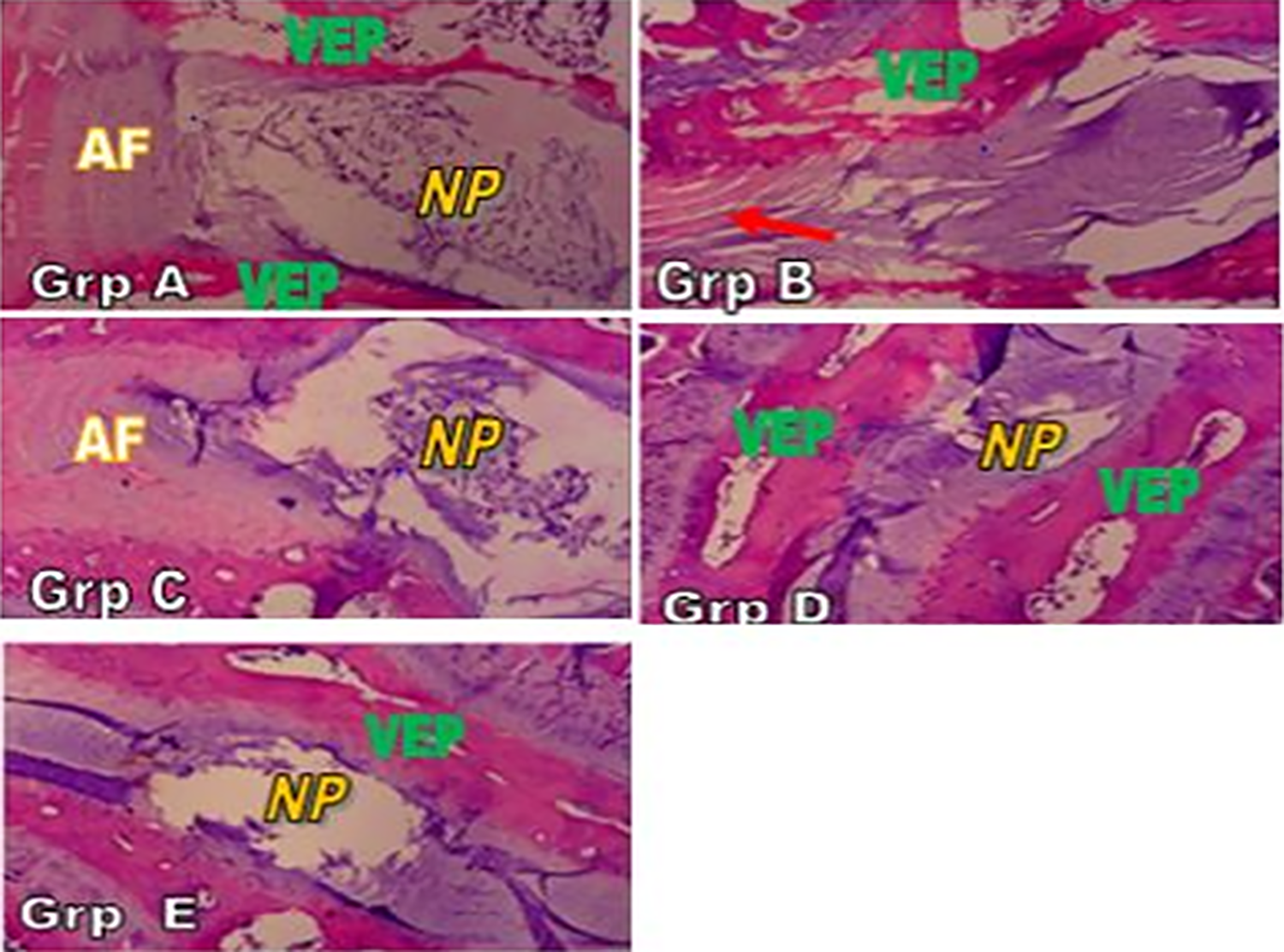
Photomicrograph of intervertebral disk (IVD) histology showing group A (nonpunctured group) with normal IVD, group B (punctured but not treated) with complete loss of cytoarchitecture and histoarchitecture, group C (punctured and treated with 600 mg/kg body weight of aloe vera gel); group D (punctured and treated with 400 mg/kg body weight of aloe vera gel); and group E (punctured and treated with 200 mg/kg body weight of aloe vera gel). Red arrow showing disorganized annulus fibrosus and loss of nucleus pulposus. Stain: hematoxylin and eosin; magnification ×40.
Effects of AVG on Organization of Collagen and Elastic Fibers in the IVD of Rabbits
Figures 2 and 3 show the micrographs of IVD sections subjected to Mason trichrome and van Gieson stains for demonstration of collagen and elastic fibers in the IVD of rabbits. The nonpunctured group (group A) showed a typical normal morphological pattern of both the collagen and elastic fibers in the AF, whereas the punctured but not treated group (group B) showed a complete disruption of the organization of the AF. The organization of the fibers was restored in the treated groups in a dose-dependent manner.
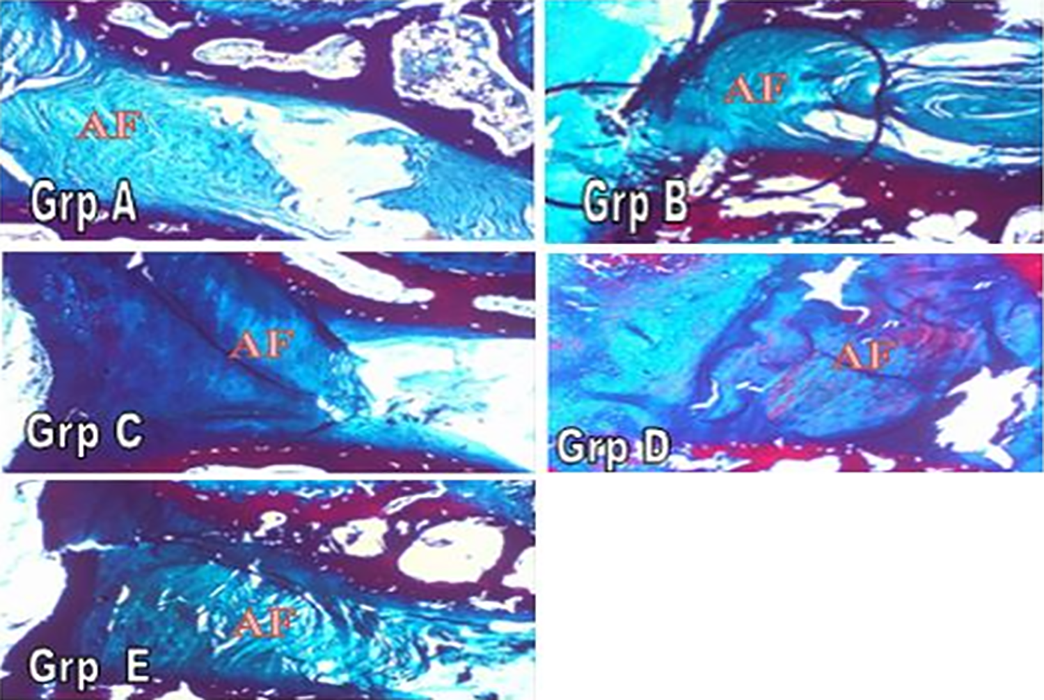
Photomicrograph of intervertebral disk collagen fibers in groups A, B, C, D, and E showing presence and organization of collagen fibers in annulus fibrosus (AF). Group B shows complete disorganization of collagen fibers, whereas groups C, D, and E show varying levels of repair when compared with group A (control). Stain: Mason trichrome; Magnification ×40.
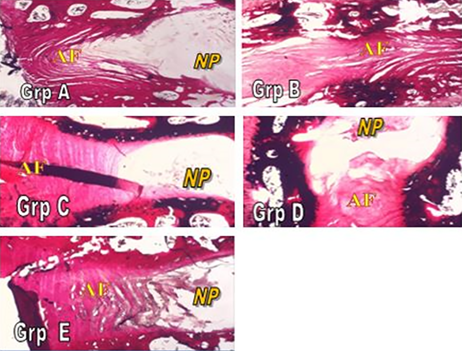
Photomicrograph of intervertebral disk elastic fibers in groups A, B, C, D, and E showing presence and organization of elastic fibers in AF. Group B shows complete disorganization of elastic fibers, whereas groups C, D, and E show varying levels of repair when compared with group A (control). Stain: Verhoeff van Gieson; magnification ×40.
Histological Grading Score for the Degree of Degeneration
The histological scores (Figure 4) showed a significantly dose-dependent response among the treated groups. There is a significant difference in the overall scores of the treated groups when compared with the control groups. The overall histological features of group C (highest dose) are comparable to that of group A (normal control), whereas groups D and E show varying levels of recovery. The negative control group (B) showed a significantly higher score in the level of degeneration of the NP compared with the group with the highest dose of AVG.
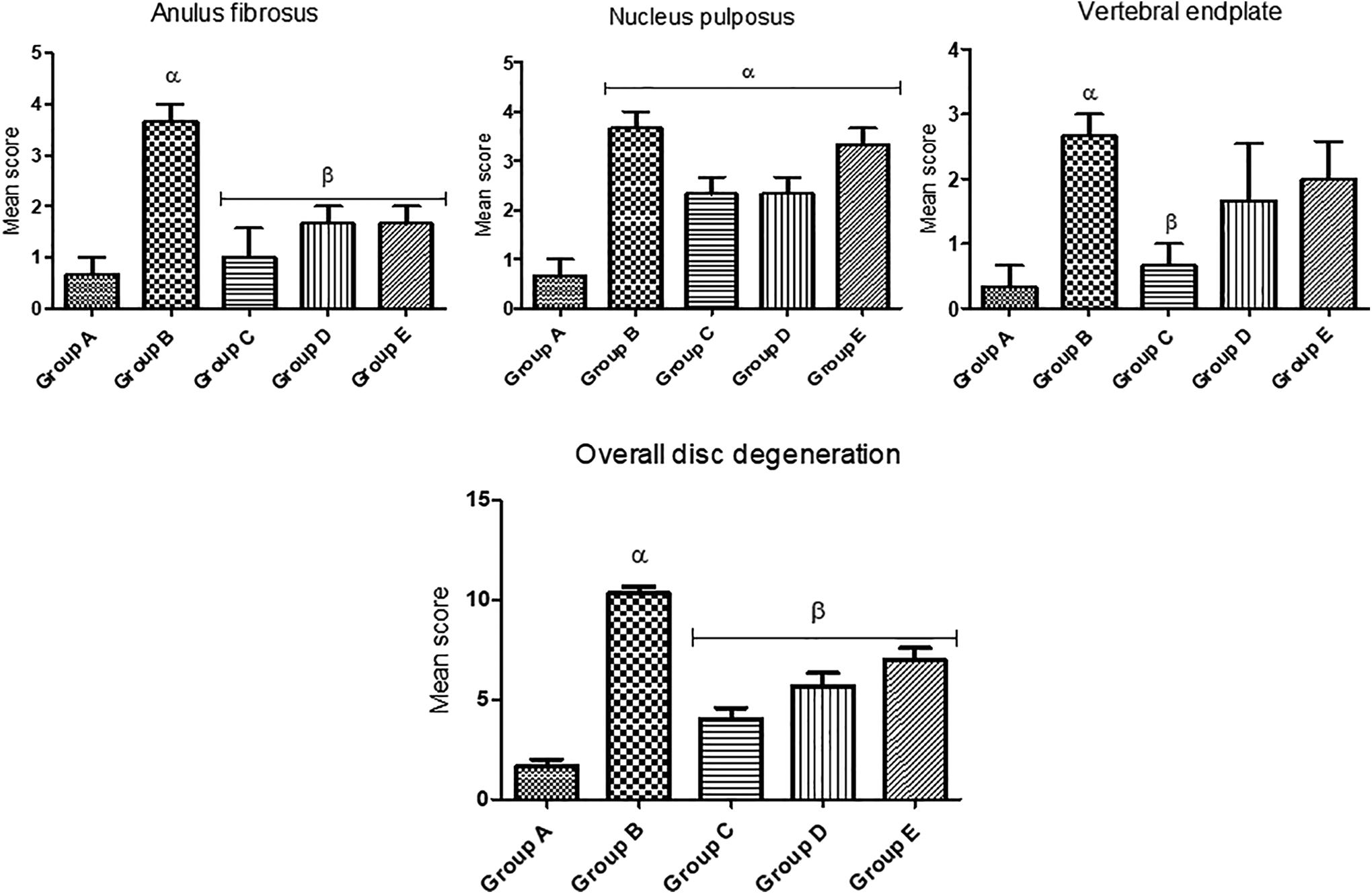
Histological grading of the level of degeneration of the intervertebral disk. Each value represents mean ± SEM; n = 5 readings. Value of P .05 was considered significant; α is significantly different from A, and β is significantly different from B.
Effect of AVG on the Cellularity of the Nucleus Pulposus of IVD
Using image J software for analysis of the chondrocyte-like cells of the nucleus pulposus across group (Figure 5), there was significant difference in the chondrocyte-like cell counts of the NP in the treated groups (C, D, and E) when compared with the punctured but not treated group (group B). The recovery of the NP also appears to be dose dependent because group E was found to be significantly different from group C.

Graph showing the total cell count per cubic millimeter in the NP. Each value represents mean ± SEM; n = 5 readings. Value of P .05 was considered significant; α is significantly different from group A, β is significantly different from group B, and γ is significantly different from group C.
Discussion
The use of an 18G needle is an effective means of inducing disk degeneration in rabbits. 8 The gross and microscopic picture consistent with degenerating IVD was observed in this study. A biomechanical model of causation was proposed in human studies as the cause of disk degeneration. 11
Histomorphological outcome is a key tool used in predicting the severity of disk degeneration. 12 This study revealed various histological alterations, ranging from discrete disruption of the nucleus pulposus, decrease in nucleus pulposus cells and lamella disorganization, and complete obliteration of its cavity in punctured rabbits. Histological evaluation of the IVD sections 6 weeks after administration of AVG revealed that the degenerative process was ameliorated. Notable progression in disk degeneration was observed in the annular punctured but saline-administered group, with most of the NP content lost and collapsed, as well as wavy or serpentine fibrocartilage lamella and a marked decrease in fibrochondrocyte-like cell of both the NP and AF, in contrast to the AVG-administered group, where we observed a resemblance to normal IVD status.
These findings are no doubt a result of the healing ability of AVG as reported in the literature. 13,14 Aloe vera accelerates wound healing by promoting the proliferation and migration of fibroblasts and keratinocytes. 14 It was also reported that aloe vera increased in a significant manner the collagen content of granulating tissue and enhanced the expression of collagen type III. 13,14
It is clear from this study that AVG has the ability to repair and remodel damaged elastic and collagen fibers. This is evident in the significant restoration of collagen and elastic fiber organization and arrangement following administration of oral AVG. The difference between the collagen and elastic components of the disk could pose a risk factor in the etiopathogenesis of disk degeneration and act as a precursor for degenerative changes, thereby causing LBP. 15
The observed effect of AVG on these important components of the disk matrix is similar. It induces both collagen and elastic fiber repair in much the same way. This may be a result of the presence of glucomannan, a mannose-rich polysaccharide, and gibberellin, a growth hormone, which can interact with growth factor receptors on the fibroblast, thereby stimulating collagen synthesis and increasing the degree of collagen cross-linking in AVG. The fibrochondrocytes in the nucleus pulposus and the annulus fibrosus is reputed for the laying down of the cellular framework of connective tissue fibers necessary for repair in a damaged disk. The observed mobilization of this important component in the treated group showed that AVG administration is effective in repairing a damaged IVD.
Conclusion
Our findings in this study showed that AVG was effective in preventing and repairing a damaged degenerating IVD in a rabbit model. It also validates the claim that the 18G needle is effective in inducing disk degeneration through annular puncture. The results of this study show that AVG could provide a viable medical therapy for LBP secondary to trauma-induced IVD degeneration. However, the effect of AVG on acute pain is not yet established.
Footnotes
Authors’ Note
Ethical approval was obtained from Health, Research and Ethics Committee (HREC) of the Institute of Public Health, Obafemi Awolowo University, Ile Ife.
Acknowledgments
The authors would like to acknowledge the department of Anatomy and Cell Biology, Obafemi Awolowo University, Ile Ife, and Mr Isaac Ifeoluwa Ogunlowo of the Department of Pharmacognosy, Obafemi Awolowo University, Ile Ife, for helping with identification and indexing of the plant material.
Declaration of Conflicting Interests
The author(s) declared no potential conflicts of interest with respect to the research, authorship, and/or publication of this article.
Funding
The author(s) received no financial support for the research, authorship, and/or publication of this article.