
Abstract
Study Design:
Literature review and transversal study.
Objective:
Advances in new technologies give the surgeons confidence to manage complex spine conditions with a lower morbidity rate. This has changed the expectations of patients and medical payers and foreshadows the shift now underway: the use of minimally invasive techniques. The ethical considerations of learning directly on patients require a change in the education and training programs.
Methods:
The education paradigm has changed, and surgical training on minimally invasive surgery of the spine (MISS) techniques should follow a “curriculum.” The assessment of skill proficiency while learning the MISS techniques must be measurable to objectively show the performance gained over time and the changes that should be performed during training. Different strategies include “ex vivo” and “in vivo” training.
Results:
We have worked on a curriculum in which the participants can perceive the growth in their knowledge through the different educational opportunities. There are 3 levels: basic, advanced, and masters.
Conclusions:
We developed an educational curriculum for MISS rationale and techniques, that showed to be effective and interesting in our region.
Keywords
Introduction
The remarkable advances of technology and instrumentation devices for spine surgery give the surgeon the certainty and confidence to manage even complex spine conditions with a lower morbidity rate. 1 Over time there is an improvement of magnification, lighting, surgical instruments, implants, retractors, navigation guide systems, and new muscle sparing techniques that enable to do more for the patient during surgery with less morbidity. 1 As a consequence, the patients’ expectations and the payers’ policy has changed. Nowadays the following is required: (1) a short hospital stay, even better if it is an ambulatory procedure; (2) rapid recovery time for early return to their activities; (3) small incisions or “Band-Aid surgery”; (4) lesser number of complications; and (5) similar or better efficacy than other surgical techniques. The changing profile of the patients and payers foreshadows the shift now underway: the use of minimally invasive techniques. Industry and spinal surgeons embody the philosophical and technical mind-set toward less invasive procedures or minimally invasive surgery of the spine (MISS).
Learning Curve
The learning curve is directly linked to the specificity of the surgical technique to be learned. Regarding the MISS technique, the duration of the learning curve is related to (1) prior surgeons’ experience and skill, (2) technical and anatomical difficulties due to the small surgical field, and (3) the quality and the repetition of training.
In the literature, the learning curve is measured by the surgical time and the occurrence of complications. 2 Both variables are evaluated according to the chronological case number. 2
The learning curve for each MISS technique is different. The lumbar microendoscopic decompression could be obtained after around 30 cases have been performed. 3,4
The learning process takes time and requires endurance, patience, and the surgeon’s persistence to achieve enough confidence and skills to perform a safe and effective procedure. 5
The ethical dilemma can be observed during the learning curve because the surgeon is not sufficiently trained and is still treating the patients. The greater number of complications perceived during the learning curve is not surprising when compared with after the surgeon becomes proficient in the technique. The surgeon may have a lower rate of complications if he or she follows an appropriate MISS curriculum. The goal is to gain expertise and skill using “flight training” before performing surgery on the patients. The training of surgeons has similarities to that of pilots. Examples of “flight training” are simulators and virtual reality software.
MISS Curriculum for Spine Surgery Training
The ethical considerations of learning directly on patients required a change in the education and training programs. This situation cannot be seen as a limiting factor for the training of residents/fellows who are performing their first surgeries, but it should be looked upon as a springboard to elaborate and provide excellence of training with a high academic level. To overcome these challenges, the spine centers adopted “ex vivo” strategies that lead to skill acquisition before going on to the next level of “in vivo” surgery. Training on cadavers is a traditional example that has become embodied in the curriculum of residency programs. This hands-on and supervised training in the cadaveric lab helps surgeons build expertise and confidence to repeat the procedure in the operating room on the patient. 6
The difference in favor of the cadaver lab training is that the procedures can be performed as many times as necessary until the skill is attained, without fearing risk to patients, complications, and medical malpractice. 7
The neuronavigation-guided simulator has been included in some curriculums of residence programs to perform the pedicle screw placement. The resident performance for pedicle screw placement has improved since this practice was instituted. 8
Assessment of Surgeon’s Skills During Ex Vivo MISS Training
The assessment of skill proficiency while learning the MISS technique must be measurable to objectively show the performance gained over time and the changes that should be performed during training. The variables that could be assessed during “ex vivo” training are
Knowledge: capacity to explain the procedure, related objectives, and the step by step methodology.
Judgment: capacity to choose the right treatment based on the acquired knowledge. This can be measured using the objective structured clinical examination (OSCE).
Dexterity: can be assessed by different methods, such as the objective structured assessment of technical skills (OSATS), the Imperial College surgical assessment device (ICSAD), minimally invasive surgical trainer (virtual reality), or combinations of the above. 9,10
Assessment of Surgeon’s Skills During MISS Training in Patient’s Surgery
The surgeon’s dexterity during MISS is usually measured according to (1) operative time (shorter as the surgeon gains experience), (2) the amount of blood loss, (3) the number of preoperative complications, (4) the need to convert to an “open” approach, (5) the time of intraoperative X-rays use, (6) the length of hospital stay, (7) radiological variables such as adequate decompression and fusion rates), and (8) the clinical and neurological outcome (neurological status recovery, pain improvement, etc).
Types of “Ex Vivo” Laboratories for MISS Training
Simulators
Simulators provide a safe setting for surgeons to improve their surgical skills recreating situations similar to real life. There are “low-fidelity” and “high-fidelity” simulators. The “low-fidelity” one is a simple physical model but allows interaction with real surgical instruments and provides proprioceptive feedback, which is a key aspect in the learning process. The “high-fidelity” simulators provide high-quality anatomical details, true-to-life consistencies, and real time feedback from the faculty. The simulators are appropriate to measure performance, that is, the number of movements made by each hand, the distance covered by each hand movement, duration of the procedure, and time used for each task. 10,11
An artificial lumbar spine prototype is available for MISS training that includes skin, muscles, bone, ligaments, and dura mater to make the training more realistic. Tubing systems simulate bone bleeding and cerebrospinal fluid leakage if the trainee could not perform an appropriate bone homeostasis or had a dural opening 12 (see Figure 1).
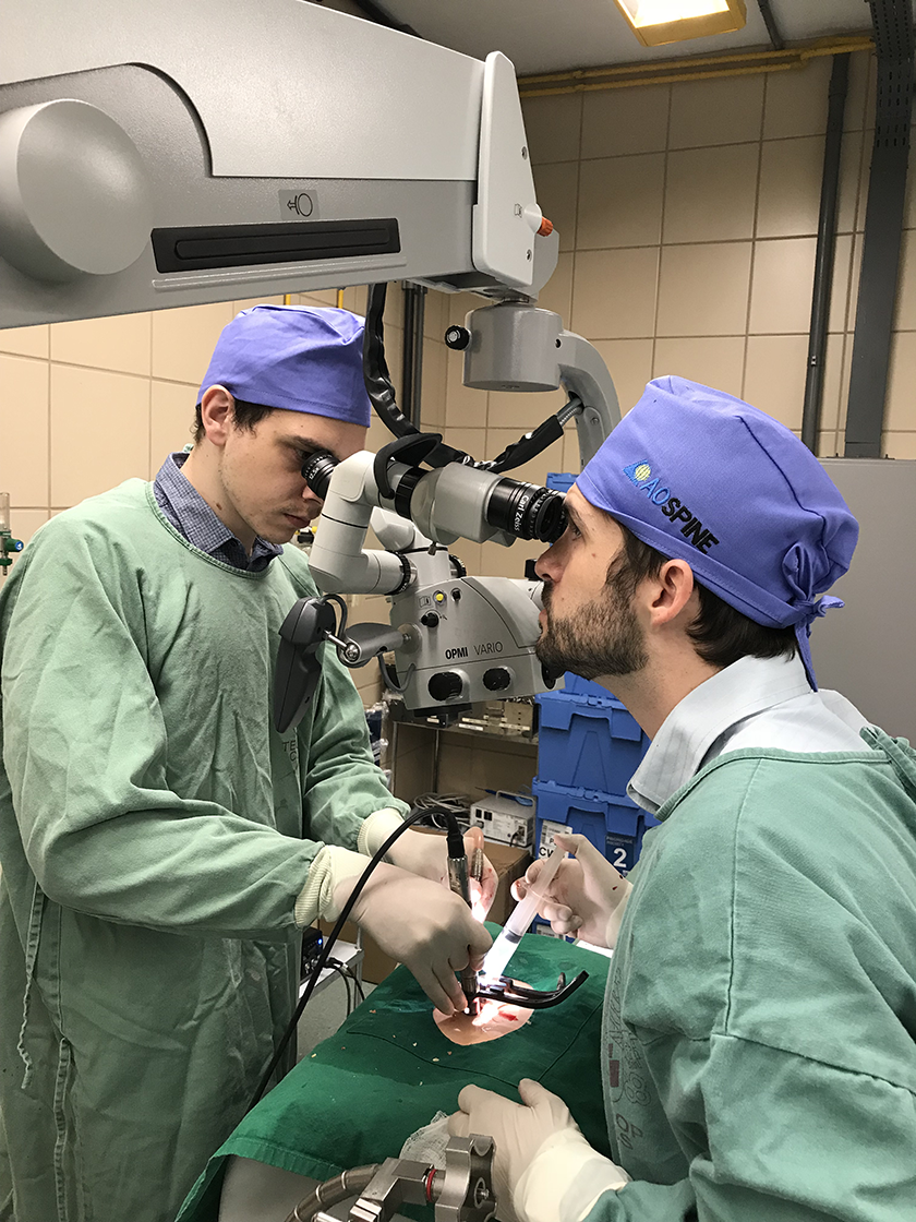
Two participants doing a tubular microdiscectomy in a Sawbone-based simulator
The training based on simulation can broaden the opportunities to use and create mixed models, including physical models like Sawbones and associated navigation systems to practice pedicle screw placement. 12
Computer-Assisted Technique
Computers are able to design very interesting learning scenarios. Virtual reality employs graphs and simulations very close to the real surgical setting. The interface includes the use of virtual surgical instruments that are identical to those used in daily practice. This technology enables a real time interaction with the creation of graphics and adds difficulties according to the level of training of each particular surgeon. 7,13
Barriers for Teaching Training in MISS
Each learning method involves particular limitations. The limitation in cadaveric or animal training includes the ethical aspects, the need for an approved structure according to the practice itself, the biological risks and the high cost.
The limitation of practice in simulators includes the high cost of the systems and the limited availability of courses, if it is compared with the large number of spine surgeons in training. It has been proven that surgeons attending computer-assisted surgery courses (CAS) were 2.2 times more likely to use navigation routinely. 14
Learning guided by tutors who share their experience and accompany surgeons in their training on MIS techniques is essential. Tutor feedback in this interaction leads to behavior and maneuver changes in attempts to improve the techniques.
The Latin American Experience
Technological advances in the field of image-guided surgery and navigation are a very useful tool for minimally invasive surgeries where the surgeon works through a narrow surgical corridor without visualization of the landmarks and considering that precision must never be sacrificed.
The initial interest in MISS began with the techniques performed later. This perhaps corresponds to the fact that most of the techniques taught in the residency program in orthopedics or in neurosurgery or to the fellows were given by surgeons trained only in posterior approaches. In this way, PLIF (posterior lumbar interbody fusion) and TLIF (transforaminal lumbar interbody fusion) are the instrumentation procedures of greatest interest for learning. However, in recent years the need to learn tubular surgery without instrumentation has arisen as the first step in MIS generating greater expertise, greater knowledge of work within tubular surgery, leading to a learning curve that allows performing surgery with instrumentation and in this way reduce complications (see Figure 2).
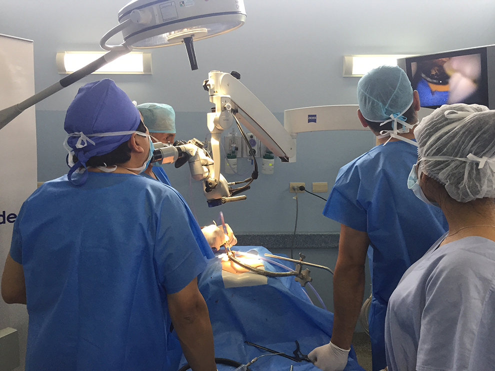
While the participant is performing a minimally invasive surgical (MIS) transforaminal lumbar interbody fusion (TLIF) procedure on a simulator, the faculty can observe and assess skills.
Currently, there is high technology in Latin America with huge progress in magnification, illumination, radiology, navigation, and osteosynthesis material, thus great interest was generated in the previous routes (ALIF [anterior lumbar interbody fusion], OLIF [oblique lumbar interbody fusion], lateral). There is a need to understand the spine over 360 degrees. Nowadays our interest in MIS education is to offer the technique that the patient needs and not only that on which the surgeon has experience. Therefore, in AO Spine Latin America, we have worked on a curriculum in which the participants can perceive the growth in their knowledge through the different educational opportunities. There are 3 levels: basic, advanced, and masters.
The basic level involves tubular microdiscectomy, placement of percutaneous screws, vertebral biopsy, and vertebroplasties. The advanced level involves TLIF, over the top, ALIF, and lateral approach. The master’s level is taught every 3 years: OLIF, ACR (anterior column realignment), high-grade listhesis, and scoliosis by MIS. All the modules have online theoretical information on each technique with the available evidence. Live videos show each surgical technique performed by the expert MIS faculty of the region. This information is reviewed before training, during training and can be reviewed continuously later. The practical education tools are simulators, cadaver lab training and live surgeries.
The assessments are made prior to training, during training and 6 months later online, when there is also a meeting with the chair and with this feedback, we analyze what percentage of adherence there is with these new techniques. In cadaver labs training, we have increased the number of participants but for a better use of the training, there are only 2 participants per table, with the same number of image intensifiers per station (see Figure 3). For the safety of the participants and their daily practice, guidelines are given for irradiation related to the C-shaped arch, since the availability of navigators in spinal surgery is increasing but it is not enough. With our education model, the MIS curriculum in AO continues to be the most interesting in the region and for the time being we are always participants in the waiting list.
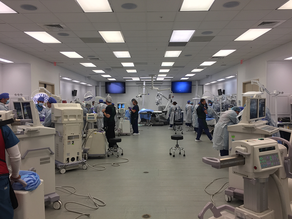
Cadaver lab with 2 participants per table with the same number of image intensifiers per station.
Footnotes
Acknowledgment
AOSpine Latin America.
Declaration of Conflicting Interests
The author(s) declared no potential conflicts of interest with respect to the research, authorship, and/or publication of this article.
Funding
The author(s) disclosed receipt of the following financial support for the research, authorship, and/or publication of this article: This supplement was supported by funding from the Carl Zeiss Meditec Group.