
Abstract
Stanniocalcin (STC), first isolated from the corpuscles of stannius of teleost fishes, was originally known for its regulation on calcium/phosphate transport. Increasing evidence demonstrates that STCs display the important function in some physiological and pathological behaviors such as calcium regulation, oxidative stress, anti-inflammation, angiogenesis, ischemia reperfusion, nerve diseases, etc. Moreover, STCs are implicated in the development and progression of multiple malignancies through promoting cell growth, proliferation, invasion, metastasis, and apoptotic escape. Some studies have shown that NF-κB upregulates STC expression, thereby activating the downstream HIF-1/ERK1/2 signaling pathway, enhancing the transcriptional activity of tumor-related factors (MMP-2/9, cyclinD1, Bcl-2, N-cadherin, etc) and contributing to tumorigenesis. Here, this brief review describes recent progress of STCs in mammalians, focused mainly on their critical functions in cancer.
Corpuscles of stannius (CS), a small endocrine gland located on the ventral surface of the kidneys of bony fishes, is extracted to purify stanniocalcins (STCs), which inhibit whole-body Ca2+ influx in the whole body, especially in gill and gut. CSs were first considered to be unique endocrine glands in fish, but were further verified to appear in mammalians, indicating that STCs may function in mammalians as calcium regulation, oxidative stress, anti-inflammation, angiogenesis, ischemia reperfusion, etc. Moreover, the relationship and molecular mechanisms of STC1 expression in cancer become the studied focus and front field. STC2 as a STC1 homolog is identified by searching for related sequences in expressed sequence tag databases. Although mammalian STC1 and STC2 are not expressed ubiquitously in tissue cells, they are distributed in a wide variety of tissues and organs including cancer, and play an important role in tumor development and progression.
Molecular structure
STC is present in all vertebrates as two isoforms, STC1 and STC2, encoded by separate genes. 1 STC1, originally described as an antihypercalcemic hormone in fish, is highly expressed in differentiated mammalian neurons. STC1 adopts a dimeric and slightly elongated structure with a high content of alpha-helices. 2 STC2, a homolog of STC1, is a 56kD glycoprotein hormone that confers calcitonin-like activity. A widespread distribution of SCT1 and STC2 is present in mammals and fish, in which the highest amount of STC mRNA is in the heart but lower amounts are found in the neural complex, branchial basket, and endostyle. STCs have a conserved pattern of 10 cysteines in all chordate, which are significant for identi-fication of amphioxus and tunicate amino acids, 14–23% identical with STC1 and STC2. The unique features of STC2, including 14 cysteines and a cluster of histidines in the C-terminal region, appear exclusively in vertebrates. 3
Physiological and pathological function
Many studies have shown that STCs play a critical role in calcium regulation, oxidative stress, anti-inflammation, angiogenesis, ischemia reperfusion, nerve diseases, and so on (Figure 1).

The physiological and pathological functions of STCs in mammals. STCs play a critical role in regulating calcium metabolism, oxidative stress, anti-inflammation, angiogenesis, ischemia reperfusion, and nerve diseases.
Calcium regulation
STC1 as a cation regulator in fish induces slightly less calcium uptake and regulates intestinal bicarbonate secretion. 4 STC2 protein is detected in the calcified lesions of several organs, and its mRNA level is significantly upregulated by inorganic phosphate (Pi). STC2 accelerates Pi-induced calcification and promotes limitation of ectopic calcification, 5 but STC1 mainly functions in modulating extracellular calcium in the stannius (CS). 1 Some reports show that immunization with STC-Ab increases the carbonate precipitate content in sea bream intestine, revealing a contribution of calcemic factors to acid-based balance. 6 CaSR-PTH-STC (calcium-sensing receptor, Parathyroid Hormone) controls of Ca2+ homeostasis in vertebrates 6 and STC1-trpm7 regulates cation homeostasis and kidney function. 7 Moreover, STCs decrease bone size, and suppress cranial intramembranous bone growth in osteoblasts, 7 but increase osteoblastic differentiation of human mesenchymal stem cells (hMSC), 7 suggesting that STCs may act as a new candidate for evaluation of osteogenic differentiation.
Oxidative stress
STC1 is an endogenous glycoprotein secreted by granulosa cells in response to oxygen deprivation. 8 Some studies indicate that intratracheal STC1 attenuates pulmonary inflammation, oxidative stress, cell apoptosis, and acute lung injury, while inhibition of STC1 increases lung injury, 9 suggesting that STC1 acts as an endogenous stress protein implicated in inflammatory modulation via inhibition of inflammatory cascade and induction of antioxidant and antiapoptosis. In addition, STC1 suppresses superoxide generation through induction of uncoupling proteins (UCPs) involved in inhibition of reactive oxygen species (ROS), and AMP-activated protein kinase (AMPK) mediates STC1-induced expression of UCP2, sirtuin 3 and protection from I/R in the kidney. 10 Another theory about oxidative stress points out that STC1 upregulates O2 generation, catalase activity, and fluorescence recovery after photobleaching (FRAP), increases Bcl-2, and slightly decreases caspase-3 expression. 11 However, STC1 is found not indispensable for induced ischemic tolerance or preserving blood-brain barrier integrity but has a slight role in stroke after recovery.
Anti-inflammatory
It has been reported that recombinant STC1 inhibits NLRP3 inflammasome activation and ROS production in macrophages, 12 where STC1 induces intracellular calcium and cell mobility, and predicts potent cytoprotective and anti-inflammatory action. 9 Abrogated STC2 during cerulein induces pancreatitis in mice via increased activation of PERK signaling, but elevated STC2 increases circulating amylase levels and maintenance of cellular junction. 13
Angiogenesis
STCs play an important role in the angiogenic process during the corpus luteum (CL) formation, 14 and regulate local angiogenesis in granulosa cells rather than in endothelial cells through the VEGF/VEGFR2 or Ang-2 signaling. 14 But they are found involved in cell viability via regulation of the caspase-3 and -7 activities in swine aortic endothelial cells. STC simulates VEGF production in granulosa cells, and simultaneously vascular endothelial growth factor (VEGF)-D enhances the expression of STC1, promoting the potent angiogenic effects. 15
Ischemia reperfusion
STC1 is over-expressed in peripheral vascular disease, and transgenic overexpression of STC1 protects against ischemia/reperfusion (I/R) kidney injury, and AVP-mediated elevations of STC-1 may be dependent on functional COX-2 activity. 16 Kidney-specific knockdown of STC1 thereby brings about severe proximal tubule injury characterized by invacuolization, decreased UCP2 expression, superoxide, cell apoptosis, and kidney failure, indicating that STC1 may be a therapeutic target for ischemia/reperfusion kidney injury.
Nerve diseases
STCs play an important role in optic nerve diseases and neuroprotective actions. STC1 increases the number of retinal ganglion cells, and reduces apoptosis and oxidative damage. 16 STC2 exerts against excitotoxic insults through the inhibition of nitric oxide and the tumor necrosis factor-α (TNF-α) and IL-1β expression. 17
STCs and cancer
Expression in cancer
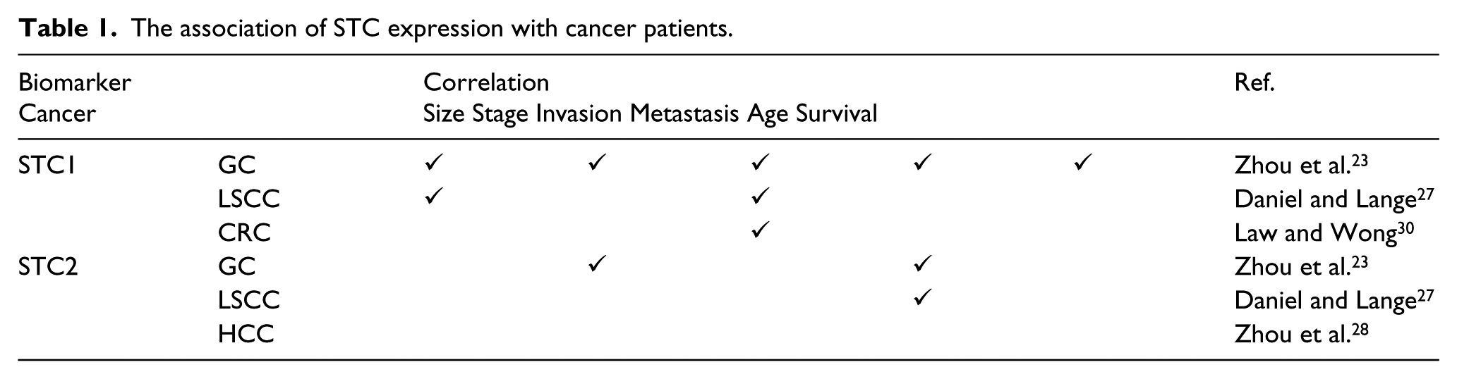
The expression of STCs is detected in almost all organ tumors in human. As indicated in Table 1, the mRNA and protein levels of STCs are markedly upregulated in esophageal squamous cell carcinoma, 18 gastric cancer,19,20 non-small cell lung cancer, 21 and ovarian cancer. 22 Overexpression of STC1 is related with T-Stage, lymphatic metastasis, clinical stage, pathological differentiation, and depth of tumor invasion. 23 The mRNA level of STC1 is adversely correlated with 2-year progression-free survival, 26 and the sensitivity and specificity of STC1 are higher than those for serum carcinoembryonic antigen and carbohydrate antigen 19-9 in patients with NSCLC. 30 In addition, the expression of STC2 is significantly correlated with age, depth of tumor invasion, lymph node metastasis, stage, venous invasion and tumor size in gastric cancer, and hepatocellular carcinoma (HCC), 24 and has low overall survival rate in patients with nasopharyngeal carcinomas and gastric cancer 25 indicating that high STC2 expression is an independent prognostic factor in gastric cancer. 23 However, few studies show that the expression of STC1 is not an independent and unfavorable prognostic factor for esophageal squamous cell carcinoma (ESCC). 27
The association of STC expression with cancer patients.
Proliferation
It is reported that overexpression of STC2 promotes growth of ovarian cancer, 26 whereas STC1 intervention improves the proliferation in renal carcinoma cells. STC2 promotes tumorigenicity and accelerates tumor growth in colon cancer. 26 Breast cancer growth is insensitive to progestins, and knockdown of STC1 inhibits proliferation of tumor cells expressing progesterone receptor (PR), suggesting that STC1 may play a potential role in therapy of breast cancer. 27
Invasion and metastasis
Overexpression of STC1 is associated with the thyroid cartilage invasion, lymphatic metastasis in laryngeal squamous cell carcinoma, 28 increases cell proliferation, migration, and colony formation in ovarian cancer cells, 26 and depends on PDGF stimulating fibroblasts to promote migration and invasion of colorectal cancer cells. 29 In addition, overexpression of STC2 correlates with lymph node metastasis and venous invasion in gastric cancer, 23 and increases motility and invasiveness of fibroblast morphology, 30 but knockdown of STC2 under hypoxic conditions reverses the migration of colon cancer. 30 However, few studies show that STC1 inhibits cell proliferation and invasion and knockdown of STC1 facilitates cell growth, migration, and invasion in cervical cancer. 31
Cell apoptosis
STC2 is identified as a potential oncogene involved in inhibiting cell death, promoting tumorigenesis in renal cell carcinoma 32 and increasing the expression of cell cycle regulatory proteins (cyclin A/D) and antiapoptotic proteins (Bcl-2), but decreasing cleavage of caspase-3/-9. The treatment with anti-STC1 monoclonal antibody or siRNA leads to elevated apoptosis and cell cycle arrest in G0/G1 phase in ovarian carcinoma cells.26,27 STC1 may be a downstream target gene of Sp1 recruiting Rb or histone deacetylase (HDAC) inhibitors involved in cellular apoptosis in human carcinoma cells.33,34
Regulatory mechanisms
Though STCs display several functions in human cancers, its regulatory mechanism is still poorly understood. It is found that STC2 can regulate the expression of cyclin D1 and activate extracellular signal-regulated kinase 1/2 (ERK1/2) promoting the tumorigenesis of HCC. 30 NF-κB as a transcri-ption factor is connected with the cancer, and directly binds to STC1 promoter activating the expression of STC1 in cervical cancer cells. 30 P53 also induces NF-κB phosphorylation and recruits acetylated histone H3 in STC1 promoter. 18 In addition, STC2 facilitates epithelial-mesenchymeal transition via upregulation of N-cadherin/vimentin and downregulation of E-cadherin, and enhances cell invasion by increase of MMP-2/-9 through ROS and ERK1/2 pathways in ovarian cancer. 30 (Figure 2).

The function and regulatory mechanisms of STCs in cancer. STCs are implicated in the development and progression of multiple malignancies through promoting cell growth, proliferation, invasion, metastasis, and apoptotic escape. NF-κB upregulates STC expression, thereby activating the downstream HIF-1/ERK1/2 signaling pathway, enhancing the transcriptional activity of tumor-related factors (MMP-2/9, cyclinD1, Bcl-2, N-cadherin, etc.) and contributing to the tumorigenesis.
Conclusions
Research in recent years has provided molecular identification of STCs in mammals at a very early stage. STCs have been reported to be involved in a growing number of physiological and pathological functions, especially in carcinogenesis.
Currently there are some reports suggesting the expression of STCs is found in many organs and cancer, yet to date, no information is available on the sequence, expression, and distribution in some cancers, such as pancreatic cancer. Apart from calcium regulation, oxidative stress, anti-inflammation, angiogenesis, ischemia reperfusion, and nerve diseases are the two organelles that are linked to the functions of STC1 and STC2, respectively. These findings resulted in further studies to investigate the involvement of STC1 and STC2 in cancer, particularly in their responses in oxidative stress and inflammatory stimulus at the tumor microenvironment. Some studies of STC biology have shown the crucial roles in tumor growth, invasion, metastasis, apoptosis, and cycle progress and reciprocal regulation with NF-kappa B, HIF-1, and ERI1/2 pathways in cancer. Ultimately these studies may highlight insights into STCs-related processes in mammals and help explore the functions and regulatory mechanisms of STCs in cancer progression.
Footnotes
Declaration of conflicting interests
The author(s) declared no potential conflicts of interest with respect to the research, authorship, and/or publication of this article.
Funding
This study was supported by the National Nature Science Foundation of China (Nos. 81302093 and 81272752).