
Abstract
Introduction
Breast cancer is a heterogeneous disease that has multiple molecular and morphological subtypes. Nonetheless, the relation between various molecular subtypes and functional characteristics of a tumor in terms of cytokine secretion remains unknown.
Methods
We studied spontaneous and mitogen-induced cytokine secretion by invasive breast carcinoma of no special type (IBC NST; cultured tumors and cultured peripheral blood cells), depending on a molecular tumor subtype (where “mitogens” means “polyclonal activators” (PA): phytohemagglutinin p, phytohemagglutinin M, concanavalin A, and Escherichia coli lipopolysaccharide). Enzyme-linked immunosorbent assays were used to determine concentrations of IL-6, IL-8, IL-10, IL-17, IL-18, IL-1β, IL-1Ra, TNF-α, IFN-γ, G-CSF, GM-CSF, VEGF, and MCP-1 in culture supernatants of the tumors and peripheral blood cells.
Results
The luminal B HER2-positive molecular subtype of IBC NST was found to feature the highest spontaneous secretion of IL-6 and IL-8 and the highest mitogen-induced secretion of IL-6, IL-8, IL-1Ra, and TNF-α by tumors; the highest mitogen-induced secretion of IL-2, IL-6, IL-8, IL-1β, TNF-α, IFN-γ, and G-CSF by peripheral blood cells; and the highest cytokine-producing potential (the ratio of mitogen-induced to spontaneous secretion) of peripheral blood cells for the secretion of IL-6, IL-8, and IL-1Ra as compared to other molecular subtypes. The triple-negative subtype of IBC NST was characterized by the lowest cytokine-producing potential of tumors for the secretion of IL-6 and IL-8 as compared to other molecular subtypes as well as a lower “stimulation index of polyclonal activators” (calculated as (cytokine secretion after incubation with PA)/(spontaneous cytokine secretion)) for IL-18 secretion as compared to luminal subtypes. The XYZ correlated with a suppressive effect of PA on cytokine secretion by tumors of the triple-negative molecular subtype.
Conclusion
Therefore, our findings indicate that in IBC NST of luminal B HER2-positive and triple-negative molecular subtypes, the cytokine network has distinctive functional features.
Introduction
Breast cancer is a heterogeneous disease that has multiple molecular and morphological subtypes. 1 The most common morphological type is invasive breast carcinoma of no special type (IBC NST), whose molecular subtypes are characterized by three main phenotypes: luminal (estrogen receptor– and/or progesterone receptor–positive), HER2-overexpressing, and triple-negative. 2 The molecular subtype is associated with the metastatic potential of breast cancer cells: the luminal tumor subtypes have a lower malignant progression rate and a better response to pharmacotherapy compared to those of the HER2-overexpressing subtype and triple-negative subtype of breast cancer. 3 According to recent studies, the progression of a luminal subtype to a more malignant subtype occurs in 30% of cases and is associated with metastatic progression of breast cancer and development of multidrug resistance.4,5 Nonetheless, the relation between various molecular subtypes and functional characteristics of a tumor in terms of cytokine secretion remains unknown. Therefore, the aim of our study was to investigate in vitro spontaneous and mitogen-induced cytokine secretion by IBC NST tumors and peripheral blood cells, depending on the molecular tumor subtype. In this article, the term “mitogens” means “polyclonal activators” (PA): phytohemagglutinin P, phytohemagglutinin M, concanavalin A, and Escherichia coli lipopolysaccharide. The choice of cytokines for study was due to their functional characteristics, in particular, their substantial role in malignant progression. The main processes taken into account in the choice of cytokines were immunosenescence (IL-1β, IL-6, IL-8, IL-10, G-CSF, and GM-CSF), chronic inflammation (IL-1β, IL-1Ra, IL-6, IL-8, IL-18, TNF-α, VEGF, and MCP-1), and immune evasion (IL-2, IL-4, IL-10, IL-17, and IFN-γ).6–8
Methods
Patients
The current study was of a retrospective nature. We studied biopsy samples of tumors and their microenvironment and peripheral blood cells from patients with grade II or III IBC NST (108 and 9 patients, respectively; 117 patients in total). Biopsies were performed from September 2018 to August 2020 and were obtained from the archives of the City Clinical Hospital No. 1 (Novosibirsk, Russia). Within 30 days of the examination, all patients underwent surgical treatment, and the final diagnosis was made on the basis of a histopathological examination. The patients were included in the study according to the following criteria: newly diagnosed with IBC NST and did not undergo neoadjuvant (preoperative) therapy. Signs of hematogenous metastasis to distant organs or concurrent endocrine, chronic, inflammatory, or infectious diseases were the exclusion criteria. According to the TNM classification of malignant tumors—which includes assessment of the primary tumor (T), regional lymph nodes (N), and distant metastases (M)—the stage of cancer was determined. Stage IA was diagnosed in 45 patients, stage IB in 31 patients, stage IIA in 16, stage IIB in 15, and stages IIIA and IIIC were diagnosed in 7 and 3 patients, respectively. The molecular tumor subtype in these patients was determined by immunohistochemical analysis of the estrogen receptor, progesterone receptor, epidermal growth factor receptor 2 (HER2), and proliferation marker Ki-67. 9 The luminal A subtype was identified in 39 patients with a mean age of 56 (36–76) years; the luminal B HER2-negative subtype was identified in 19 patients with a mean age of 54 (23–74) years; the luminal B HER2-positive subtype was identified in 32 patients with mean age of 58 (39–77) years; the HER2-overexpressing subtype was identified in 10 patients with a mean age of 53 (40–69) years; and the triple-negative subtype was identified in 17 patients with mean age of 59 (38–75) years. At the time of the study, metastases in regional lymph nodes were present in 9 patients with the luminal A subtype, 10 patients with the luminal B HER2-negative subtype, 13 patients with the luminal B HER2-positive subtype, 3 patients with the HER2-overexpressing subtype, and 5 patients with the triple-negative subtype.
Immunohistochemical analysis
Biopsy samples of IBC NST were fixed in neutral formalin, dehydrated, and embedded in paraffin. Paraffin sections of IBC NST were dewaxed and rehydrated with xylene and ethanol according to the standard technique. Expression levels of HER2, estrogen receptor, progesterone receptor, and Ki-67 in the IBC NST samples were evaluated in accordance with the procedures and evaluation criteria 9 recommended for the identification of molecular subtypes of breast cancer (Gallen International Expert Consensus, 2011), using monoclonal antibodies (Ventana Med. System Inc., USA): anti-HER2 (4B5; 790-2991), anti-ER (SP1; 790-4325), anti-PR (1 × 10²; 790-4296), and Ki-67 (790-4286). The tumor tissue sections were additionally stained with hematoxylin and eosin and embedded in balsam.
Evaluation of spontaneous and mitogen-induced cytokine secretion
Spontaneous and mitogen-induced secretion of cytokines by cultured tumors and cultured peripheral blood cells from IBC NST patients was analyzed using the Cytokine-Stimul-Best Kit (AO Vector-Best, Russia). For each tumor, 8 mm3 biopsy samples 10 were placed in two vials (one sample per vial), one of which contained 1 mL of the DMEM-F12 culture medium, and the other contained PA (2 μg/mL phytohemagglutinin P, 2 μg/mL phytohemagglutinin M, 4 μg/mL concanavalin A, and 2 μg/mL lipopolysaccharide from E. coli) in the same volume of the medium. The vials were incubated at 37°C for 72 h. The tumor sample was removed from each vial, and the remaining cells were precipitated by centrifugation at 900 × g for 15 min to obtain a supernatant, which was analyzed by the enzyme-linked immunosorbent assays.
After vein sampling, 1 mL of whole blood was placed into vial no. 1 containing 4 mL of the DMEM-F12 medium, 2.5 U/ml heparin, 100 μg/mL gentamicin, and 0.6 mg/mL
The cytokine-producing potential of IBC NST tumors and peripheral blood cells were assessed based on the stimulation index of polyclonal activators (SIPA), which was calculated using the formula A/B, where A is cytokine secretion after incubation with PA and B is spontaneous cytokine secretion. Stimulation index of polyclonal activators is expressed in arbitrary units (a.u.). Stimulation index of polyclonal activators ≤ 1.0 a.u. was interpreted as a suppressive effect of PA on cytokine secretion, and SIPA ≥ 1.1 was interpreted as a stimulatory effect.
Statistical analysis
This analysis of the results was performed in the SPSS Statistics software, v22.0 for Windows. Independent groups were compared by the Kruskal–Wallis test, followed by intergroup comparison via the Mann–Whitney U test. Statistical significance of differences between data expressed as a percentage was determined by Fisher’s exact test. To evaluate the measured cytokine data, we performed principal component analysis (PCA) by means of the Kaiser–Meyer–Olkin measure of sampling adequacy and Bartlett’s test of sphericity. The Spearman rank correlation coefficient (R) and its statistical significance (p) were calculated for some variables. The differences between groups were considered statistically significant at p < 0.05. A heatmap was constructed using the Morpheus platform (https://software.broadinstitute.org/morpheus/). The data are presented as a median and interquartile range.
Results
Spontaneous cytokine secretion by cultured IBC NST tumors, depending on the molecular tumor subtype. Q: an interquartile range, where 50 denotes a median.

IBC NST: invasive breast carcinoma of no special type.
As for the other molecular subtypes, cultured IBC NST tumors of the luminal B HER2-negative subtype showed the lowest spontaneous IL-10 secretion, and the luminal A subtype manifested the lowest spontaneous secretion of IL-18, TNF-α, GM-CSF, VEGF, and MCP-1 as compared to the other subtypes. It is known that (i) VEGF is a key mediator of angiogenesis in both nominally healthy people and patients with malignant tumors 17 ; (ii) IL-18 creates an immunosuppressive environment for the tumor 18 ; and (iii) TNF-α, GM-CSF, and MCP-1 induce the synthesis of matrix metalloproteinases and activation of epithelial–mesenchymal transition. 11 Therefore, their low spontaneous secretion by the luminal A subtype may be one of the reasons for the relatively slow malignant progression typical for this molecular subtype.3,4 The cultured tumors of the triple-negative molecular subtype featured the highest spontaneous secretion of IL-1β in our study.
Mitogen-induced cytokine secretion by IBC NST, depending on the molecular tumor subtype. Q: an interquartile range, where 50 denotes a median.

IBC NST: invasive breast carcinoma of no special type.
Evaluation of the cytokine-producing potential (i.e., SIPA) of IBC NST allowed us to identify the features specific for the triple-negative molecular subtype and for the HER2-overexpressing subtype (Figure 1). The triple-negative subtype of IBC NST manifested lower SIPA for the secretion of IL-6 and IL-8 by the cultured tumors as compared to the other molecular subtypes. The triple-negative subtype and HER2-overexpressing subtype had lower SIPA for the secretion of IL-18 by the cultured tumors as compared to luminal subtypes. The obtained differences indicate a suppressive effect of the tested set of mitogens (PA) on cytokine secretion by cultured IBC NST tumors of these molecular subtypes. IL-18 is known to have immunosuppressive activity in breast cancer by regulating natural killer (NK) cell subsets and inducing PD-1 expression on mature NK cells; IL-18 promotes drug resistance to chemotherapeutic agents.
18
IL-6 and IL-8 are pleiotropic cytokines that can exert an immunosuppressive effect via recruitment and regulation of biological activity of myeloid-derived suppressor cells in the tumor microenvironment.19,20 It is possible that IL-6, IL-8, and IL-18 together form an immunosuppressive network in IBC NST of the triple-negative molecular subtype. SIPA for cytokine secretion by cultured IBC NST tumors, depending on the molecular tumor subtype. Bars correspond to median ± interquartile range. ∗P = 2 × 10⁻², ∗∗P = 1 × 10⁻² as compared to the other molecular subtypes, #P = 1 × 10⁻², ##P = 1 × 10⁻² as compared to the luminal molecular subtypes. SIPA: stimulation index of polyclonal activators; IBC NST: invasive breast carcinoma of no special type.
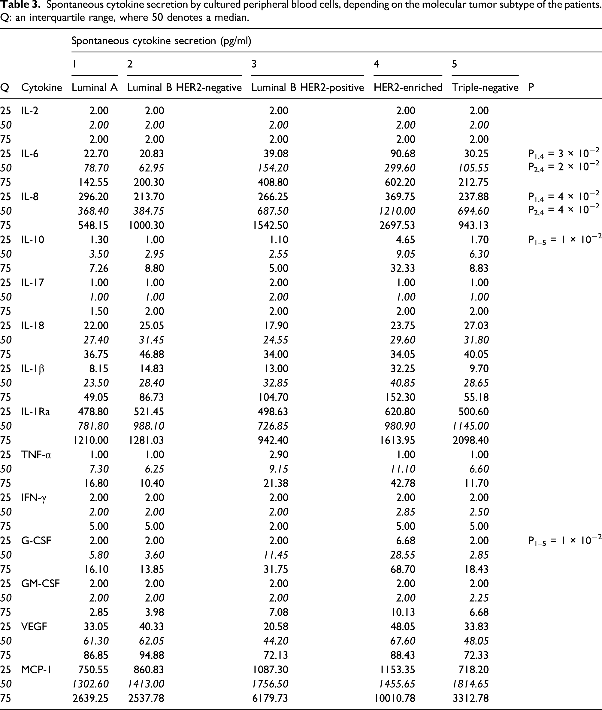
Spontaneous cytokine secretion by cultured peripheral blood cells, depending on the molecular tumor subtype of the patients. Q: an interquartile range, where 50 denotes a median.
Mitogen-induced cytokine secretion by cultured peripheral blood cells, depending on the molecular tumor subtype of IBC NST. Q: an interquartile range, where 50 denotes a median.

IBC NST: invasive breast carcinoma of no special type.
As for the cytokine-producing potential of the cultured peripheral blood cells from patients with IBC NST, specific features were noted in the luminal B HER2-positive subtype and HER2-overexpressing subtype (Figure 2). We found that the luminal B HER2-positive molecular subtype is characterized by higher SIPAs for the secretion of IL-6, IL-8, and IL-1Ra by the cultured peripheral blood cells as compared to the other subtypes. The HER2-overexpressing subtype proved to have lower SIPA for IL-10, TNF-α, and G-CSF secretion relative to the other subtypes. SIPA for cytokine secretion by cultured peripheral blood cells, depending on the molecular tumor subtype of IBC NST. Bars correspond to median ± interquartile range. ∗P1–5 = 8 × 10⁻³, ∗∗P1–5 = 1 × 10⁻³, ∗∗∗P1–5 = 3 × 10⁻³, #P1–5 = 1 × 10⁻², ##P1–5 = 1 × 10⁻², ###P1–5 = 2 × 10⁻² as compared to the other molecular subtypes. SIPA: stimulation index of polyclonal activators; IBC NST: invasive breast carcinoma of no special type.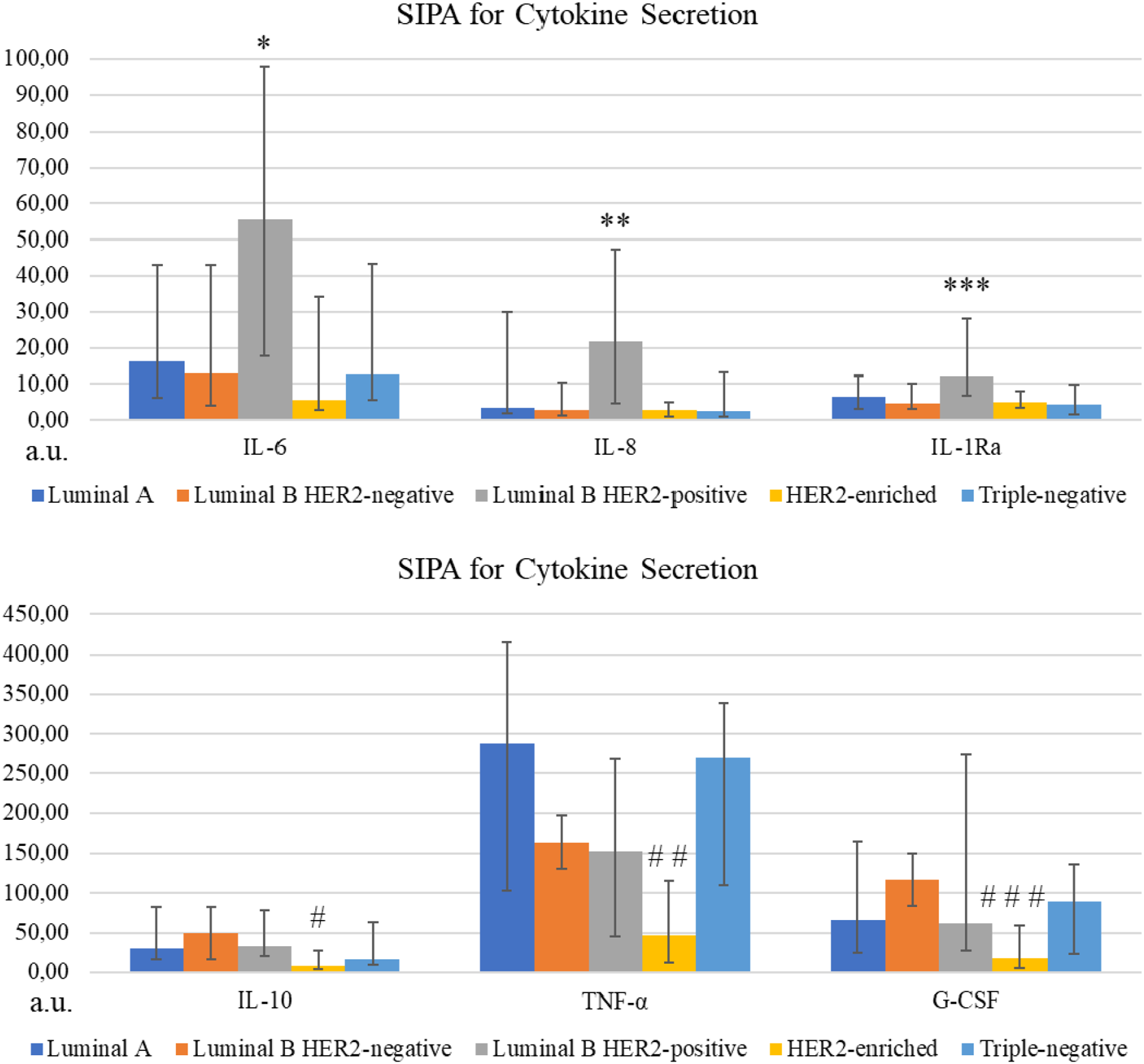
On the basis of our findings and the fact that IL-6 and IL-8 potentiate each other’s biological effects,21,22 we tested an index consisting of the mathematical product of IL-6 SIPA and IL-8 SIPA for the cultured peripheral blood cells. We found that 75.00% of cases of IL-6 SIPA × IL-8 SIPA ≥ 251.00 a.u. correspond to the luminal B HER2-positive molecular subtype, and 74.35% of cases with ≤ 250.00 a.u. correspond to the other molecular subtypes (p = 0.006 according to Fisher’s exact test).
PCA of cytokine secretion by cultured IBC NST tumors. KMO = 0.667, p > 10−10.

PCA: principal component analysis; KMO: Kaiser–Meyer–Olkin; IBC NST: invasive breast carcinoma of no special type.
PCA of cytokine secretion by cultured peripheral blood cells from IBC NST patients. KMO = 0.723, p > 10−10.

PCA: principal component analysis; KMO: Kaiser–Meyer–Olkin; IBC NST: invasive breast carcinoma of no special type.
Next, we evaluated the correlations between PCA groups of secreted cytokines and markers of molecular subtypes (Figure 3). Secretion of IL-6, IL-8, IL-10, IL-1Ra, G-CSF, GM-CSF, and MCP-1 by the cultured tumors correlated with the expression of HER2, which is characteristic of the luminal B HER2-positive and HER2-enriched molecular subtypes. Similarly, secretion of IL-6, IL-8, G-CSF, GM-CSF, and MCP-1 by the cultured peripheral blood cells correlated with the expression of HER2. These results indicated that there were specific patterns of cytokine secretion in patients with luminal B HER2-positive and HER2-enriched molecular subtypes. Probably, these groups of cytokines form a cytokine network that maintains HER2 expression in the IBC NST tumor. Correlations between markers of molecular subtypes and the groups of cytokines secreted by a cultured tumor (A) or by cultured peripheral blood cells (B) from invasive breast carcinoma of no special type patients.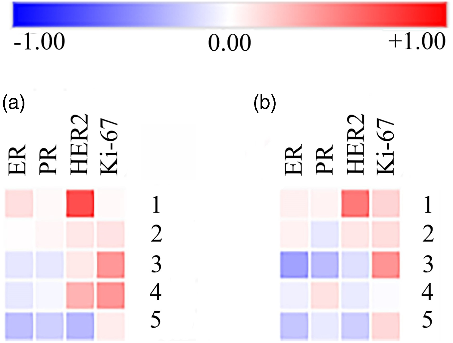
Correlations between spontaneous cytokine secretion by cultured tumors and by cultured peripheral blood cells, depending on the molecular tumor subtype of IBC NST.
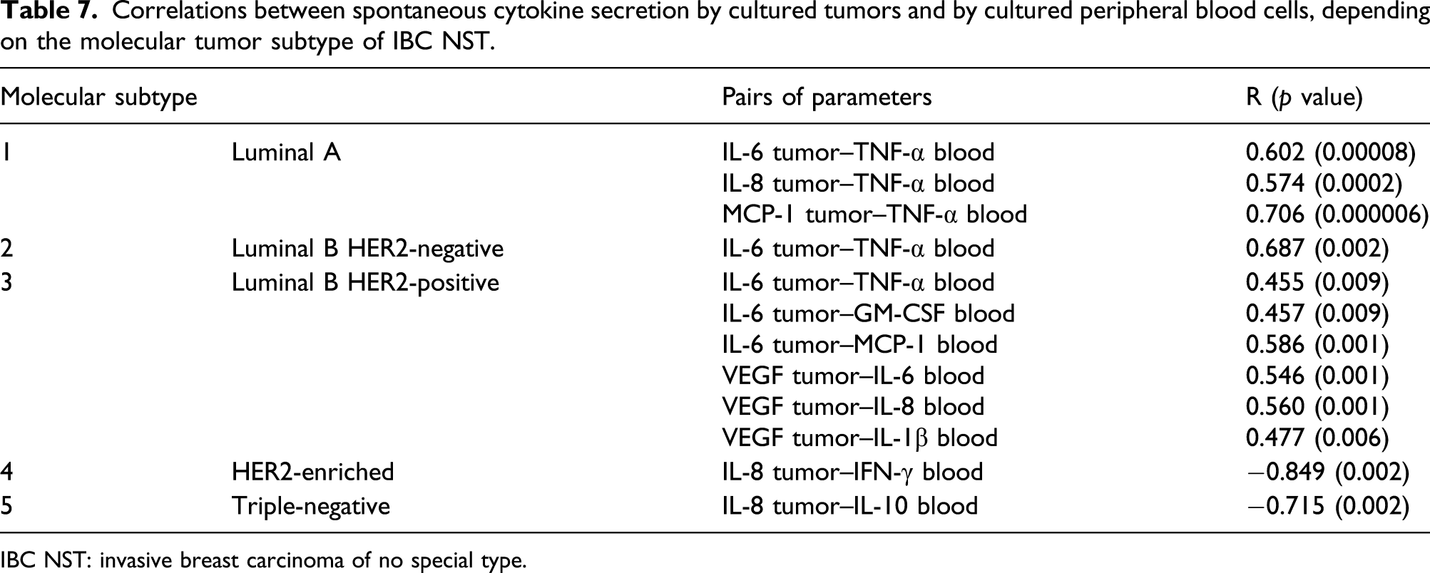
IBC NST: invasive breast carcinoma of no special type.
Discussion
Cytokines play the most important role in malignant transformation of cells and tumor progression. 6 Depending on an immunobiological environment, cytokines can have either a stimulatory or suppressive effect on malignant cells. 7 The antitumor immune response is known to be determined by a combination of intracellular and extracellular interactions between tumor cells and the cells of the tumor microenvironment, resulting in a complex cytokine network.6,7 Furthermore, it is peripheral blood leukocytes that constitute a functional reserve for tumor microenvironment cells. 8 Heterogeneity of malignant tumors is known to be based on marked intratumoral diversity of XYZ that ensures various levels of cytokine secretion; these levels are specific to each particular tumor. 23 Although the currently known molecular subtypes of breast cancer determine the treatment strategy, they do not allow interpretation of a tumor’s immunobiological environment, whose functional characteristics include cytokines.9,24
The present study revealed that the luminal B HER2-positive molecular subtype of IBC NST differs significantly from the other subtypes not only by high spontaneous but also by high mitogen-induced secretion of cytokines by the cultured tumor as well as by high mitogen-induced secretion by cultured peripheral blood cells and a high cytokine-producing potential of peripheral blood cells. Cytokines IL-6 and IL-8 are most specific for the luminal B HER2-positive molecular subtype. IL-6 and IL-8 can directly or indirectly affect the expression of estrogen and progesterone receptors and of receptor HER2 by tumor cells in IBC NST of the luminal B HER2-positive subtype because these cytokines potentiate each other’s biological effects and affect the activity of steroidogenesis enzymes.5,12–14 IL-6 is reported to increase transcription of the CYP19 gene encoding aromatase: an enzyme that transforms androgens into estrogens in adipose tissue.
13
Overexpression of HER2 initiates the HER2–IL-6–STAT3 signaling cascade, which promotes the development of resistance to anti-HER2 therapy through self-renewal of breast cancer stem cells.15,16 Based on the published data and our results, it can be assumed that the high spontaneous secretion of IL-6 and IL-8 by cultured tumors of the luminal B HER2-positive subtype is caused by the activity of cells comprising the tumor, including its microenvironment; it is also possible that these cytokines can contribute to high expression of estrogen and progesterone receptors and receptor HER2, which defines the luminal B HER2-positive subtype (Figure 4(a)). The evaluation of the cytokine-producing potential of IBC NST allowed us to identify specific features of the triple-negative molecular subtype, for example, lowest SIPAs for the secretion of IL-6 and IL-8 as well as lower SIPA for IL-18 secretion relative to luminal subtypes. The observed differences point to a suppressive effect of PA on the secretion of cytokines by IBC NST of this molecular subtype. It is possible that IL-6, IL-8, and IL-18 together form an immunosuppressive network in IBC NST of the triple-negative molecular subtype (Figure 4(b)) because IL-6, IL-8, and IL-18 are known to exert immunosuppressive action on cells constituting the tumor microenvironment and to enable the development of multidrug resistance.18–20 The cytokine network in luminal B HER2-positive (A) and triple-negative (B) molecular subtypes of breast cancer.
From the results of this study, it can be deduced that for patients with the luminal B HER2-positive molecular subtype of breast cancer, an anticytokine therapy based on inhibitors of IL-6 and IL-8 receptors may be effective. According to the literature, IL-6 receptor inhibitors suppress bone metastases in a breast cancer model. 25 Concerning the triple-negative molecular subtype of breast cancer, it is probable that an anticytokine treatment with inhibitors of IL-6, IL-8, and IL-18 receptors will suppress the intratumoral immunosuppressive network through, among other mechanisms, nonspecific suppression of PD-L1 expression. 18
There are some limitations to this study. First, power calculation for estimation of the sample size needed for this study was not done because this study was limited by the number of patients who signed the agreement to participate and by the terms of this agreement itself. Second, due to high variation within the study population, the results close to the p = 0.05 cutoff should be interpreted with caution. On the other hand, the strength of this study on IBC NST is not only the analysis of spontaneous and mitogen-induced secretion of cytokines but also the SIPA calculation, which eliminates the error associated with differences in the number of cytokine-producing cells. Additionally, this study comprehensively covers the secretion of cytokines, both in cultured tumors and cultured peripheral blood cells, thereby making it possible to elucidate specific features of the cytokine network functioning in various molecular subtypes of IBC NST.
Conclusion
Spontaneous and mitogen-induced secretion of cytokines by cultured tumors and cultured peripheral blood cells from IBC NST patients is associated with molecular subtypes of breast cancer. The luminal B HER2-positive molecular subtype shows (i) the highest spontaneous secretion of IL-6 and IL-8 and the highest mitogen-induced secretion of IL-6, IL-8, IL-1Ra, and TNF-α by the cultured tumors; (ii) the highest mitogen-induced secretion of IL-2, IL-6, IL-8, IL-1β, TNF-α, IFN-γ, and G-CSF by the cultured peripheral blood cells; and (iii) the highest cytokine-producing potential (mitogen-induced to spontaneous secretion ratio) of the cultured peripheral blood cells for the secretion of IL-6, IL-8, and IL-1Ra as compared to other molecular subtypes. It is possible that these features are associated with the modulatory activity of these cytokines toward the synthesis of enzymes involved in estrogen and progesterone metabolism and are based on the interaction of these cytokines with estrogen and progesterone receptors and with receptor HER2. The triple-negative subtype of IBC NST has the lowest cytokine-producing potential of the cultured tumors for the secretion of IL-6 and IL-8 as compared to the other molecular subtypes as well as lower SIPA values for IL-18 secretion as compared to luminal subtypes. XYZ is associated with a suppressive effect of the tested set of mitogens (PA) on cytokine secretion by the cultured tumors of the triple-negative molecular subtype. Probably, the cytokines produced by the cultured tumors, namely, IL-6, IL-8, and IL-18, together form an intratumoral immunosuppressive network in IBC NST of the triple-negative molecular subtype. Therefore, our findings indicate that the cytokine network has distinctive functional features in IBC NST of luminal B HER2-positive and triple-negative molecular subtypes.
Compliance with ethical standards
The study was conducted in accordance with the Helsinki Declaration of the World Medical Association. All recommendations of ICMJE were followed. All participants of the study gave their voluntary informed consent to participate in the study. The study protocol was approved (Protocol No. 2016-3) by the Ethics Committee of the Institute of Molecular Biology and Biophysics, a subdivision of the Federal Research Center of Fundamental and Translational Medicine (Novosibirsk, Russia).
Footnotes
Declaration of conflicting interests
The author(s) declared no potential conflicts of interest with respect to the research, authorship, and/or publication of this article.
Funding
The author(s) disclosed receipt of the following financial support for the research, authorship, and/or publication of this article: The study was financed as part of a state assignment (No. АААА-А18-118030790008-7) by the Ministry of Health of the Russian Federation.
Ethics approval
Ethical approval for this study was obtained from the Institute of Molecular Biology and Biophysics, a subdivision of the Federal Research Center of Fundamental and Translational Medicine (Protocol No. 2016-3).
Informed consent
Written informed consent was obtained from all subjects before the study.
Trial registration
Molecular markers of breast cancer and the influence of Human Leukemia Differentiation Factor on the metastatic potential: No. АААА-А18-118030790008-7.