
Abstract
Background
Dual-energy contrast-enhanced mammography (DECEM) is an advanced breast imaging technique of digital mammography.
Purpose
To assess the total radiation dose received from complete DECEM using different combinations of exposure parameters for low- and high-energy images.
Materials and methods
A dedicated phantom with three different concentrations of iodine inserts was used. Each iodine insert was 10 mm in diameter and concentration of 1.0 mgI/cm3, 2.0 mgI/cm3, and 4.0 mgI/cm3. The phantom was exposed at varying kVp levels. Mean glandular dose (MGD) was estimated. Contrast to noise ratio (CNR) and figure of merit (FOM) of the iodine inserts were used to assess the image quality.
Results
The optimum CNR of the recombined images was obtained by using 28 kVp + 49 kVp tube voltage combination for 50 mm thickness, 50% fibroglandular phantom only with a 26% dose increase compared to the highest voltages (32 kVp + 49 kVp) that can be used for low energy (LE) and high energy (HE) imaging. The CNR value was increased with increasing iodine concentration (R 2 > 0.99).
Conclusion
The use of as low as possible tube voltage for the LE imaging of standard 50% fibroglandular–50% adipose, 50 mm thickness breast while using the highest tube voltage for HE imaging has reduced the MGD while keeping optimum image quality.
Introduction
The conspicuity of the lesion is reduced in dense breasts when imaged with conventional mammography. 1 Therefore, at the next level, mammography uses iodinated contrast media to enhance breast cancer. 2 The technique uses the advantage of differential attenuation at the k absorption edge of iodine (33.169 keV). 3 Dual-energy contrast-enhanced mammography (DECEM) is an advanced breast imaging technique of digital mammography.4,5 In dual-energy technique, two images were acquired at low-energy and high-energy levels by using different target filter combinations.6,7 Low-energy (LE) and high-energy (HE) images are obtained after the administration of iodine-based contrast media intravenously. 8 These two images are recombined to create a final image, known as recombined image which clearly demonstrates contrast-enhanced mass within the breast due to different contrast enhancement within the normal breast tissues and the tumor areas.6,8
Low-energy images are acquired as standard mammograms with low energies which are below the k-edge of iodine (x-ray energy range from 26 kVp to 31 kVp).7,9 LE images are similar to the 2-dimensional full-field digital mammography (2D-FFDM) and are used to interpret the structural changes of the breast. 10 HE images are acquired in the energy range 45–49 kVp to ensure that the x-ray beam energy is above the k-edge of iodine and demonstrate only contrast media uptake. 11 According to the previously published research, radiation dose to the breast is higher in complete DECEM due to the use of two exposures per view.12–15 Yakoumakis et al. confirmed that the low-energy imaging mainly contributes to the total radiation dose received from the complete DECEM procedure. 16
Only few studies are available regarding the optimization of radiation dose in CESM. A study conducted by Nishikawa et al. focused on the possibility of exposure dose reduction in CESM using dual-energy subtraction technique. 17 Phantom was used to optimize the scan protocol and parameter setting for reducing radiation dose without image degradation. They acquired the images using fully automated mode and manual mode and evaluated the image quality by image noise, contrast, and exposure dose. This study result reported that the average glandular dose (AGD) was able to reduce to 1.41 mGy from 1.96 mGy by setting manual mode. This study results suggested that it is possible to reduce the exposure dose by using manual mode instead of fully automated mode when performed CESM in clinical practice.
The aim of this research study is to use different combinations of energy levels for LE and HE images to make a recombined image and then assess the radiation dose received from the complete DECEM procedure without degrading the image quality of LE image and the final recombined image.
Materials and Methods
Exposure factors used for LE and HE imaging of 50 mm thickness 50% fibroglandular–50% adipose phantom.

Mean glandular dose was calculated by using a method published by Dance et al.
18
Respective conversion coefficients were obtained by interpolating and extrapolating from the coefficients developed by Dance
Image quality of LE and recombined images was calculated using the Fiji Image J software platform. The area of iodine inserts was selected as the region of interest (ROI) to calculate the mean pixel value of the iodine inserts (SI). The same ROI was used to measure the mean pixel value of the background (SB). Standard deviations (SD) for all ROI measurements of iodine inserts (I) and the background (B) were obtained. The CNR was calculated according to the equation given by the European mammography protocol.
19

The FOM was calculated to evaluate the image quality with respect to the radiation dose delivered to the breast, and it is defined as follows

Results
The accurate g and c factors were calculated by interpolating and extrapolating of Dance's coefficients.
g-factor calculation for LE exposure: For 28 kV, 0.552 HVL, g-factor; 0.285, for 30 kV, 0.567 HVL, g-factor; 0.293, and for 32 kV, 0.584 HVL, g-factor; 0.302.
g-factor calculation for HE exposure: For 45 kV, 0.699 HVL, g-factor; 0.367, for 47 kV, 0.709 HVL, g-factor; 0.373, and for 49 kV, 0.719 HVL, g-factor; 0.379.
c-factor of 50% fibroglandularity breast is 1.000 for any thickness of the breast while it varied for 100% adipose and 100% fibroglandular breasts.
c-factor for 50 mm, 100% adipose phantom LE exposure: at 28 kV; 1.228, at 30 kV; 1.224, and at 32 kV; 1.220.
c-factor for 50 mm, 100% adipose phantom HE exposure: at 45 kV; 1.182, at 47 kV; 1.178, and at 49 kV; 1.174.
Dance reported s-factor for W/Rh target/filter combination which was 1.042.
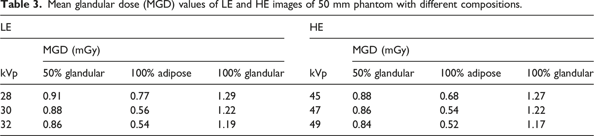
Mean glandular dose (MGD) values of LE and HE images of 50 mm phantom with different compositions.
Total MGD values of recombined images according to different phantom compositions.

Calculated CNR values of three iodine inserts with different concentrations of recombined images.

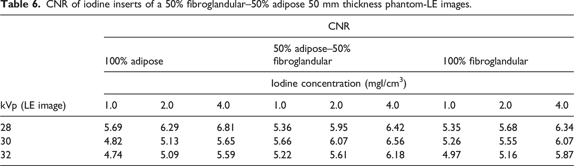
CNR of iodine inserts of a 50% fibroglandular–50% adipose 50 mm thickness phantom-LE images.
In the present study, FOM values were not given relevant to 0 mGy and reported values given for the estimated total MGD values. FOM of the recombined images was plotted as a function of tube voltage used for HE exposure. Graphs were plotted for three different breast equivalent compositions to demonstrate the variation of FOM values according to different breast equivalent compositions. Each line plotted on the graph corresponds to the tube voltages used for LE exposure
Discussion
Different combinations of kVp used to create recombined images in the study.

The minimum MGD was observed with 32 kVp LE + 49 kVp HE (1.70 mGy) while the maximum was observed with 28 kVp + 45 kVp HE (1.79 mGy) for 50 mm thickness 50% fibroglandular phantom. The tube voltage combinations which gave minimum and maximum mean MGD for 100% adipose and 100% glandular phantom were the same as for 50% fibroglandular phantom (Table 3 and Table 4; Figure 1). The maximum CNR value (2.40) was observed with 4.0 mgI/cm3 iodine insert of 50% fibroglandular phantom for 28 kVp LE + 45 kVp HE voltage combination. But, CNR was highest (2.59) with 4.0 mgI/cm3 iodine insert of the 100% adipose phantom for 30 kVp LE + 45 kVp HE while CNR was highest (2.61) with 4.0 mgI/cm3 iodine insert of the 100% fibroglandular phantom for 30 kVp LE + 45 kVp HE (Table 5). Total MGD of complete dual-energy contrast-enhanced mammography for 50 mm thickness, 50% fibroglandular–50% adipose phantom at different kVp combinations.
The CNR values of the iodine inserts in recombined images were decreased with increasing the selected tube voltage for LE and HE imaging. Hence, the total MGD increased 53% and 29% when using 28 kVp LE + 45 kVp HE and 28 kVp LE + 49 kVp HE compared to the highest kVp combination (32 kVp LE + 49 kVp HE) for 50% fibroglandular phantom, respectively (Table 3 and Table 4). Although LE images consist of iodine, they are similar to the 2D-FFDM images. Therefore, LE images should meet the optimum image quality standards. According to the results of Table 6, maximum CNR values were observed at 30 kVp for 50% fibroglandular phantom. The CNR values between 28 kVp and 30 kVp were not significantly different, and all those values are within the standard range as mentioned in Toroi et al. 22 . Therefore, it is possible to use 28 kVp or 30 kVp to obtain LE images of 50% fibroglandular standard breast. However, to increase the sensitivity of the recombined image, the optimum kVp for the LE image of 50% fibroglandular phantom is 28 kVp. However, to keep the radiation dose to the breast as low as possible, it is better to use the highest kVp to obtain HE images (49 kVp). Hence, 28 kVp + 49 kVp tube voltage combination produced recombined images without the loss of CNR with less dose increasing (29%) compared to the radiation dose received from 32 kVp LE + 49 kVp HE.
The study results for the 50% fibroglandular phantom confirmed that the FOM value reduced gradually with increasing HE tube voltage for each LE tube voltage and maximum values reported with 28 kVp LE tube voltage (Figure 2(a)–(c)). According to the t-test results (p > .05), FOM values at 45 kVp and 49 kVp HE were not significantly different for any of the LE tube voltages. Therefore, FOM results of recombined images confirmed to use the 28 kVp LE + 49 kVp HE for the standard 50% fibroglandular phantom to reduce the radiation dose without degrading the image quality. Figure of merit (FOM) values of 4.0 mgI/cm3 iodine inserts of a 50 mm thickness (a) 50% fibroglandular–50% adipose, (b) 100% adipose, and (c) 100% glandular phantom recombined images.
Although maximum CNR was observed with 30 kVp LE + 45 kVp HE for 100% adipose phantom, the radiation dose can be reduced by 11% and 13% when using 30 kVp LE + 47 kVp HE and 30 kVp LE + 49 kVp HE, respectively, without the loss of image quality. FOM values for 100% adipose phantom were high at 30 kVp LE imaging. Radiation dose for 100% glandular phantom at 28 kVp LE + 45 kVp HE and 28 kVp LE + 47 kVp HE exceeded the dose limit reported in European reference frame (EUREF) guidelines (Table 3). Although both CNR and the FOM values were high for the 28 kVp LE + 47 kVp HE combination, it is not possible to use for the 100% glandular phantom. Therefore, 32 kVp LE + 49 kVp HE with ≥2.0 mgI/cm3 iodine concentration is the optimum combination for 100% glandular phantom (Table 4 and Figure 2c).
Pearson correlation test found that the CNR value of the iodine inserts in recombined images had a strong positive relationship with the concentration of iodine (r = 0.925). It was found that a linear fit to the calculated CNR value of the iodine inserts of recombined images resulted in R
2
values superior to the 0.99 (Figure 3) and it was comparable to the Dromain et al. According to Dromain et al., this linear fit can be used to detect minimal iodine concentration of a lesion by using Rose criteria
20
. Contrast to noise ratio (CNR) as a function of the concentration of iodine inserts of the recombined image (a) 28 kVp LE + 49 kVp HE optimized voltage combination and (b) 28 kVp LE + 45 kVp HE voltage combination which gives the highest CNR.
Our study has several limitations. The tube voltage optimization was done only for 50 mm thickness and three different breast equivalent phantoms. This evaluation should be extended to the clinically available range of breast thicknesses and breast glandularities before these tube voltages set it for clinical use. In addition, the study recommended to evaluate the LE image quality visually according to EUREF criteria for implication of practice.
In conclusion, the use of as low as possible tube voltage for the LE imaging of standard 50% fibroglandular breast while using the highest tube voltage for HE imaging has reduced the MGD while keeping optimum image quality. The best match between the radiation dose and image quality was found in the recombined images.. Moreover, CESM procedure can be performed with ≥2.0 mgI/cm3 iodine concentration to obtain a better CNR value in the recombined images of three breast compositions even at high voltages when radiation dose needs to be the main consideration.
Footnotes
Acknowledgments
We gratefully acknowledge the staff of the mammography unit at the Juntendo University Shizuoka Hospital, Japan for their immense support.
Declaration of Conflicting Interests
The author(s) declared no potential conflicts of interest with respect to the research, authorship, and/or publication of this article.
Funding
The author(s) disclosed receipt of the following financial support for the research, authorship, and/or publication of this article: The first author was supported by Tokyo Human Resources Fund for City Diplomacy Scholarship. But no specific grants from funding agencies outside the University.
Ethics Approval
Ethical committee approval is not required since this article does not contain any procedures involving human participants.