
Abstract
Background
Widespread abdominal imaging has led to a substantial increase in the detection of incidentalomas. Currently, an increasing number of centers offer surveillance of the pancreas to individuals at high risk (IARs) of pancreatic ductal adenocarcinoma (PDAC).
Objective
The aims of this study were to evaluate the frequency and type of incidental findings in a magnetic resonance imaging (MRI)-based surveillance program for IARs for PDAC, and to discuss the benefit of detecting these lesions.
Methods
The outcome of MRI screening was reviewed in 568 individuals from three long-term pancreas surveillance programs conducted at three large European expert centers. All MRIs were studied in detail for the presence of incidental lesions.
Results
The most common lesions were liver cysts, renal cysts and liver hemangioma, which together comprised 75% of all lesions. Only five (0.9%) patients underwent surgery for a benign lesion. Cancer was detected in 11 patients (1.9%); early detection of tumors was beneficial in at least five cases.
Conclusion
The present study demonstrates that extrapancreatic incidentaloma is a common finding in IARs for PDAC, but rarely requires additional treatment. CDKN2A-p16-Leiden mutation carriers were the only patient group found to harbor a substantial number of cancers, and detection resulted in benefit in several cases.
Keywords
Introduction
The widespread use of magnetic resonance imaging (MRI) and computed tomography (CT) has led to a substantial increase in the detection of incidental findings, more commonly referred to as incidentalomas. The most frequent and extensively described incidentalomas found with abdominal imaging are adrenal masses, liver cysts and renal cysts. The clinical significance of these lesions is often unknown. The management of an incidentaloma depends on the site, size and type of the lesion. Several guidelines have been published with detailed recommendations for management of these lesions.1–5 Experience has shown that with additional imaging and subsequent surgical intervention, most lesions prove to be benign. Currently, an increasing number of centers offer surveillance of the pancreas to individuals at high risk (IARs) of pancreatic ductal adenocarcinoma (PDAC), usually involving MRI and/or endoscopic ultrasonography (EUS).6–9 These IARs can be subdivided into two groups: (1) patients with an underlying gene defect associated with a high risk of PDAC, most commonly BRCA2 or CDKN2A mutations, and (2) patients with a positive family history of PDAC, also known as familial pancreatic cancer (FPC). Detection of extrapancreatic incidental lesions in these high-risk groups may offer benefit if the lesion is (pre)malignant. However, if only benign lesions are found, additional imaging and surgical intervention might be a burden, especially in high-risk groups that already undergo surveillance for multiple cancers.
In the present study, we evaluated the frequency of extrapancreatic incidentalomas in large, long-term, prospective surveillance programs for PDAC at three European expert centers. The aims of this study were (1) to evaluate the occurrence and type of extrapancreatic incidental findings in these surveillance programs, and (2) to assess the benefit of detecting these lesions.
Methods
Characteristics of participants (n = 568) in pancreas surveillance programs in three European expert centers.
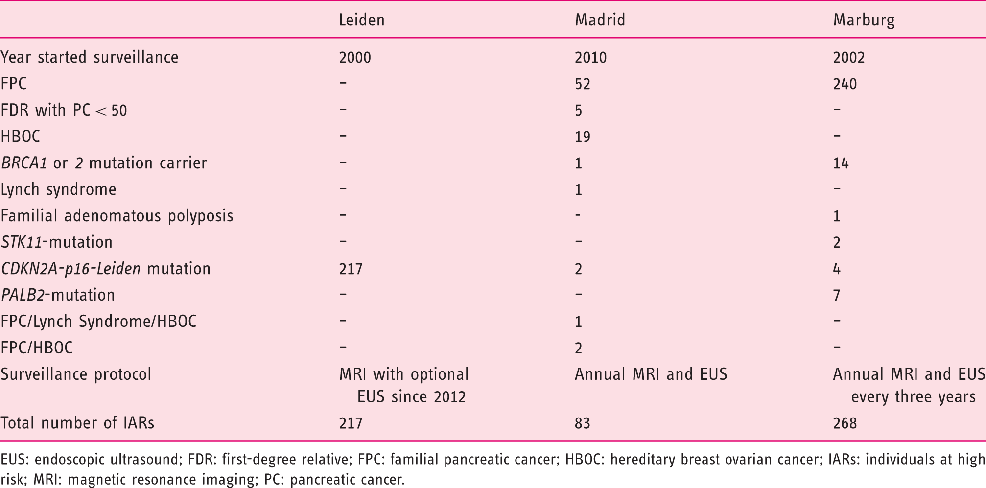
EUS: endoscopic ultrasound; FDR: first-degree relative; FPC: familial pancreatic cancer; HBOC: hereditary breast ovarian cancer; IARs: individuals at high risk; MRI: magnetic resonance imaging; PC: pancreatic cancer.
All MRIs were studied in detail for the presence of incidental findings including cysts, solid lesions, focal nodular hyperplasia (FNH), hemangioma and cancers. For all patients with an incidental lesion, further information was collected on whether additional imaging, intervention or surgery was performed. The observation time was from the start of a screening program up to 1 January 2018. The study was approved by the ethics committees of the respective centers.
Oral or written informed consent was received from all patients. The study protocol conforms to the ethical guidelines of the 1975 Declaration of Helsinki.
Results
Leiden, the Netherlands
Total number of incidental extrapancreatic lesions in the three European cohorts.
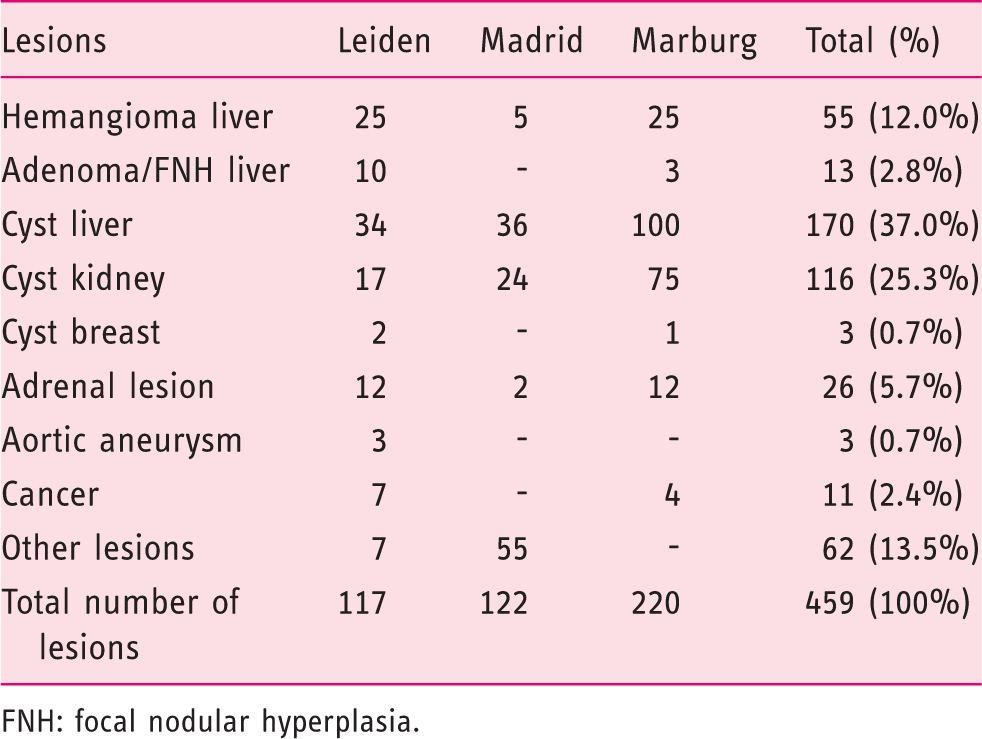
FNH: focal nodular hyperplasia.
Incidentalomas in the adrenal glands (adrenaloma) were identified in 12 cases (10.3%). In two of the 12 cases the lesion was removed during pancreatic surgery for a solid lesion. The first patient was a 40-year-old homozygote p16-Leiden carrier with a solid lesion in the uncinate process of the pancreas, together with a mass in the right adrenal gland detected on the first MRI. CT confirmed both lesions and defined the adrenal mass as an adrenaloma of 3.5 cm. A pancreaticoduodenectomy was performed and the adrenal mass was resected. Pathological examination revealed a PDAC and an adrenal adenoma without evidence of malignancy.
The second patient was a 66-year-old woman who came for her first MRI scan. The MRI showed a mass in the adrenal gland of 2.4 cm, together with a 1 cm hypovascular mass in the uncinate process. Subsequent CT confirmed both lesions but could not define the adrenal mass. The patient underwent a pancreaticoduodenectomy and an adrenalectomy. Pathological examination showed an intraductal papillary mucinous neoplasm with low-grade dysplasia, and an adrenaloma of 2.4 cm with adrenocortical hyperplasia.
Characteristics of extrapancreatic cancers (or metastatic disease) detected by the Leiden program for PDAC.
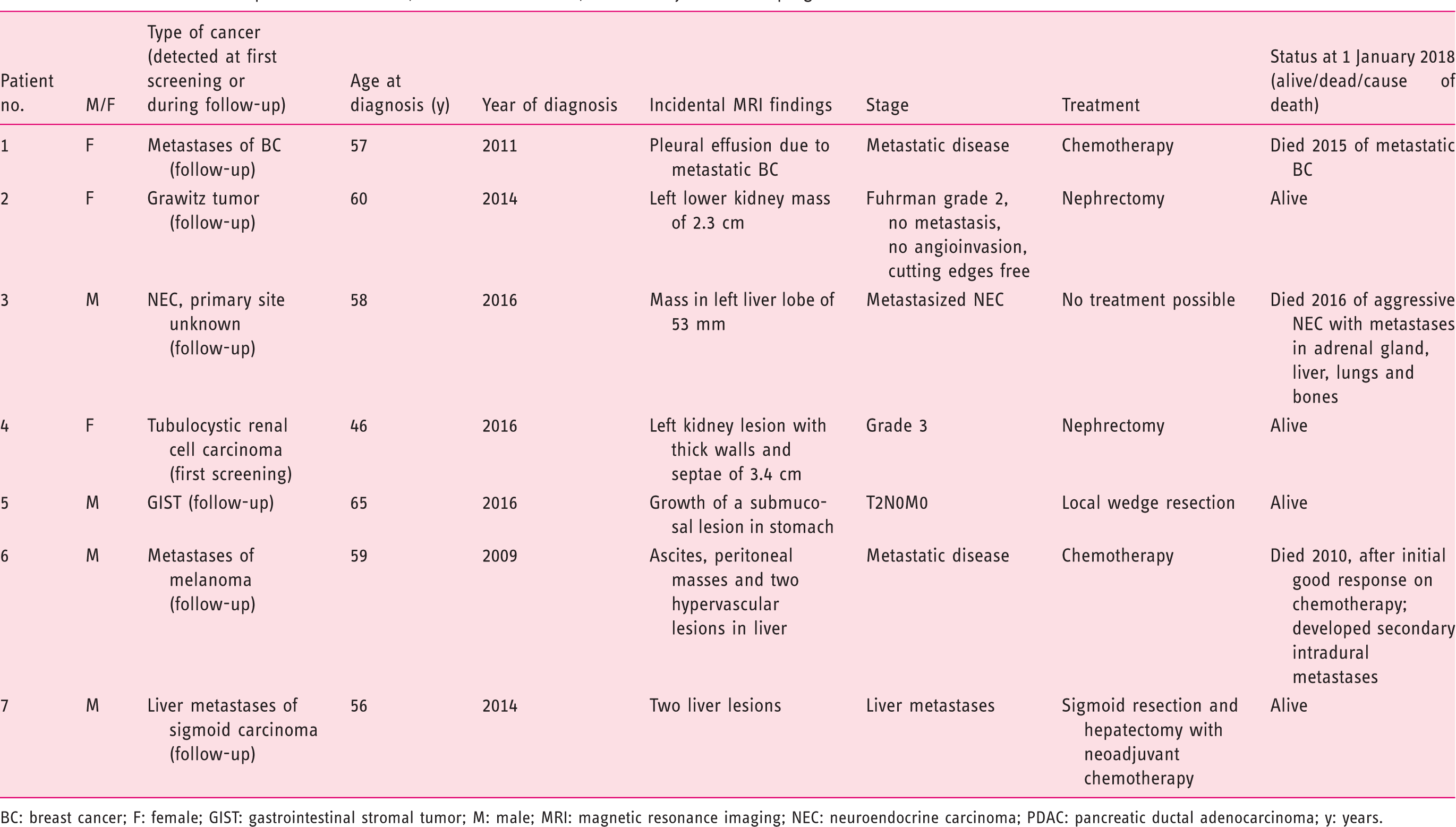
BC: breast cancer; F: female; GIST: gastrointestinal stromal tumor; M: male; MRI: magnetic resonance imaging; NEC: neuroendocrine carcinoma; PDAC: pancreatic ductal adenocarcinoma; y: years.
Madrid, Spain
Eighty-three IARs were under surveillance, consisting of 37 men (44.6%) and 46 women (55.4%). The analyzed cohort included a number of high-risk groups. Forty-two belonged to FPC families, five individuals had a first-degree relative with PDAC younger than 50 years, 19 belonged to a hereditary breast ovarian cancer (HBOC) family, one individual was a BRCA2 carrier, one belonged to a Lynch syndrome family, two had a CDKN2A-p16-Leiden mutation, one belonged to a family with evidence of combined FPC, Lynch syndrome and HBOC, and two belonged to a family with mixed FPC/HBOC. The mean age at start of surveillance was 50 years (range, 29–81 years), with a median follow-up time of 2.9 years (range, 0.1–6.7 years). In total, 122 incidental lesions were detected in 83 individuals (Table 2). Liver cysts (29.5%) were the most commonly found lesions and renal cysts were the second most common finding (19.7%).
In none of the patients was surgical management required. There was one patient who required additional imaging after a solid renal tumor was found (0.8%), but the lesion was characterized as an angiolipoma.
Marburg, Germany
Of the 268 IARs under surveillance in Marburg during the study, 109 were men (40.7%) and 159 were women (59.3%). Average age at start of screening was 48 years (range, 25–75 years) and the median follow-up time was three years (range, 0.1–14.6 years). The cohort included 240 individuals with FPC, four BRCA1 mutation carriers, 10 BRCA2 carriers, seven PALB2 mutation carriers, four CDKN2A/p16-Leiden mutation carriers, two STK11 mutation carriers and one patient with familial adenomatous polyposis with PDAC. A total of 220 lesions were identified in the 268 patients (Table 2). The most common findings were cysts in the liver (45.5%) or kidney (34.1%). Adrenaloma were observed in 12 cases (5.4%). Liver cysts in two patients and a renal cyst in one patient (1.1% of all patients) required surgical removal.
Regarding the need for additional investigations, the two patients who had surgery for liver lesions had an additional contrast-enhanced ultrasonography. Another 47-year-old man had an additional gastroscopy because EUS gave a suspicion of a MALT (mucosa-associated lymphoma tissue) lymphoma, which was a peptic ulcer. In a 43-year-old woman, a mammography was performed because MRI showed contrast-enhancing lesions in both breasts. Mammography diagnosed fibroadenomas. In another 51-year-old female patient, a 53 × 50 mm solid liver lesion on MRI was further evaluated by contrast-enhanced ultrasonography, which confirmed a hemangioma.
Characteristics of extrapancreatic cancers (or metastatic disease) detected by the German program for PDAC.
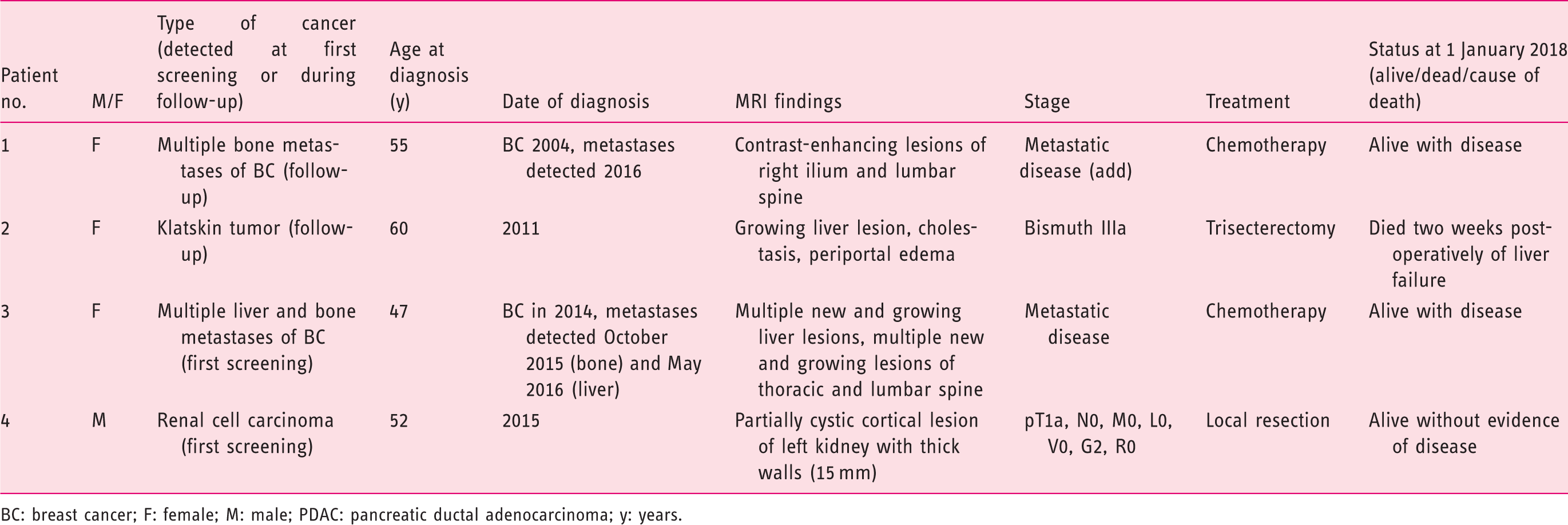
BC: breast cancer; F: female; M: male; PDAC: pancreatic ductal adenocarcinoma; y: years.
The second patient was a 60-year-old woman undergoing MRI surveillance in 2011. The MRI showed a lesion in the liver, with cholestasis and periportal edema. This lesion turned out to be a bile duct carcinoma (Bismuth stage IIIa). The patient underwent extended liver resection but unfortunately died two weeks later of postoperative liver failure.
The third patient was a 48-year-old female patient who underwent surgery because of breast cancer in 2014. A year later the patient underwent pancreatic cancer screening. MRI showed multiple lesions of the thoracic and lumbar spine, which proved to be bone metastases of the breast cancer. One year later, liver metastases were detected. The patient is still alive.
The detection of the cancers (or metastatic cancers) by the PDAC screening program did not result in a cure for any of these three patients.
The fourth patient, a 52-year-old man, showed a 15 mm, partially cystic cortical lesion of the left kidney with thick walls. This lesion was resected and turned out to be a renal cell carcinoma. The patient is alive without evidence of disease at last follow-up.
Discussion
The present study shows that MRI-based pancreas surveillance programs for PDAC result in the detection of a large number of incidental lesions. The most commonly found lesions were liver cysts, renal cysts and liver hemangioma, which together accounted for 74% of all incidental lesions, followed by adrenal incidentaloma in 6% of patients. Only five (0.9%) patients underwent surgery for a benign lesion: two patients for a liver cyst, one for a renal cyst and two for an adrenal incidentaloma.
Cancer was detected in 11 patients (1.9%), including seven CDKN2A-p16-Leiden mutation carriers, and metastatic disease was detected in six of the 11 patients. Early detection of tumors was beneficial in at least five of the patients.
Several studies have reported frequencies of incidental findings detected during abdominal imaging. One study reported the rate of incidental findings of whole-body MRI in 148 healthy control participants. 11 The most frequently found abnormalities were renal cysts (42.9%), gallstones (12.2%) and liver cysts/hemangioma (10.2%). In a similar study whole-body MRI was performed in 118 healthy individuals. 12 A total of 106 incidental lesions were found in the 83 individuals with an abnormality, the most common lesions being renal cysts (16.0%), liver hemangioma (12.3%) and liver cysts (11.3%). These findings are in agreement with our findings for benign lesions. However, the rate of incidentally detected cancers in the subgroup of CDKN2A-p16-Leiden mutation carriers was much higher.
What was the benefit of the detection of incidental lesions in our study? Although incidental findings were frequent, only 0.9% of the total group of IARs underwent a surgical intervention for a lesion, which was then found to be benign in all cases. A primary cancer, metastases of a previous cancer or a new cancer was detected in 1.9%. By contrast, in the Leiden cohort of CDKN2A-p16-Leiden mutation carriers, extrapancreatic cancer was detected in a substantial proportion of patients (seven patients out of 217 (3.2%)). The early detection of cancers in seven mutation carriers allowed curative resection of renal cancers in two patients, a gastric stromal tumor in one patient and colonic resection (and early start of chemotherapy) in one patient with CRC. In the German cohort, the detection of a renal cell carcinoma allowed curative resection. In addition, the identification of metastatic breast cancer in two patients allowed the early start of chemotherapy.
Strengths of the current study include the substantial size of the study group, the wide variation of high-risk groups and the long follow-up time. A possible limitation was that we are not informed about which definitions were used for a significant incidentaloma in the three expert centers and which guidelines for their management.
What are the clinical implications of our findings? First, it is important to inform all participants at the start of the surveillance program about the possibility of detecting incidental lesions. Based on our findings, it might be explained to patients that lesions are almost always harmless and will not require additional treatment. However, carriers of a CDKN2A-p16-Leiden mutation should be told that cancer might be detected outside the pancreas in a small proportion of patients.
To improve the investigation of the pancreas, there is currently a trend toward restricting MRI scanning to the pancreas only. However, to avoid missing cancers located outside the pancreas in CDKN2A-p16-Leiden mutation carriers, MRI assessment should include at least one scan of all abdominal organs.
In summary, the present study demonstrates that incidentaloma is a common finding in IARs for PDAC, but rarely requires additional treatment. CDKN2A-p16-Leiden mutation carriers were the only patient group found to harbor a substantial number of cancers, and detection resulted in benefit in several cases.
Footnotes
Acknowledgments
We thank Prof A. Mahnken for reading the MRIs, and Thomas Gress, Christian Bauer and Tobias Grote for comparing EUS and MRI results. We are thankful for the grant support of the Deutsche Krebshilfe (no. 111092) and a generous donation from the Gauff-Foundation.
Declaration of conflicting interests
None declared.
Ethics approval
The study protocol conforms to the ethical guidelines of the 1975 Declaration of Helsinki, and was approved by the ethics committees of the respective participating centers.
Funding
This work was supported by a grant from the Deutsche Krebshilfe (no. 111092) to DKB and EPS, and a donation from the Gauff-Foundation to DKB.
Informed consent
Oral or written informed consent was received from all patients.
D.K.B. and H.F.A.V. share senior authorship of this work.