
Abstract
Goltz syndrome is a rare condition characterized by thinning of the skin, which leads to the herniation of fat and results in both skin and systemic abnormalities. The primary cause of this syndrome is the mutation of the PORCN gene, which is associated with the X chromosome. A newborn baby was admitted to the neonatal intensive care unit due to skeletal and skin abnormalities. The major findings in this patient included anophthalmia, microform cleft lip, subcutaneous fat herniation, and split foot. An abdominal ultrasound examination revealed a solitary right kidney and an echocardiogram showed patent ductus arteriosus. The patient was treated for neonatal sepsis, and the family received counseling about the disease. We report this case because of its exceptional rarity.
Introduction
Focal dermal hypoplasia (FDH) (Goltz Syndrome) is an X-linked dominant multisystem disorder that is fatal in utero in males. The primary cause is the PORCN gene mutation associated with the X chromosome, justifying the predominance of females. 1
The main feature of Goltz syndrome is patchy dermal hypoplasia with the herniation of fat through defects in the dermis. Associated systemic abnormalities are commonly reported, such as musculoskeletal defects, gastrointestinal and central nervous system malformations, cardiac defects, and renal and eye anomalies.2,3 Less than 300 cases have been reported in the literature. Here, we report a preterm neonate with multisystem anomalies.
Case report
A 20-min neonate from the labor ward was admitted to the neonatal intensive care unit (NICU) after presenting skeletal deformity and skin lesions.
She was born to a 26-year-old Para III mother and had regular prenatal check-ups. She underwent regular pregnancy screenings and received a negative test result. Pregnancy was uneventful and delivered via spontaneous vaginal delivery to affect a female neonate with Apgar scores of 7 and 8 in the first and fifth minutes, respectively.
On examination, vital signs were within the normal range. The weight measured 1600 g, the height was 41 cm, and the head circumference was 28 cm. Head and neck examinations revealed anophthalmia of the left eye and microform cleft lip (Figure 1). Dermatologic examination revealed hypo- and hyper-pigmented subcutaneous skin lesions with fat herniation (Figure 1). A 3 × 4 cm abdominal wall defect was notable on the abdomen (Figure 2).

Anophthalmia of the left eye, microform cleft lip, subcutaneous fat herniation of the skin, split foot, and clinodactyly of the right index toe.
A 3 × 4 cm abdominal wall defect (omphalocele).
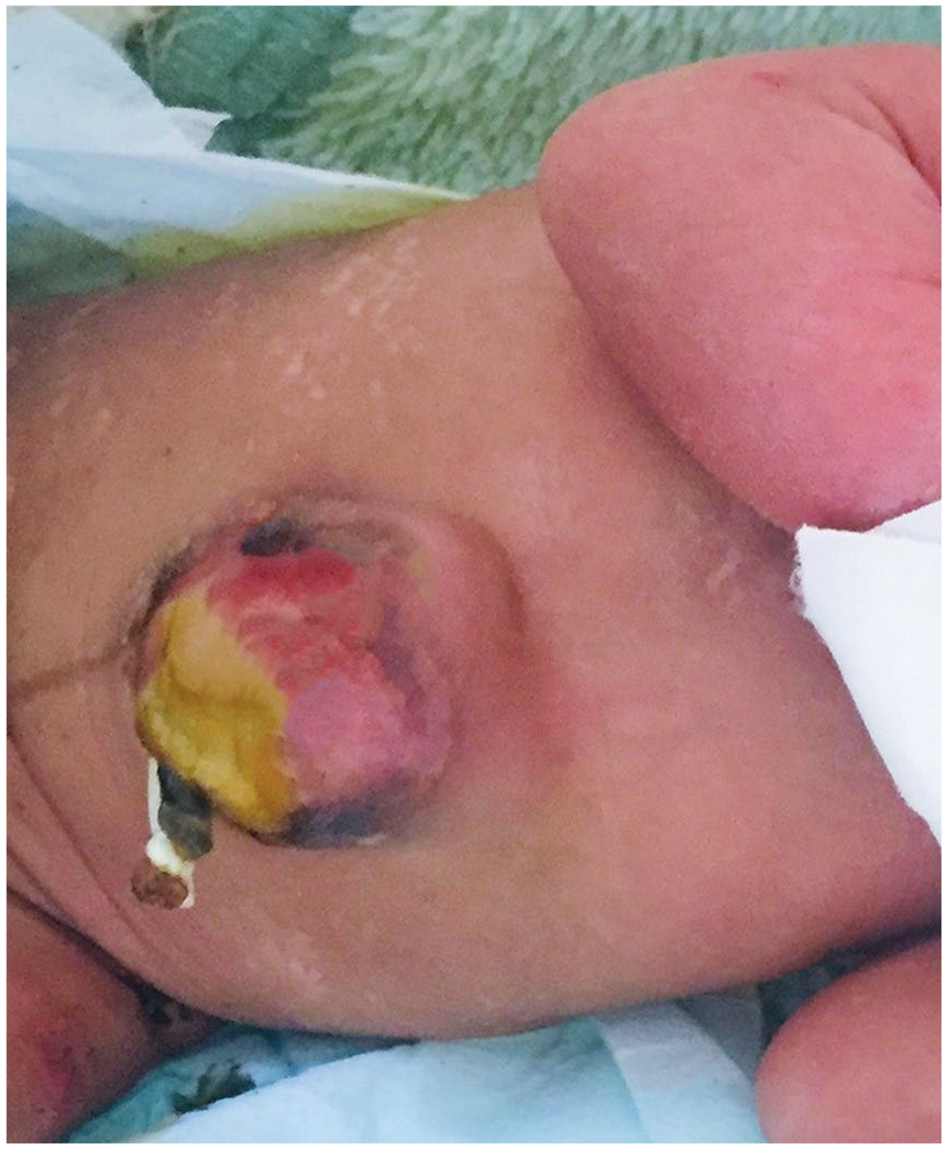
Moreover, there was erythematous atrophic skin ulceration with crusted edges on the posterior aspect of the right lower leg (Figure 3). Examination of lower extremities showed split foot and clinodactyly of the right index toe (Figure 1). On primitive neonatal reflexes, Moro was complete, Grasp was strong, but Suckling was unsustained. With the above finding, the patient was investigated with routine laboratory tests and showed normal. An abdominal ultrasound examination showed the presence of a single kidney on the right side. Echocardiogram evaluation revealed patent ductus arteriosus. Cranial ultrasonography exhibited no abnormalities. With the diagnosis of preterm + low birth weight + Goltz syndrome + early-onset neonatal sepsis, the patient was started with first-line antibiotics for neonatal sepsis management. Trophic feeding (20 mL/kg) was initiated and increased daily until the patient tolerated full feeding. After 5 days of NICU stay, the patient was discharged from the hospital and appointed to cardiac, dermatology, and pediatric surgery clinic. Unfortunately, the patient could not attend the follow-up for transport issues.

Erythematous atrophic skin ulceration with crusted edges.
Discussion
FDH is a rare syndrome characterized by thinning of the skin, resulting in fat herniation with cutaneous and systemic abnormalities. The disease exhibits various clinical features, including developmental skin defects with ocular, dental, and skeletal anomalies.
A systematic review of 159 cases of FDH from 33 countries reported that most of the cases were from Europe, North America, and Asia than in Africa, South America, and Oceana. In Africa, only a total of five cases have been found in the literature. Skin involvement was noted in more than 95% of the cases. The most frequent cutaneous lesions were atrophy, pigmentation, fat herniation, nail dystrophy, alopecia, and papilloma.4,5 These dermatologic manifestations aligned with our findings except for the alopecia and papilloma. The skin lesions might extend further, as seen in a neonate who has the extension of the lesions within 1-month duration to involve larger areas of the body with a more characteristic appearance. 6
Regarding skeletal deformities, it is another prominent feature of FDH. This constitutes syndactyly, polydactyly, clinodactyly, split foot, and limb malformations. Other unusual skeletal and facial features include costovertebral abnormalities, split sternum, and fibrous dysplasia of bone and facial cleft.4,7–10 One of the common eye abnormalities in Goltz syndrome is anophthalmia and microphthalmia. Other less common eye anomalies in the literature are corneal abnormalities, retinal detachment, and coloboma of the eyes.4,6 The present case had anophthalmia of the left eye on ophthalmologic examination. Bhatia et al. and Sarma et al. reported FDH cases with abdominal wall defects. The reason might be related to the skin defect associated with FDH, which results in abdominal organs herniating out easily. In addition, they found postoperative concerns such as abdominal wall dehiscence.11,12 As reported in the literature, kidney abnormalities vary from one patient to another. Reddy and Laufer 8 and Han et al. 13 found FDH cases with unilateral kidneys. Both patients fulfilled the diagnostic criteria for Goltz syndrome. These findings agree with our case, which showed a solitary right kidney. Since children are growing, some features are yet to appear. Examples include oral manifestations such as squamous papilloma, gingival hyperplasia, delayed tooth eruption, malocclusion, and absence of lingual frenulum. 14
Straightforward diagnosis of these patients seems challenging. Wang et al. suggested a potential standard for the diagnosis of FDH, which is a typical finding of skin or PORCN mutation plus anyone of three clinical symptoms, including skeletal system abnormalities, ocular and oral abnormalities. Although it lacks proof, Gysin et al. proposed that in the context of typical skin changes, the presence of visible Blaschko lines on teeth in the form of vertical grooves is nearly pathognomonic for FDH.4,15 Genetic analysis was not done in the present case, but the patient fulfilled all clinical criteria found in published papers. Management of these patients requires a holistic approach with the involvement of relevant specialists. The future trajectory of research should be focused on enhancing the therapeutic options for FDH.
Conclusion
In summary, this case demonstrated the common features of the disease such as limb, skin, and eye anomalies. Furthermore, our case exhibited the presence of a single kidney, a rare but documented occurrence.
Footnotes
Acknowledgements
None.
Author contributions
A.A.M. contributed to the study conception, search-related literature as well as writing the whole manuscript. H.H.H., D.S., and A.H. were involved in the study design, literature review, and critical revision of the manuscript.
Declaration of conflicting interests
The author(s) declared no potential conflicts of interest with respect to the research, authorship, and/or publication of this article.
Funding
The author(s) received no financial support for the research, authorship, and/or publication of this article.
Ethical approval
A case report is described in the article. No further approval from the Ethics Committee was necessary.
Informed consent
Written informed consent was obtained from a legally authorized representative for anonymized patient information to be published in this article.