
Abstract
Dermatofibrosarcoma protuberans is a malignant tumor of the soft tissue which is characterized by local recurrence with an exceptional metastasize, and for this reason, patients with dermatofibrosarcoma protuberans require a long-term follow-up. This clinical case presents a 41-year-old patient, with no pathological history, who has a malignant tumor of the chest wall, with a pulmonary nodule, which appears on the chest computed tomography scan like a well-rounded parenchymal nodule measuring 15 mm in diameter. This case was discussed in the multidisciplinary meeting which concluded that it is a dermatofibrosarcoma protuberans with metastasis of the lung. In the context of these data, it is thought that the pulmonary nodule is of metastatic origin according to the decision of the multidisciplinary meeting, while the final diagnosis was a hamartochondroma, which is a benign tumor of the lung. The interest of this clinical case is to discuss the possible diagnoses of the pulmonary nodule in the context of malignant wall tumor.
Introduction
Dermatofibrosarcoma protuberans is a rare chest wall malignant tumor which most frequently affects the dermis and subcutis 1 and is recognized by the local recurrence. Its incidence represents 0.1% of malignant tumors of soft tissues. 2 Pulmonary hamartochondroma is the most common benign tumor of the lung. The association of the two tumors is not exceptional (benign lung tumor and malignant tumor of the chest wall), but the appearance of the hamartochondroma in the form of a pulmonary nodule has made it possible to present this clinical case for discussion of the diagnosis of this nodule. Is it a metastatic nodule or a nodule of another etiology? Our goal is to answer this question in this manuscript.
Case report
This clinical case presents a 41-year-old patient, with any pathological history, who consults for a tumefaction of the left posterior thoracic wall evolving in a context of conservation of the general state. The clinical examination finds a hard mass, budding, fixed to the deep plane (Figure 1). The patient consulted a dermatologist who performed a biopsy of the mass returning in favor of a dermatofibrosarcoma protuberans. The chest computed tomography (CT) scan objectified in addition to the tumor of the chest wall the presence in the upper left lobe of a well-rounded parenchymal nodule measuring 15 mm in diameter (Figures 2 and 3). Positron emission tomography (PET) scan was not requested because of its unavailability in our region. This case was discussed in the multidisciplinary meeting which concluded that it is a dermatofibrosarcoma protuberans with metastasis of the lung. The decision was to operate the patient. Wide resection of the tumor at 3 cm from its banks was performed, after the patient has benefited of a posterolateral thoracotomy for the pulmonary nodule whose macroscopic appearance has objectified cartilaginous consistency suggestive of a hamartochondroma, and a wedge resection was practiced. Histological study showed tumoral proliferation with fasciculate and storiform architecture. The tumor cells are spindle-shaped with elongated nuclei, increased size, fine chromatin and eosinophilic cytoplasm poorly limited. This proliferation infiltrates the dermis and hypodermis. For the pulmonary nodule, it was a lesion well limited and consists essentially of mature cartilaginous lobules surrounded by fibro-adipose tissue. All that were dissociated by slots were covered with a respiratory type epithelium, cylindrical ciliated and pseudostratified. The follow-up to now is 8 months, without any local recurrence or distant metastasizes.

Dermatofibrosarcoma protuberans of the left posterior thoracic wall, hard and budding.

Thoracic CT showed a cutaneous tumor process in the posterior thoracic wall compatible with dermatofibrosarcoma protuberans.
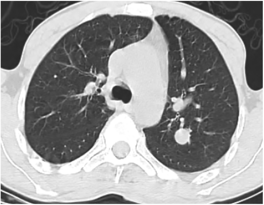
Thoracic CT in parenchymal window showed pulmonary nodule of the left upper lobe solid measuring 15 mm.
Discussion
The association of dermatofibrosarcoma and pulmonary hamartochondroma is not exceptional on the medical side. In this case, dermatofibrosarcoma protuberans of chest wall was associated with a pulmonary nodule, and the first question which was asked was, “What is the diagnosis of this nodule? Is it a metastatic nodule of the dermatofibrosarcoma protuberans or just a benign nodule?” According to the multidisciplinary committee, the presence of the tumor of the chest wall and the features of the pulmonary nodule (well-rounded nodule, measuring 15 mm), the latter was considered as a metastasis site of the dermatofibrosarcoma protuberans. However, as a result, histological study of this nodule was in favor of a hamartochondroma and a wedge resection was performed. Dermatofibrosarcoma protuberans is a rare tumor, but not exceptional, that has been identified for the first time by Jean Darier and Marcel Ferrand as a real clinical and histological entity. 3 Its tendency to local recurrence requires extensive surgical resection. 4 Therefore, metastasizes to the regional lymph node or distant site (lung, bone, etc.) remain rare and are found in less than 6% of the cases, and for this particularity are classified as a sarcoma of intermediate grade malignancy.5–10 The basic treatment of dermatofibrosarcoma protuberans remains surgical excision, with margins of 3–5 cm to avoid local recurrence. 11 Radiotherapy is indicated in case of incomplete excision, and chemotherapy is the reference treatment in metastatic cases.11,12
On the contrary, hamartochondroma is the most frequent lung benign tumor. 13 It is a tumor derived from the peribronchial mesenchymal tissue constituted in an absolute disorder and in variable proportions of cartilage, connective tissue, fat, smooth muscle and respiratory epithelium. 3 The multidisciplinary meeting concluded that the parenchymal nodule is likely a metastasize of the chest wall tumor, since the features of the nodule were not in favor of other etiological orientations. Radiologically, pulmonary hamartochondroma is usually of heterogeneous density, sometimes with a popcorn appearance. In our case, the signs that are in favor of a pulmonary metastasis are especially the presence of the dermatofibrosarcoma protuberans, the solid character of the nodule, with a homogeneous density, without greasy density, and the central character of the nodule. However, the main points are with a benign nodule: the well-defined contours, the absence of local recurrence of dermatofibrosarcoma since the rate of metastasis increases after local recurrence. Also, it must be pointed out that the patient did not benefit from a PET CT to clearly specify the benign or malignant nature of the pulmonary nodule because of the unavailability in our region.
Conclusion
The principle of the management of a pulmonary nodule is to not ignore a malignant nodule at a curable stage, and to not carry out invasive explorations for a benign nodule. In this clinical case, association of dermatofibrosarcoma protuberans with pulmonary hamartochondroma has allowed to discuss the diagnosis of pulmonary nodule that remains generally of variable etiologies.
Footnotes
Author contributions
All the authors contributed substantially to the authorship of this manuscript.
Declaration of conflicting interests
The author(s) declared no potential conflicts of interest with respect to the research, authorship, and/or publication of this article.
Ethical approval
All procedures performed in studies involving human participants were in accordance with the ethical standards of the institutional and/or national research committee and with the 1964 Helsinki Declaration and its later amendments or comparable ethical standards.
Funding
The author(s) received no financial support for the research, authorship, and/or publication of this article.
Informed consent
Written informed consent was obtained from the patient for their anonymized information to be published in this article.