
Abstract
Ciliated muconodular papillary tumors are benign lesions located in the peripheral lung field. Recent studies revealed BRAF and epidermal growth factor receptor gene mutations and anaplastic lymphoma kinase gene rearrangement. Five ciliated muconodular papillary tumors were screened for the BRAF V600E and EGFR mutations via polymerase chain reaction. Immunohistochemical analysis was performed for the detection of the BRAF V600E and anaplastic lymphoma kinase proteins, as well as other markers including phosphorylated extracellular signal-regulated protein kinase. Three tumors (60%) harbored the BRAF V600E mutation. Immunohistochemical analysis confirmed this mutation in all of the tumor cell types. EGFR mutation and immunoactivity of the anaplastic lymphoma kinase protein were not detected. Phosphorylated extracellular signal-regulated protein kinase was negative both in the cytoplasm and nucleus of the BRAF V600E–positive tumors. Mucin 1, mucin 4, thyroid transcription factor 1, and cytokeratin 7 were positive, and mucin 5AC was partially positive, whereas napsin A and cytokeratin 20 were negative. Ciliated muconodular papillary tumor may originate from the terminal bronchioles, and the status of ERK activation reflects its benign behavior.
Keywords
Introduction
A ciliated muconodular papillary tumor (CMPT) is characterized by papillary proliferation of ciliated columnar cells, goblet cells, and basal cells. Of the approximate 30 cases of CMPTs reported in the English literature,1–9 no recurrence or metastasis has been reported. In a study of 10 CMPTs, Kamata et al. 3 revealed that 50% harbored a BRAF mutation, and 30% had an epidermal growth factor receptor (EGFR) mutation. Other studies reported CMPTs with AKT mutations, anaplastic lymphoma kinase (ALK) rearrangements, and KRAS mutations.2,5,6,9 These results indicate that a CMPT is a neoplastic lesion.
Extracellular signal-regulated kinase (ERK) is a component of the mitogen-activated protein kinase (MAPK) pathway and activated by phosphorylation and nuclear translocation. ERK activation has been suggested to play a role in the pathogenesis and progression of various cancers. The BRAF V600E mutation is reported to activate the MAPK pathway and promote cell proliferation. A previous study reported a poorer prognosis of ERK-activated colon cancer than of colon cancer without ERK activation. 10 However, to the best of our knowledge, no report has yet addressed the status of ERK activation in CMPT.
In this study, BRAF V600E and EGFR mutations were screened in five CMPTs resected at our hospital. Immunohistochemical (IHC) analysis of the BRAF V600E mutation and ALK was also performed. Moreover, immunostaining of phosphorylated extracellular signal-regulated kinase (p-ERK) was performed to reveal the role of the MAPK pathway in the pathogenesis of CMPT. Tumor origin was also estimated by IHC staining of mucin core proteins and diagnostic marker proteins of lung cancer. This study is conducted independently and does not constitute any other larger studies.
Case section
Patient characteristics
The characteristics of five patients (2 male, 3 females) are shown in Table 1. All tumors were single and less than 18 mm in diameter. No recurrence or metastasis was observed during follow-up examinations conducted from 0.5 to 6 years. Three patients had a history of malignancy.
Patient characteristics.

LUL: left upper lobe; LLL: left lower lobe; RLL: right lower lobe.
Histological analysis of CMPT
All tumors consisted ciliated columnar cells, mucinous cells, and basal cells arranged in papillary and glandular structures (Figure 1(a) and (b)), consistent with the features of CMPTs noted in previous reports. A transitional zone from normal bronchioles with 0.2 mm diameters to CMPT was observed in patient 3 (Figure 1(e)).
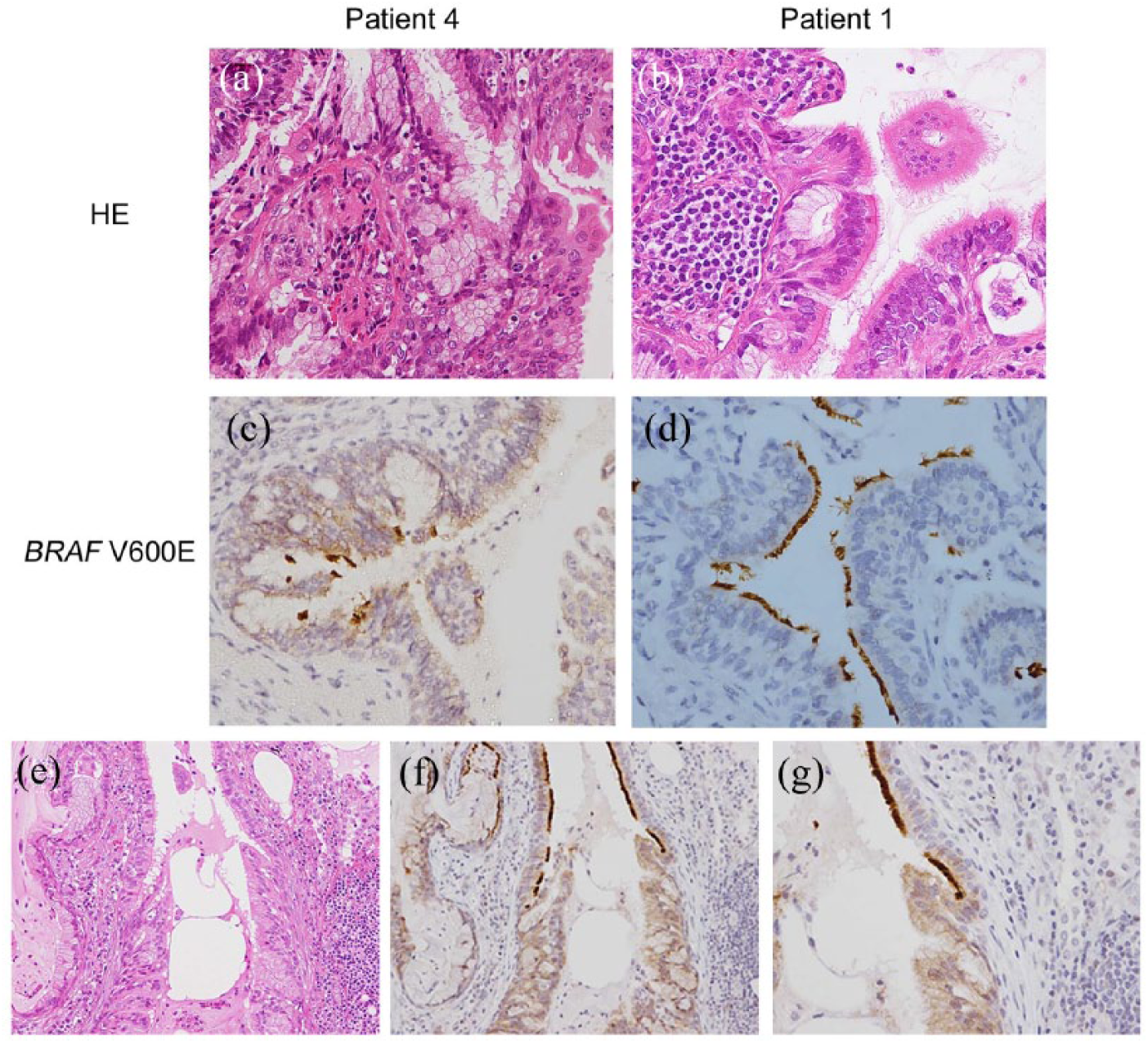
Histological analysis of CMPT and IHC analysis of the BRAF V600E mutation. Ciliated columnar cells, mucinous cells, and basal cells formed papillary and glandular structures (a, b). IHC analysis of BRAF V600E with VE1 antibody in patients 4 and 1 (c, d). (b) A transitional zone between the normal bronchioles and tumor was observed in patient 3 (e). Cytoplasmic staining was stronger for CMPT than for the normal bronchioles in the transitional zone of patient 3 ((f) and (g): high magnification).
IHC analysis of BRAF V600E, ALK, and p-ERK
Immunostaining for BRAF V600E was positive in tumors from patients 3, 4, and 5 (Figure 1(c) and (d) and Table 2). All three types of tumor cells were stained. The cilia of adjacent bronchioles were also stained. In the transitional zone from normal bronchiolar epithelium to CMPT, cytoplasmic staining of CMPT contrasted with that of the bronchiolar epithelium (Figure 1(f) and (g)).
Immunohistochemical analysis.

TTF-1: thyroid transcription factor 1; MUC: mucin; CK: cytokeratin; p-ERK: phosphorylated extracellular signal-regulated kinase; b: basal cells; c: ciliated columnar cells; m: mucinous cells; −: negative; +−: mild; +: moderate; ++: diffuse.
IHC staining of all five tumors were negative for the ALK protein.
Staining for p-ERK was negative in both the cytoplasm and nucleus of the BRAF V600E–positive tumor cells (Figure 2(a) and Table 2). However, in BRAF V600E–negative tumors, some nuclei of the mucinous cells were positive for p-ERK (Figure 2(b) and Table 2).

IHC analysis of phosphorylated ERK. A representative BRAF mutation–positive case (patient 3, (a)) and a negative case (patient 1, (b)) are presented.
IHC analysis of mucin core proteins and lung cancer markers
The results of IHC analysis for mucin core proteins and lung cancer–related markers are shown in Table 2. All tumors were positive for MUC1 and MUC4, whereas some columnar and mucinous cells were positive for MUC5AC. The tumors were also positive for thyroid transcription factor 1 (TTF-1) and cytokeratin 7 (CK7) but negative for napsin A and cytokeratin 20 (CK20).
Gene mutation analysis by polymerase chain reaction
The DNA extracted from dissected tumors was screened for the BRAF V600E mutation. Three tumors that were positive for the BRAF V600E mutation by IHC analysis harbored the BRAF V600E mutation (patients 3, 4, and 5; Figure 3). Isolated bronchioles of patient 5 were also examined by laser capture microdissection, which showed that all were negative for the BRAF V600E mutation (data not shown). EGFR mutations were also screened using extracted DNA from formalin-fixed, paraffin-embedded tissues according to the polymerase chain reaction-based method described previously. 11 All tumors were negative for EGFR mutations.
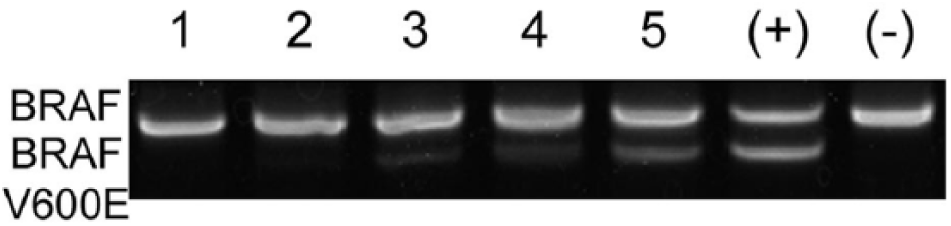
BRAF V600E analysis by the LH method. The BRAF V600E mutation was detected in patients 3, 4, and 5. (+): positive control; (−): negative control.
Discussion
In this study, three of five CMPTs harbored the BRAF V600E mutation, whereas the others were negative for the BRAF V600E mutation, EGFR mutations, and ALK gene rearrangements. A past study reported that 50% of CMPTs harbored a BRAF mutation, but no mutations of 50 screened genes were correlated to cell proliferation. 3 In this study, screening was limited to the BRAF V600E mutation. EGFR mutations, including one detected in a past study, were also screened. Other than ALK expression by IHC analysis, no other genetic analysis was performed. Therefore, tumors with no detected mutations in this study may have mutations of BRAF and/or other genes, such as AKT and KRAS, as previously reported.5,9 Other gene mutations may coexist even in tumors positive for the BRAF V600E mutation.
IHC analysis with the VE1 monoclonal antibody revealed that the cytoplasm and cilia of CMPTs and the normal bronchiole epithelium were positive for the BRAF V600E mutation. However, genetic analysis to identify other mutations of the bronchiole was not performed. A previous report indicated that the VE1 antibody cross-reacted with the dynein protein in cilia, including that in the bronchial epithelium. 12 Therefore, the positive staining of cilia was considered to be a false positive. Meanwhile, cytoplasmic staining was clearly stronger for the CMPTs than for the bronchial epithelium at the transitional zone, suggesting that the BRAF V600E mutation can be identified in CMPTs by cytoplasmic staining.
Histological analysis of CMPTs revealed a papillary structure originating from the central airway. A transitional zone of normal bronchioles (0.2 mm in diameter) to CMPT was also observed in patient 2. Approximately, all cells were positive for MUC1 and MUC4, and partially positive for MUC5AC. The former two proteins are reportedly expressed in the epithelium of the central and peripheral airways, whereas the expression of the latter protein is decreased in peripheral bronchioles less than 1 mm in diameter. 13 All CMPTs were negative for napsin A, which is known to be expressed in type II alveolar epithelium. BRAF mutations are reported to exist in less than 3% of lung adenocarcinomas. 14 The prevalence of BRAF mutations of adenocarcinoma is extremely low compared with that of CMPT in the present and a previous study. 3 According to the results of IHC and gene mutation analyses, a CMPT may originate from peripheral bronchioles, which is different from lung adenocarcinoma.
The status of ERK activation in other cancers reportedly varies. In contrast, a previous study reported that colon cancer cells with a BRAF mutation had a lower level of nuclear positivity and higher level of cytoplasmic positivity for p-ERK compared with those of colon cancer cells with wild-type BRAF. 15 Dual-specificity phosphatases participate in a negative feedback mechanism responsible for the dephosphorylation of nuclear p-ERK. This system may explain the lack of nuclear staining of p-ERK in colon cancer cells. In this study, p-ERK was negative in the cases in which BRAF V600E was detected suggesting the involvement of other mechanisms in the de-activation of the MAPK cascade in CMPTs. In any case, the status of p-ERK activation may explain the benign nature of a CMPT.
Conclusion
We presented five CMPTs and three of them harbored the BRAF V600E mutation. Histological and IHC analyses indicated a bronchiolar origin of CMPTs. Negative staining of nuclear p-ERK may reflect the restricted ability of proliferation signaling pathways.
Footnotes
Acknowledgements
Declaration of conflicting interests
The authors declared no potential conflicts of interest with respect to the research, authorship, and/or publication of this article.
Funding
The authors received no financial support for the research, authorship, and/or publication of this article.