
Abstract
Transradial artery approach as primary access for transcatheter diagnosis and intervention is associated with lower risk of bleeding and major vascular complications, improved patient comfort and shorter time to hemostasis and ambulation than femoral one. Patient’s adequate hand collateral perfusion, assessed by the Barbeau test, must be depicted prior to transradial artery approach in order to assess any absolute contraindication (D waveform). We describe the distal transradial artery approach, recently proposed for coronary interventions, used in emergency to embolize an intestinal bleeding in an 84-year-old woman and a left pectoralis major muscle bleeding in an 83-year-old woman, both with high risk of bleeding for femoral approach and contraindication for transradial artery approach (Barbeau D waveform).
Keywords
Introduction
The use of radial artery (RA) as the primary access for transcatheter diagnosis and intervention is associated with considerable reduction in the risk of vascular and bleeding complications, including easier accomplishment of postprocedural hemostasis, than femoral approach (FA).1,2 Other advantages of transradial arterial access (TRA), over transfemoral one, are shorter patient resumption leading to instant ambulation, with reduction-related costs and increased patient gratification. Furthermore, TRA may be advantageous in obese patients. 2 Some studies in literature have demonstrated the feasibility and safety of TRA also for non-coronary vascular interventions, such as uterine artery embolization, arterial renal disease and transarterial chemoembolization. 3
Before attempting TRA, it is fundamental to evaluate collateral perfusion to the hand through Barbeau test, in order to avoid ischemic hand complications. 3 A pulse oximetry is put on the patient’s thumb and the procedure starts with contemporary radial and ulnar artery occlusion. For 2 min following the release of the compression on the ulnar artery, the pulse should be measured and the wave must be recorded through the pulse oximetry. Depending on the type of waveform, registered ulnopalmar patency includes the following four types: (1) no damping of the pulse tracing immediately after compression, (2) damping of pulse tracing, (3) loss of pulse tracing followed by recovery within 2 min and (4) loss of pulse tracing without recovery within 2 min.2–4 Barbeau D waveform is the only true contraindication of TRA.2,3 It relieves high risk of hand ischemia in case of radial obstructive complication secondary to poor ulnar compensation.
Although TRA has many advantages, sometimes it can be difficult or impossible to reach not only in case of Barbeau D waveform but also for a small RA (⩽2 mm), anatomical variations (RA loops, tortuosity, hypoplasia) and in patients with a dialysis fistula.2,5 For these reasons, it would be desirable to find another arterial access having the advantages of radial access when the latter is not feasible.
Very recently, the distal transradial artery approach, from the anatomical snuffbox (AS), on the dorsal side of the hand has been proposed. 6 The puncture is distal from the branch supplying the superficial palmar arch. An occlusion at this site potentially maintains antegrade flow through the superficial palmar arch, preventing ischemia and hand disability. 6
Case report
Case 1
An 84-year-old woman with a history of arterial hypertension, type II diabetes mellitus, atrial fibrillation and chronic heart failure recently underwent to percutaneous transluminal coronary angioplasty (PTCA) and valvuloplasty for which she was taking anticoagulation (Sintrom 4 mg) and double antiplatelet therapy (clopidrogrel 75 mg + ASA 100 mg). Some days later, she presented melena and shock requiring blood transfusion and resuscitation maneuvers (hemoglobin (Hbg): 6.6 g/dL; platelet (PLT): 112 × 10³ µL). Once she was hemodynamically stabilized, a gastroscopy was performed which showed normal findings and a colonoscopy which showed digested blood in the colon with many clots within ascending colon; hence, an urgent angiography and eventually embolization were requested.
Because of high value of coagulation test (international normalized ratio (INR): 2.8; prothrombin time (PT): 33.6 s), the patient had a high risk of bleeding and vascular complications related to the FA, so we performed a TRA. The right RA was inaccessible because it was occluded by the previous PTCA. We performed the Barbeau test showing a type D waveform (Figure 1), which is a contraindication to TRA. Both high risk of bleeding to FA and the contraindication to the TRA forced us to find another possible arterial access with less risk of complications. We decide to evaluate with ultrasound (US) examination the left distal radial artery (DRA), in the AS, that was of sufficient diameter (2 mm). Patient gave her informed consent before the interventional procedures.
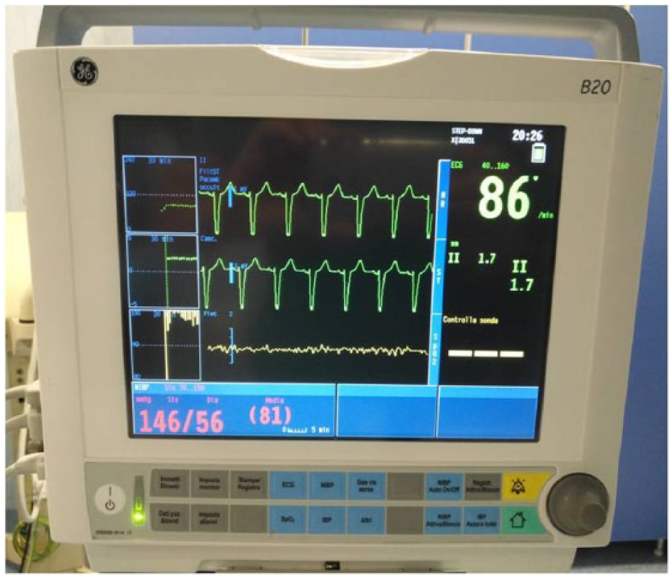
Barbeau test shows a D waveform recorded through the pulse oximeter. It demonstrates a loss of pulse tracing without recovery within 2 min (yellow line at the bottom).
A subcutaneous local anesthesia consisting of an injection of a solution of 1 mL of lidocaine 2% and 100 μg of nitroglycerin was injected in order to prevent arterial spasms. The DRA access was obtained under US guidance and Seldinger technique with a 21-gauge micropuncture needle and a 0.018-in. wire. After a 4-French sheath was inserted (Figure 2), another bolus of nitroglycerin (200 μg) and heparin (2000 IU) were administered intra-arterially on the basis of our institutional protocol. The superior mesenteric artery angiography showed ectatic vessels and early venous opacification indicating an ileal angiodysplasia. Then, a microcatheter was introduced for a superselective angiography of the ileo-colic artery. In order to stop the active bleeding, we performed an ileo-colic artery embolization using Spongostan and coil. A final diagnostic angiogram demonstrated the success of the procedure. Hemostasis was obtained with the TR band injecting 7 cm 3 of air, deflating 1 cm 3 every 5 min on the basis of our protocol for RA (Figure 3).

The 4 F sheath placed in the distal radial artery at the site of the anatomical snuffbox.

(a) TR band placement at the site of the vascular access, (b) insufflation of air, (c) removal of the sheath and (d) final result in the absence of bleeding.
The procedure of embolization was safely and successfully performed without any complications or discomfort for the operator and the patient. No RA occlusions occurred.
A color Doppler examination of the left RA that showed the patency of the vessel was performed the following day. Evaluation of the access site and a color Doppler examination of the left RA were repeated before discharge and during the follow-up at 1, 3 and 6 months.
Case 2
An 83-year-old woman with a history of autoimmune thrombocytopenia, arterial hypertension and type II diabetes mellitus was admitted to the hospital with extensive hematoma of the left hemithorax following trauma. Blood tests revealed anemia (Hbg: 10 g/dL) and thrombocytopenia (PLT: 24 × 10³ µL). A computed tomographic (CT) scan of the thorax proved contrast blush in the region of left pectoralis major muscle; hence, an urgent angiography and embolization were requested.
Because of thrombocytopenia, the patient had a high risk of bleeding and vascular complications related to the FA, so we decided to perform a TRA, but the Barbeau test showed a type D waveform. DRA diameter (2 mm) was suitable for puncture at US examination. The patient gave her informed consent before the interventional procedures.
The DRA access was obtained with the same technique already described. A diagnostic angiography of left axillary and subclavian arteries was performed using a 100-cm 4-French Berenstein catheter. It showed contrast extravasation of left lateral thoracic artery (LLTA). A microcatheter was introduced for a superselective angiography of the LLTA and we used Spongostan to embolize the LLTA. A final diagnostic angiogram demonstrated the success of the procedure. An occlusive pressure of the DRA was obtained in the same way as described. The evaluation of the RA patency, using a color Doppler examination, was performed in the same way as in case 1, which gave similar results.
Discussion
An accurate and profound knowledge of vascular anatomy of the hand related to possible variations is mandatory for selecting patients suitable for such vascular access in order to obviate disappointing unexpected complications. 5
RA and ulnar artery are widely interconnected at the level of the hand through the superficial and deep palmar arches, commonly with good hemodynamic compensation.5,7 This compensation is evaluated by Barbeau test.
We can find the DRA in AS that is a depression located in the radial part of the wrist; it is laterally limited by the tendons of abductor pollicis longus and extensor pollicis brevis muscles and medially limited by the tendon of extensor pollicis longus muscle. 8
The site of puncture of DRA, in AS, is distal from the origin of the palmar carpal branch, the superficial palmar branch and the dorsal carpal branch.5–7 So this access is distal from the origin of some branches that determine the main perfusion of the hand; this means that in case of occlusion, theoretically, the RA feeding flow to the hand is not totally compromised.
The most frequent complication described in the literature, even if rare, is the occlusion of the RA in the snuffbox, which can be prevented through administration of nitroglycerin; however, for the reasons described above, antegrade flow is maintained via the superficial palmar arch, preventing ischemia and hand disability. 6 Other complications described, very rare, are hematoma of wrist, edema, numbness, dissection, arteriovenous fistula and aneurysm. 6
In these two cases described, we used the distal transradial artery approach in patients with a high risk of bleeding and vascular complications related to the FA and contraindication to the TRA, but for its advantages and the low complication rate, more studies should be carried out to use this new technique in all patients with sufficient diameter of DRA.
Conclusion
In case of indication for TRA and type D waveform at Barbeau test, especially in emergency, the DRA represents a potential safe option for arterial access.
It is a feasible and safe technique that combines the benefits of radial access in cases of contraindications to it; indeed, DRA is distal to the origin of the branches that determine the main perfusion of the hand.
Footnotes
Declaration of conflicting interests
The author(s) declared no potential conflicts of interest with respect to the research, authorship and/or publication of this article.
Ethical approval
Our institution does not require ethical approval for reporting individual cases or case series. All procedures performed in studies involving human participants were in accordance with the ethical standards of the institutional and/or national research committee and with the 1964 Helsinki declaration and its later amendments or comparable ethical standards.
Funding
The author(s) received no financial support for the research, authorship and/or publication of this article.
Informed consent
Written informed consent was obtained from the patients for their anonymized information to be published in this article. Informed consent was obtained from all individual participants included in the study.