
Abstract
Objective:
We aimed to assess performance parameters in a Hawassa Tuberculosis Culture Laboratory, in the Sidama Regional Public Health Institute.
Methods:
A cross-sectional survey was conducted between 27 October 2020 and 31 May 2021, on 439 clinical specimens. The specimens were processed using standard procedures, and the final suspension was inoculated into a Microbial Growth Indicator Tube and Lowenstein–Jensen media slant. Ziehl–Neelsen staining and the Bioline test kit were used to identify and confirm Mycobacterium tuberculosis. The data were analyzed using the IBM Statistical Package for Social Sciences (SPSS, version 26).
Results:
Out of a total of 439 specimens that were processed, the recovery rates for smear-positive specimens were 61% (25 out of 41) and 58.5% (24 out of 41) for the Mycobacterial Growth Indicator Tube, and the Lowenstein–Jensen methods, respectively. For smear-negative samples, the recovery rates were 4.5% (18 out of 398) for both methods. Only 4 (0.9%) specimens were rejected. The mean turnaround times to detect mycobacteria from smear-positive samples were 14 and 32 days for the Mycobacterial Growth Indicator Tube and Lowenstein–Jensen methods, respectively. The standard deviations were ±6.3 days and ±9.7 days, respectively. For smear-negative samples, the mean turnaround times were 17.7 and 31 days for the Mycobacterial Growth Indicator Tube and Lowenstein–Jensen methods, respectively. The standard deviations were ±9.2 days and ±9.6 days, respectively. The contamination rates for the Mycobacterial Growth Indicator Tube and Lowenstein–Jensen methods were 9.8% (43 out of 439) and 9.6% (42 out of 439), respectively. The detection rate of nontuberculosis mycobacteria was 1.4% (6 out of 439).
Conclusion:
It demands attention to improve the low recovery rate among smear-negative cultures and culture contamination rates.
Keywords
Introduction
Tuberculosis (TB) is a communicable disease that is a major cause of public health problems and one of the leading causes of death globally. About one-third of the global population is estimated to have been infected with TB, but the vast majority of people will not develop active TB disease. In the absence of treatment, the death rate from the disease is high (about 50%). 1
The emergence of drug-resistant forms of TB, which need more resources to detect, treat, and monitor carefully, is one of the top challenges the country and the globe at large are facing nowadays.2,3
Cigarette smoking, living in a rural area, a sputum smear result positive at baseline, being unemployed, previous contact with TB-infected individuals, previous TB treatment, a history of hospital admission, noncompliance with anti-TB treatment, a HIV positive status, and insufficient instruction about the treatment are associated risk factors with the emergence of MDR-TB.4,5
An accurate diagnosis of TB requires that Mycobacterium TB bacteria be detected and sensitivity to anti-TB drugs determined. The quality of all activities should be controlled by using quality indicators (QIs) to ensure that the tests fulfill their purpose. 6
Substandard laboratory services have critical implications for the program, including the inability to diagnose people with the active disease who will keep on transmitting the TB bacilli to the community, leading to unnecessary anti-TB therapy for people free of the disease and extended therapy or the medication being halted early. 7
Quality indicators, or parameters, are an empirical measure of laboratory practices. They are established measures to determine how well an organization meets operational and performance expectations. 8
Keeping track of QIs is an important process that measures how well a set of inherent characteristics fulfills performance requirements. These indicators also validate how effectively a laboratory meets the requirements for the quality of its testing processes.
To establish QIs, first, it’s important to define the numerator and denominator precisely. Once QIs are in place, it’s essential to continuously monitor them, including trend graphs, frequency histograms, and flow charts, and detect any deviations, and, if necessary corrective measures should be taken. 9
It is not easy to monitor laboratory quality without performance measurement metrics. Quality parameters serve as the laboratory’s excellence dashboard by creating the basis for accountability, quality enhancement, prioritization, and transparency in the health delivery system. 10
The recommended QIs for each WHO-approved method will vary based on factors such as the local situation and the patient population tested. Laboratories should monitor indicators and establish baseline performance and acceptable ranges. Documentation of corrective actions and subsequent improvement and normalization of laboratory indicators are critical components of quality assurance. In this regard, the recommended QIs for the mycobacteriology laboratories are the number and proportion of diagnostic specimens, that were culture positive Mycobacterium tuberculosis complex (MTBC) and nontuberculosis mycobacterium (NTM) combined, the number and proportion of diagnostic specimens that were MTBC positive, the number and proportion of diagnostic acid-fast bacilli (AFB) smear-positive specimens that were culture positive for MTBC, the number and proportion of diagnostic AFB smear-negative specimens that were culture positive for MTBC, the number and proportion of contaminated cultures leading to uninterruptable results and laboratory turnaround time.11,12
A particular laboratory should establish appropriate QIs locally, and data should be collected and analyzed monthly. The information generated about the QIs should be weighed against the pre-set targets. 13
Quality indicators frequently used in mycobacteriology laboratories are specimen rejection rate, culture contamination rate, turn-around time, number and proportion Mycobacterium tuberculosis (MTB) detected, number and proportion Rifampicin-resistance detected, MTB detected and Rifampicin indeterminate, MTB not detected, error rate, invalid rate and rate of no result. The information should be collected and analyzed on a regular basis.12,14
In this regard, the skill of establishing and assessing critically selected QIs for mycobacteriology laboratory carrying out TB diagnosis and treatment follow-up service is limited; thus, this has posed the importance of studying in this area. 15
Methods
Study design and study period
Facility-based prospective cross-sectional survey was conducted from 27 October 2020 to 31 May 2021.
Study area
The study was conducted in Sidama Regional Public Health Institute mycobacteriology laboratory, in Hawassa, Ethiopia. Hawassa City is located 275 km from Addis Ababa, the capital of Ethiopia.
The laboratory serves as the inter-regional reference center to Sidama, South, and Central Ethiopia, and the Southeastern part of the Oromia region.
The areas where the laboratory providing the services exist, astronomically roughly, between 40.43-80.58 North Latitude and 340.88-390.14 East longitude, it is bordered by Kenya in the South, Sudan, in the Southwest, Gambella region in the northwest, and surrounded by Oromia region in the northwest and northeast directions.
There were a total of 744 health facilities, 79 hospitals, and 665 health centers in the catchment area where the mycobacteriology laboratory provided the services. Among these health facilities, 63 were gene Xpert sites and 9 were treatment-initiating centers for MDR-TB. All nongene Xpert sites perform AFB microscopy or refer samples to gene Xpert sites to diagnose TB. Patients, who tested positive for rifampicin resistance by gene Xpert, were referred to the respective treatment-initiating center. The treatment-initiating center will inform patients to provide sputum specimens before the initiation of treatment, and then every month.
The specimens collected at the nine treatment-initiating centers were sent to the reference laboratory of the Sidama Regional Public Health Institute for TB culture and drug susceptibility testing.
The nine treatment-initiating centers, Yirgalem General Hospital from the Sidama Region, Jinka and Arbaminch General Hospitals and Dilla University Teaching Hospital from the South Ethiopia region, Butajira and Nigist Eleni Mohamed Memorial Hospitals from Central Ethiopia, and Bule Hora, Yabello, and Adola General Hospitals from Oromia region were referring samples to Regional mycobacteriology laboratory in Hawassa.
Study population
Sample size and sampling technique
Single population proportion formula was used to calculate the sample size (n)
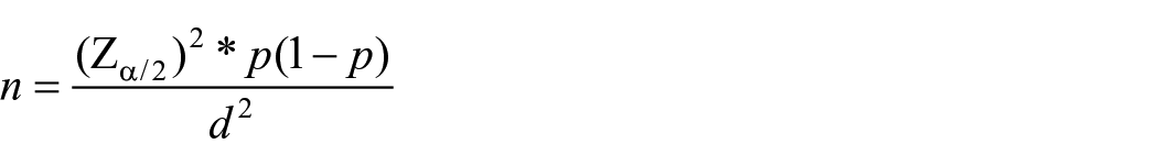
Considering the following assumptions: Zα/2 = significance level at α = 0.05,
p = prevalence = 0.5 or 50% prevalence was taken as there was no previous study for QIs of TB culture and margin of the error, d = 0.05, the total sample size was calculated as
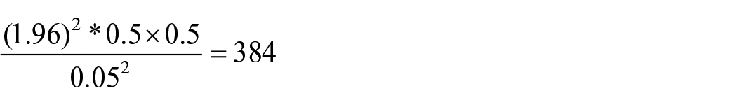
A total of 439 sample size was used including 15% nonresponse rate. A simple-random sampling technique was used to select 439 out of the 1500 specimens referred for MDR-TB culture and drug susceptibility testing to Hawassa mycobacteriology laboratory during the study period.
Inclusion and exclusion criteria
All cases referred to the Hawassa mycobacteriology culture laboratory for baseline culture and treatment monitoring during the study period were included in the study group, except the extra-pulmonary TB cases were excluded.
Data collection
The sputum specimens were collected from TB/MDR-TB cases by trained lab personnel at referring facilities or TB/MDR-TB treatment-initiating centers. First, the presumptive TB cases were screened at the health facilities level, during their routine health care, among those cases who were presumptive TB and/ or MDR-TB cases were requested to give sputum samples for Xpert MTB/RIF assay. Those who were MTB-positive and rifampicin-resistance-positive were linked to their respective treatment-initiating centers.
The treatment-initiating centers had requested a patient to give a specimen for baseline culture testing and collected before anti-TB treatment had been initiated; then every month, to monitor treatment outcomes.
The specimens were triple packaged, and referred through postal service or courier to the mycobacteriology culture laboratory in Hawassa, Sidama, Ethiopia. On arrival, the reception unit checked on the package, any leakage or breakage, requisition form, and then, kept a record of the specimen and delivered it to the mycobacteriology lab. 16
Demographic and clinical data were collected from requisition forms accompanying the specimens, and then, specimens were handled according to the “Sample management Standard operating procedure (SOP).”
Specimens that were not transported in the cold chain with ice packs, volume less than 2 ml, leaking specimens, specimens collected in inappropriate containers, and specimens delivered after 5 days of the collection were recorded and rejected.16,17
The number of specimens rejected was used as the numerator, to assess the specimen rejection rate. The accepted specimens were delivered to the mycobacteriology laboratory immediately and were processed for culture testing based on the requisition placed by physicians, following the SOP of the laboratory.
Specimen processing and inoculation
Specimens were treated with the N-acetyl-L-cysteine sodium-hydroxide (NALC-NaOH) method and centrifuged at 3000 revolutions per minute (rpm). The supernatant was decanted and the sediment was re-suspended with 1 ml of phosphate buffer and inoculated into Lowenstein–Jensen (LJ) and microbial growth indicator tube (MGIT). 13
Smear from the sediment had been prepared, adding a drop of sediment to the clean slide using a Pasteur pipette. The smear was allowed to air dry and stained by the Ziehl–Neelsen technique and examined to see how well the decontamination process was undergone. This was achieved by examining microscopically for bacteria or fungi at 100 HPF. 18
Specimens were processed by the sodium hydroxide and N-acetyl-l-cysteine (NaOH/NALC) method and concentrated by centrifugation at 3000 rpm for 15 min.
The supernatant was discarded, and the sediment was re-suspended with sterile phosphate buffer to a final volume of 2 ml. This suspension was then used for making smears for acid-fast staining and for inoculation into the MGIT culture tube and LJ slant. 18
While the delay was unavoidable, unprocessed specimens were stored at 2°C–8°C for 2 days.
The duration between login and logout time at the reception unit Register was used to assess turnaround times. Laboratory data were collected using, formats, registers, and Software provided data for MGIT.
Lowenstein–Jensen culture
After all specimens decontaminated 2–3 drops of the sediment were inoculated into LJ slants. 18
BACTEC MGIT 960 system
Before the inoculation, the MGIT growth supplement was added to the MGIT medium.
Then 0.5 ml of the sediment was inoculated to the MGIT tube. 18
Smear examination
Smears were prepared from the concentrated specimens, stained by the Zeil–Nelsen staining technique and examined for AFB to check the complete removal of contaminants other than mycobacteria during the decontamination process. 18
The reference range/acceptance criteria for LJ and MGIT media were inoculated with H37Rv incubated at 370°C is 2+ and 3+ within 21 days 23. 18
Quality assurance
We have ensured the quality of the data using standardized data-gathering tools, pretesting of the forms and worksheets on 5% of participants, training and supervising the data collectors to be able to collect the accurate data, and ensured that all laboratory examinations were carried out as per the standard laboratory protocols.
Statistical analysis
The data were analyzed using IBM Statistical package for social sciences (SPSS, version 26). The computed values of the indicators were compared against the preset targets.
Results
Sociodemographic and clinical characteristics of the participants
A total of 439 specimens from TB/MDR-TB patients were included in the Solid/LJ and Liquid/MGIT Culture methods QIs assessment. Of these, 291 (66.3 %) specimens were from males and 148 (33.7 %) specimens were from female patients.
The mean age of the study participants was 30 years (standard deviation 12.6 ± 3) and the majority of participants age were within the age range of 15–24 years 146 (33.3%) and 25–34 years 130 (29.6%). The majority of the participants were from Yirgalem general hospital referring to site 184 (41.9%). HIV-status of participants who were reactive is 195 (44.4%), nonreactive 78 (17.8%), and unknown 166 (37.8%). TB treatment history of participants was on first line 212 (48.3%), second line 40 (9.1%), first and second line 10 (2.3%), new 133 (30.3%), and unknown 44 (10%). Based on the patient registration group majority of the participants were new 174 (39.6%). The main reason for laboratory testing was for follow-up 355 (80.9%) (Table 1).
Demographic and clinical characteristics of patients tested at mycobacteriology laboratory, Hawassa, Sidama, Ethiopia, from 27 October 2020 to 31 May 2021 (n = 439).

Quality indicator for the pre-examination phase
Specimen rejection rate
A total of four specimens were rejected during the study period. Two specimens were without a request form, and the other two requests were without a sample. The specimen rejection rate calculated was 4 (0.9%).
Quality indicators for the examination phase
The recovery rates from smear-positive samples were 25/41 (61%) and 24/41 (58.5%) for the MGIT and LJ methods, respectively, while the recovery rate from smear-negative samples was 18/398 (4.5%) for both methods. The contamination rates were 45/439 (10.2%) and 42/439 (9.6%) for liquid and solid cultures, respectively. The NTM detection rate was 6/439 (1.4%) (Table 2).
Smear and culture test results at Hawassa mycobacteriology laboratory, Sidama, Ethiopia from 27 October 2020 to 31 May 2021 (n = 439).

Turnaround time for BACTEC MGIT 960 and Lowenstein–Jensen methods
The mean turnaround time or time to detect (TTD) mycobacteria from smear-positive samples were 14 and 32 with SD ± 6.3 and 9.7 days for MGIT and LJ methods, respectively. Turnaround time for smear-negative samples was 17.7 and 31 with SD ± 9.2 and 9.6 days for MGIT and LJ methods respectively.
Discussions
The QIs that were included in this study have been believed to help in evaluating and monitoring the performance of mycobacteriology laboratory services and also to develop continuous improvement plans to achieve an accredited laboratory. In this study, we have assessed five key QI parameters, which are discussed below and relevant to TB Culture laboratory service in resource-limited settings.
Specimen rejection has significant consequences on patient care, including patient discomfort and delay in result availability leading to delayed treatment; therefore, routine monitoring of specimen rejection rate is critical to control a trend. 19
The specimen rejection rate for the mycobacteriology laboratory in the current study setting (0.9%), which was higher than (0.43%) the overall lab rejection rate and lower than the (1.43%) specific rejection rate for TB culture compared to the previous study finding at Amhara Public Health Institute, Ethiopia. 20
In the current study, the recovery rate for smear-positive samples was 25/41 (61%) and 24/41 (58.5 %) for the MGIT and LJ methods, respectively. which is significantly lower than the previous study findings 87.4% (97/111) and (66.7% (74/111), respectively). 6
The recovery rate from smear-negative samples was 18/398 (4.5%) for both methods. This finding was also much lower than the previous study reports of 13.4% (108/797) and 17.4% (139/797) for LJ and MGIT methods, respectively. 6
In this study, the overall recovery rate for both methods was 9.6% (42/439). This finding is also much lower than the previous study report that revealed, the overall recovery rate for MGIT and LJ methods were 26% and 20%, respectively. 6
The recovery rate of mycobacteria may be influenced by various factors, including the population evaluated (confirmed cases or TB suspects) and the type of facility performing the evaluation. The laboratory should consider these variables when determining baseline recovery rates. 11
The incidence of culture contamination with bacteria (other than mycobacteria) varies from laboratory to laboratory depending upon several factors. According to the Centers for Disease Prevention and Control (CDC) guidelines, up to a 5% contamination rate is acceptable in cultures of clinical specimens on LJ media.
A general recommendation is that 5% + 2% is acceptable for all media types. However, for MGIT, slightly higher contamination may be accepted (up to 7%–8%).
Too harsh decontamination process may be indicated with a very low contamination rate (<3%) finding, which would also affect the growth of mycobacteria and may reduce the positivity rate and increase the time-to-detection of positive mycobacterial culture.
On the other hand, a higher contamination rate (above 8%) may be due to improper or under-decontamination of specimen; very mucoid specimens that are hard to liquefy may result in high contamination; long storage and transportation time of the specimen after collection. In such situations, especially in hot weather, bacteria tend to overgrow and then are hard to kill by routine decontamination procedure or use of nonsterile materials such as pipettes, tubes. 21
A recent study has unveiled that the contamination rate for the MGIT and LJ culture methods were 43/439(9.8%) and 42/439 (9.6%), respectively. This was a greater contamination rate compared to the CDC target (7%–8% for MGIT and up to 5% for LJ). 17
In the recent study, contamination rate (9.8%) was within the standard target of (8%–10% for MGIT), on the other hand, the contamination rate is significantly higher (9.6%) than the target (3%–5%) for the LJ culture. 13
The current study has uncovered a significantly lower contamination rate (9.8%) for MGIT compared to the previous study that reported (a 15%) contamination rate, with a greater (9.6%) contamination rate than the similar study (9.3%) for the LJ method. 6
The current contamination rate for the LJ method (9.6%) was also much greater than the study finding from India (5.5%) 12
Non-TB mycobacteria are opportunist pathogens that have a specific role in lymphadenitis, skin, and respiratory infections. Treatment of the disease due to these bacteria is difficult since these bacteria are resistant to a large scale of antibiotics including anti-TB drugs.
This is because non-TB mycobacteria morphologically are similar to mycobacterium TB as a result, and non-TB mycobacteria are mistakenly treated by ineffective anti-TB drugs. To avoid this problem measuring the nonmycobacterium TB rate in the laboratory has a significant contribution to patient care. 17
This study has uncovered that the NTM identification rate was 6/439 (1.4%), which was much lower than the previous studies carried out in Iran (5.01%) 18 and India (4.2%). 21
An accurate and reliable diagnosis of drug-resistant TB requires that mycobacterium TB be isolated, drug susceptibility results be completed and results conveyed to the clinician. Prompt turnaround time of laboratory results is of paramount importance for rapid diagnosis and appropriate treatment of drug-resistant TB. 2
In the current study, the mean turnaround time for smear-positive samples was (14 and 32 days) for MGIT and LJ methods, respectively; on the other hand, the turnaround time for smear-negative samples was (17.7 and 31 days) for MGIT and LJ, respectively.
The mean time to detect significant growth of mycobacteria from smear-positive samples by MGIT and LJ methods (14 and 32 days, respectively, were longer as compared to the (8–10 and 21 days) of the GLI target for MGIT and LJ methods. 13
On the other hand, the current study findings revealed a shorter turnaround time to detect significant growth of mycobacteria from smear-positive samples by MGIT (14 days), but a longer time for the LJ method (32 days) compared to the previous study findings (16 and 31 days) for MGIT and LJ methods, respectively. 6
The recent study findings of the turnaround time for smear-negative samples (17.7 and 31 days) for MGIT and LJ methods, respectively, were shorter compared to the previous study findings of (20 and 36 days) for MGIT and LJ methods, respectively. 6
While compared to the standard targets of (14–42 and 28–56 days) for the MGIT and LJ methods, respectively, the current study findings were within the target range.
Limitations of the study
The performance of referring health facilities during patient orientations and sample collections were not addressed.
The study did not include QIs for phenotypic drug susceptibility testing and Line probe assay (Genotype MTB-DR-plus) technique because the technique was newly established and had low sample volume at the time of data collection.
Conclusions
The study found that most QIs, such as the rejection rate of specimens, the time it takes to deliver results, and the detection rates of non-TB mycobacteria, were within the acceptable standard range. However, the culture contamination rate was higher than the target range. It is strongly recommended that measures be taken to reduce contamination rates to a range of 3%–5% for solid cultures. Additionally, the study found very low culture recovery rates, which require further investigation. Therefore, we recommend that the Ministry of Health, Public Health Institutes, and Public Health laboratories review and investigate key performance indicators that are relevant to the early detection of TB cases, treatment monitoring, and overall improvement of the services.
Supplemental Material
sj-docx-1-smo-10.1177_20503121241274716 – Supplemental material for Status of quality indicators in a mycobacteriology culture laboratory, Hawassa, Sidama, Ethiopia
Supplemental material, sj-docx-1-smo-10.1177_20503121241274716 for Status of quality indicators in a mycobacteriology culture laboratory, Hawassa, Sidama, Ethiopia by Wolde Abreham Geda, Tariku Lambiyo Anticho and Moges Desta Ormago in SAGE Open Medicine
Footnotes
Acknowledgements
We would like to acknowledge the School of Medical laboratory Science and staff including Mr. Siraj Husein, Mr. Mekonen Girma, and Mr. Dawit Yihdego for their guidance and support. We would also like to thank the leadership and staff of the Hawassa Regional laboratory, in particular Mr. Teshome Befikadu (MPH), Section Supervisor, Mrs. Zeineba Temam (MSc.), Mr. Addis Molla (BSc), Mrs. Roza Nasir (MSc.), and Mr. Belete Habte (BSc.) Mycobacteriology lab experts for their unreserved support. I am also so grateful to the Sidama, Central, south Ethiopia and Oromia regions Health bureaus and public Health institutes for their collaboration.
Author’s contribution
WAG, conceived the concept. WAG, MDO, and TLA have contributed significantly to the design, or acquisition of data, or analysis and interpretation of data. All authors have participated in drafting, reviewing, and/or revising the manuscript and have approved its submission.
Data availability
The data related to the study are available within the manuscript and will be accessible through Sidama Public Health Institute upon reasonable request.
Declaration of conflicting interests
The author(s) declared no potential conflicts of interest with respect to the research, authorship, and/or publication of this article.
Funding
The author(s) received no financial support for the research, authorship, and/or publication of this article.
Ethical consideration
We got the ethical clearance and approval from the institutional review board (IRB) of Hawassa University, College of Medicine and Health Sciences, with Reference number IRB/030/13, and a support letter from the School of Medical Laboratory Science of the University. We submitted Ethical clearance with the support letter to the Sidama Public Health Institute. We also got permission from the Institute to carry out the study. We took verbal informed consent from the study participants, and assent for the minors from their Legally Authorized Representative before data collection. The study team discussed with all stakeholders to ensure transparency on study purposes and benefits of the study to the participants and others, in improving the quality of the diagnostic services, the absence of any risk caused by participating in this study, and the right of participants to withdraw from the study at any time, whenever concerns arise. The checklist was de-identified and kept securely in locked cabinets; in addition, the database was password-protected to keep the confidentiality of the patients. We conducted the study as per the 1964 Declaration of Helsinki.
Informed consent
We obtained the ethical clearance and approval from the institutional review board (IRB) of Hawassa University, College of Medicine and Health Sciences, with Reference number IRB/030/13. In addition, we got verbal informed consent from the study participants and assent for the minor subjects from their Legally Authorized Representative before the data collection.
Supplemental material
Supplemental material for this article is available online.
References
Supplementary Material
Please find the following supplemental material available below.
For Open Access articles published under a Creative Commons License, all supplemental material carries the same license as the article it is associated with.
For non-Open Access articles published, all supplemental material carries a non-exclusive license, and permission requests for re-use of supplemental material or any part of supplemental material shall be sent directly to the copyright owner as specified in the copyright notice associated with the article.