
Abstract
Objective:
Morphometry of frontal sinuses has been studied extensively in many countries. However, these findings cannot be generalized due to genetic and environmental factors affecting the skeletal structure. The main aim of this study was to morphometrically analyze the frontal sinuses in patients who underwent non-contrast computed tomography (NCCT) skull imaging at Teaching Hospital Peradeniya and to assess its association with age and sex.
Methods:
This retrospective study included 300 NCCT skull images obtained by a multi-slice computed tomography(MSCT) scanner at the Teaching Hospital Peradeniya. This age- and sex-matched sample was selected using consecutive sampling and consists of seven age groups (20–29, 30–39, 40–49, 50–59, 60–69, 70–79, and 80–89). Right and left frontal sinus values (length, height, width of each sinus, and total width) were measured using the RadiaAnt DICOM Viewer 2020.2.3 software, and age and sex were obtained from the CT images. The association among the variables was analyzed using the t-test and binary logistic regression model.
Results:
In our study sample, 59% (n = 177) were male and the mean age was 57.94 years. The means of left side sinus values were larger than right sinus values, which was statistically significant. A statistically significant association was seen between frontal sinus parameters and sex; those of males were higher than that of females. No significant association between the sinus size and age was observed.
Conclusion:
Morphometric analysis of frontal sinuses using NCCT images is useful for the sex differentiation of unknown bodies for medico-legal purposes. Furthermore, the mean sinus values specific to the current study population will also be helpful in ethnic differentiation.
Introduction
Person identification is an important aspect of forensic medicine, for which the use of various techniques has been discussed in the literature. Instances, such as the discovery of unidentified human skeletal remains, putrefied dead bodies where facial recognition is difficult, bodies with extensive injuries and burns causing disfigurement, etc., mandate special techniques for person identification. Furthermore, accurate identification will also assist in crime reconstruction and assist in missing person investigations. Therefore, the development of accurate, reliable, and objective tools and methods for person identification necessitates considerable attention.
Some proven methods employed by forensic scientists and legal authorities for person identification are fingerprinting, DNA analysis, and odontology.1–3 However, in instances where these methods cannot be used, an anthropometric assessment of frontal sinuses will be useful. 4 Four reasons that validate the use of frontal sinuses for person identification have been discussed in the literature. First is the universally accepted individual variability, which was proven by Christensen using mathematical models. The second is structural stability maintained throughout the lifespan. The third is resilience with the ability to withstand significant trauma, burns, postmortem decomposition, etc. Lastly, it is one of the most imaged areas of the body for diagnostic purposes, thus increasing the availability of antemortem records that can be used for comparison. 4
The frontal sinus is part of the paranasal sinuses located in the frontal bone. It is a paired, irregular-shaped, air-filled cavity, lined by mucosa. The left and right sinuses are frequently observed to be asymmetrical in size, mainly due to the irregular and asymmetrical placement of the septum separating the two sinuses. 5 This is an important factor in determining the unique nature of the frontal sinuses, making them highly variable among individuals. This makes it extremely valuable to be used as a tool for person identification. 5
Therefore, this study morphometrically analyzed the frontal sinuses, employing non-contrast computed tomography (NCCT) skull images of patients presenting to a tertiary care hospital in Sri Lanka, to identify the frontal sinus patterns specific to the study population that will be useful for person identification.
Materials and methods
This retrospective study was conducted using stored digital NCCT skull images taken by a multi-slice computed tomography scanner (Siemens SOMATOM Emotion 16, Germany) at the Teaching Hospital Peradeniya, Sri Lanka between June 2020 and September 2021. The slice number and slice thickness were 16 and 1 mm, respectively. All digital images of patients between 20 and 90 years of age were considered for the study. Criteria for the exclusion of images were as follows:
absent and single sinus
bony erosions due to chronic sinusitis
craniofacial fractures and deformities
poor quality due to motion artifacts
rotation of more than 15°
Ethical clearance was exempted by the ethics review committee at the Faculty of Medicine, University of Peradeniya (protocol number: 2021/EC/41). Consent was not obtained as this requirement was waived by the local ethics committee.
Using data from available similar research in the literature, 6 300 NCCT images were included in this retrospective study between June 2020 and September 2021. The study sample was categorized into seven main age categories 20–29, 30–39, 40–49, 50–59. 60–69, 70–79, and 80–89 years. Demographic data including age and sex were obtained from the CT images. The CT images were analyzed using the RadiaAnt DICOM Viewer 2020.2.3 software. Axial, coronal, and sagittal planes with a bone window of the CT images were used to obtain sinus measurements (Figure 1). Any visible pneumatization of the frontal bone divided into two cavities by a bony septum was considered the frontal sinus.
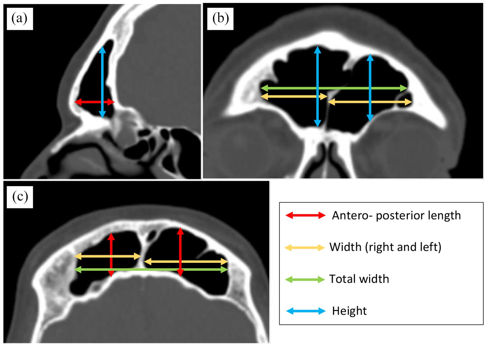
The bone window of NCCT skull images for obtaining measurements: (a) Sagittal view for obtaining length and height measurements, (b) coronal view for obtaining width and height measurements, and (c) axial view for obtaining length and width measurements.
The measurements obtained were as follows:
Right sinus height—maximum vertical distance between superior and inferior borders
Right sinus width—maximum horizontal distance between medial and lateral borders
Right sinus length—maximum horizontal distance between anterior and posterior borders
Left sinus height—maximum vertical distance between superior and inferior borders
Left sinus width—maximum horizontal distance between medial and lateral borders
Left sinus length—maximum horizontal distance between anterior and posterior borders
Total sinus width—maximum horizontal distance between the lateral borders of the two sinuses
All the measurements were obtained in millimeters, with a precision of 0.01 mm.
For each sinus measurement, two views were used and the mean value was taken.
Axial and sagittal views for length measurements
Coronal and sagittal views for height measurements
Axial and coronal views for width measurements
All the measurements were obtained by a person with adequate technical experience, under the supervision and cross-checked by a board-certified Consultant Radiologist with more than 10 years of work experience.
Statistical analysis
The data were entered into an excel sheet and were analyzed using JASP version 0.14.1.0. 7 Initially, descriptive statistical methods were used to describe the collected data. The mean length, height, and width measurements of the frontal sinuses were analyzed using analysis of variance (ANOVA) to assess its association with age. 8 The association of the measurements with sex was analyzed using the t-tests. 9 Furthermore, to find out the variables (sinus measurements) which were independently associated with the sex of the person, a binary logistic regression model was used. 10
Results
The study sample of 300 was categorized into seven age groups (Table 1). It was further sub-categorized as male and female. The sample distribution in relation to age and sex is demonstrated in Table 1.
Age and sex distribution among the study population.

The mean age of the study population was 57.94 years. In the total study population, a male predominance was observed with 177 males and 123 females. The mean ages in the male and female groups were 57.29 and 58.87 years, respectively.
The mean measurements of the sinuses were obtained (from two views for each measurement) (Table 2). The left sinus measurements were observed to be larger than the right (Figure 2). This difference was statistically significant.
Summary of the frontal sinus measurements.
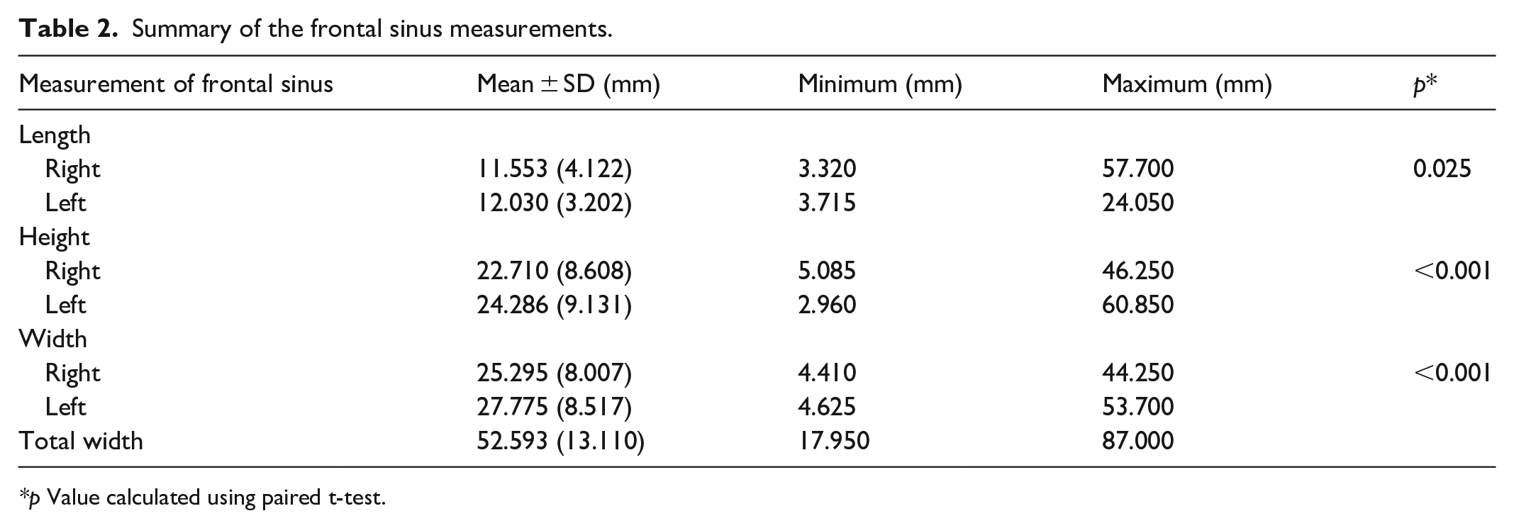
p Value calculated using paired t-test.

Variation between right and left frontal sinus measurements.
A statistically significant variation of all the sinus measurements with sex was observed (Table 3). All the mean sinus measurements were statistically higher than females which was statistically significant.
Variation of frontal sinus measurements with sex.

p Value calculated using Student’s t-test.
MRSL, mean right sinus length; MLSL, mean left sinus length; MRSH, mean right sinus height; MLSH, mean left sinus height; MRSW, mean right sinus width; MLSW, mean left sinus width; MTSW, mean total sinus width; SD, standard deviation.
WELCH test demonstrated homogeneity of the study population by age enabling the use of ANOVA statistical test to assess the association between sinus measurements and the age (Table 4). However, none of the sinus measurements demonstrated a statistically significant association with the age of the patients.
Variation of frontal sinus measurements with age.

p Value calculated using ANOVA test.
ANOVA: analysis of variance; MRSL: mean right sinus length; MLSL: mean left sinus length; MRSH: mean right sinus height; MLSH: mean left sinus height; MRSW: mean right sinus width; MLSW: mean left sinus width; MTSW: mean total sinus width; SD: standard deviation.
To find out the sinus measurements which were independently associated with the sex of the person, binary logistic regression model was used. Results of the final logistic regression results identified, mean left sinus length (p < 0.001) and mean left sinus width (p = 0.021) measurements were highly associated with the sex of the person.
Discussion
The development of the frontal sinus begins in the fetus around fourth to fifth week of gestation and continues up to early adult life. It reaches the maximum size by the second decade and remains constant thereafter.11,12
According to the literature, the variability of frontal sinuses is observed even among monozygotic twins.13,14 This uniqueness of the frontal sinus has been akin to that of fingerprints. 15 Contributed by genetic and environmental factors, frontal sinus is recognized to be highly variable among different individuals. 16 A descriptive study conducted among an Indian population demonstrated the uniqueness and asymmetry of the frontal sinuses. 17 These findings further reinforce the validity of employing frontal sinus patterns for person identification.
In the literature, specific frontal sinus patterns in relation to age, sex, etc. have been studied in specific ethnic groups. However, due to environmental, racial, and genetic factors affecting skeletal development, the same tools cannot be applied universally. 18 Although extensively studied in the literature, due to limited resources, lack of expertise and cost, this technique is not widely used or researched in developing countries like Sri Lanka.
With the development and increased availability of digital radiographic techniques, employment of frontal sinus pattern analysis for person identification has gained significant recognition. NCCT is considered the method of choice for imaging paranasal sinuses. It offers the added advantage of being able to manipulate images three-dimensionally. 19
According to the literature, variation in frontal sinus size with the ethnicity/race is observed. In a study assessing frontal sinus measurements using CT in an Asian population, the mean adult sinus width, height, and depth were obtained as 27.9, 52.8, and 21.6 cm, respectively. 20 All the mean sinus measurements in this study are higher compared to the present study population. However, in another study conducted in a Turkish population the mean measurements were observed to be similar to those of this study. This study observed left side sinus measurements to be greater than the right, which was also confirmed by the Turkish study. 6 However, a study done in France did not observe a statistically significant difference between the right and left frontal sinus measurements. 21
In the current study population, none of the frontal sinus measurements had a statistically significant association with age. This further confirms that after the second decade, when the development of frontal sinuses completes, the sinus size remains static throughout life. A study conducted in Greece also concluded that frontal sinus is not a good predictor of age. 22 Another study conducted in a German population observed that the frontal sinus development is completed by 18 years in males and 15–16 years in females, remaining static thereafter. 23 This static nature was further confirmed by a study comparing skull radiographs performed 6–8 months apart. 24 A study conducted in France using NCCT images also did not observe a statistically significant correlation with the age of the individual. 21
According to this study, morphometric analysis of frontal sinuses demonstrated a significant association with the sex of the individual. All the measurements (length, height, and width) were found to be larger in the males compared to females. Furthermore, final logistic regression analysis demonstrated a high association of MLSL and MLSW values with sex. Therefore, these parameters have the potential to be used to develop a predictor model for accurate sex determination.
This sexual dimorphism of frontal sinuses was also observed in a study conducted in a Greek population. 22 Another study conducted in Germany also observed the final size of the frontal sinus to be 13.4–17.1% smaller in females compared to males. 23 An Iranian study using paranasal CT scans observed the average volumes of each right and left sinuses to be higher in males compared to females. 25 Another study in France observed a significant sexual dimorphism in relation to the total frontal sinus volume. 21
Contrary to these findings, several studies in other ethnic groups have failed to demonstrate a statistically significant association of frontal sinus size with age and sex. In a recent study, the correlation of frontal sinus metrics with sex was assessed in an Indian population using skull radiographs. 26 Based on the mathematical model, the ability to predict female sex using the right, left, and total sinus areas were calculated as 60.9%, 55.2%, and 55.2% respectively. In males, the mean values of variables were calculated to be much greater. It was concluded that the frontal sinus area measurement is unpredictable for sex determination. 26
Limitations
Several limitations identified by the authors were as follows. This study was conducted in a cohort limited to a single tertiary care center in Sri Lanka; therefore, the results will not reflect the general population. Also, the sample size was not calculated. Future studies including several centers and larger sample size will be beneficial in formulating a more generalized conclusion as well as developing a predictor model. Furthermore, in this study, only height, width, and length parameters were used. In future research, sinus volume and shape may be studied in relation to age and sex.
Conclusion
Morphometric analysis of frontal sinuses using NCCT images is useful for the sex differentiation of unknown bodies for medico-legal purposes. Furthermore, the mean sinus values specific to the current study population will also help in ethnic differentiation.
Footnotes
Acknowledgements
The authors are grateful for the contribution made by Mrs. D.M.S. Dissanayake and Mr. A.A. Chinthana of the Department of Radiology, Teaching Hospital Peradeniya during the process of data collection.
Author contributions
Study conception and design—CW, AV, JU, SK
Data collection—CW, JU
Analysis and interpretation of results—CW, JU, AV
Draft manuscript preparation—CW, AV, SK, JU
Reviewed results and approved final manuscript—CW, AV, SK, JU
Declaration of conflicts of interest
The author(s) declared no potential conflicts of interest with respect to the research, authorship, and/or publication of this article.
Ethical approval
This retrospective study was exempted from ethical clearance by the ethics review committee at the Faculty of Medicine, University of Peradeniya (protocol number: 2021/EC/41). Consent was not obtained as this requirement was waived by the local ethics committee.
Ethical approval
Ethical approval for this study was waived by the ethics review committee at the Faculty of Medicine, University of Peradeniya (protocol number: 2021/EC/41) because this study does not involve human subjects and only uses digitally stored NCCT images.
Funding
The author(s) received no financial support for the research, authorship, and/or publication of this article.
Note
Informed consent was not sought for this study because the need for informed consent was waived off by the review board due to the retrospective nature of the study.
Informed consent
Consent was not obtained as this requirement was waived by the local ethics committee.
Trial registration
Not applicable.