
Abstract
Invasive fungal infections are an important cause of morbidity and mortality in hematopoietic stem cell transplant and solid organ transplant recipients. Evolving transplant modalities and techniques, complex and extensive immunosuppressant strategies, and the increased use of broad spectrum antifungal prophylaxis has greatly impacted the epidemiology and temporal pattern of invasive fungal infections in the transplant population. The goal of this article is to provide an up-to-date review of the most commonly encountered invasive fungal infections seen in transplant recipients, including epidemiology, risk factors, clinical features, diagnostic dilemmas, management and their overall influence on outcomes.
Introduction
Recent advances and improvements in medical therapeutics, chemotherapy, and organ transplantation methodology have substantially reduced the overall morbidity and mortality associated with transplantation. However, along with these improvements, a variety of opportunistic infections frequently caused by relatively avirulent organisms have emerged. Critically ill, immunocompromised patients, especially those who have undergone transplants, are the prime targets for these opportunistic fungal infections, primarily due to Candida and Aspergillus spp. This increase is multifactorial in origin and reflects increased recognition as well as a growing population of patients at risk.
Candidiasis
Candida spp. are ubiquitous fungi and are the most common fungal pathogens that affect humans [Vazquez and Sobel, 2011; Pfaller and Diekema, 2007]. The growing problem of systemic candidiasis reflects the enormous increase in the pool of patients at risk and the increased opportunity that exists for Candida spp. to invade tissues normally resistant to invasion. Candida spp. are true opportunistic pathogens that exploit recent technological advances to gain access to the circulation and deep tissues. Candida spp. are the most common cause of fungal infection affecting immunocompromised patients and are currently the fourth most common pathogen recovered from blood cultures [Pfaller and Diekema, 2007].
Epidemiology
Candida spp. produce a wide spectrum of diseases, ranging from superficial mucocutaneous disease to invasive illnesses, such as hepatosplenic candidiasis and systemic candidiasis [Vazquez and Sobel, 2011; Pfaller and Diekema, 2007]. Management of invasive candidiasis remains severely hampered by delays in diagnosis and the lack of reliable diagnostic methods that allow detection of both fungemia and tissue invasion by Candida spp. [Pappas, 2006; Pappas et al. 2003].
Candida spp. Why should they be identified?

HIV, human immunodeficiency virus.
Candida spp. contain their own set of well recognized virulence factors. Although not well characterized, several virulence factors may contribute to their ability to cause infection [Yang, 2003]. As with most fungal infections, host defects play a significant role in the development of candidal infections. Numerous host defects have been associated with candidal infections. Risk factors associated with candidemia and systemic candidiasis include granulocytopenia, HSCT, solid organ transplants (SOTs) (kidney and liver), total parenteral hyperalimentation, solid neoplasm, corticosteroids, broad-spectrum antibiotics, prolonged intensive care unit stay, prolonged hospitalization, mechanical ventilation for over 3 days, pancreatitis, severe trauma, recent surgery (especially gastrointestinal tract), central venous catheters, and hemodialysis [Vazquez and Sobel, 2011; Pappas, 2006].
Clinical manifestation
Manifestations of invasive candidiasis.
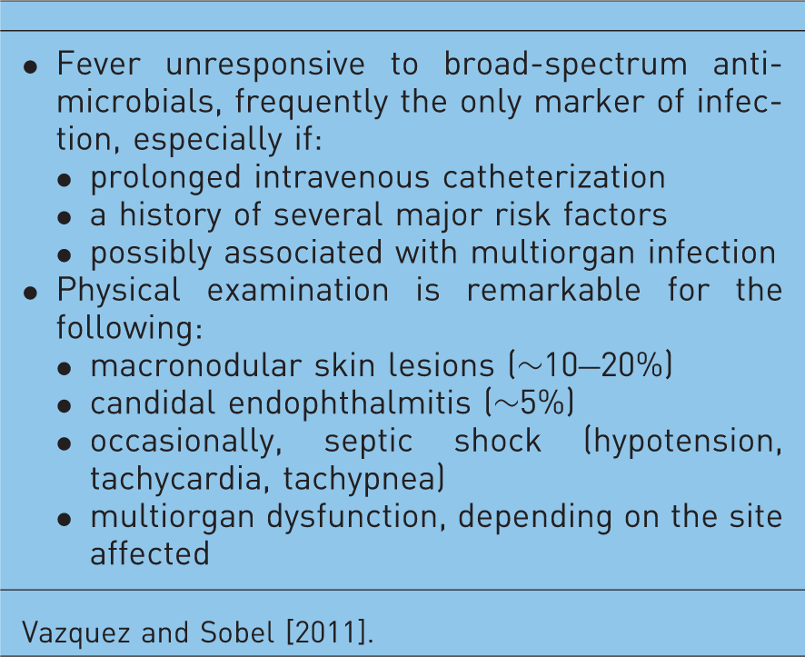
Systemic candidiasis may be divided into two different categories: candidemia without organ involvement and disseminated candidiasis (organ infection by Candida spp.). Deep organ infections due to Candida spp. are generally observed as part of the disseminated candidiasis syndromes, which may be associated with either single- or multiorgan involvement. The patient's history commonly reveals the following: several days of fever that is unresponsive to broad-spectrum antimicrobials (frequently the only marker of infection), prolonged intravenous catheterization, and several key risk factors. Physical examination is remarkable for the following: fever, macronodular skin lesions (approximately 10%), candidal endophthalmitis (approximately 5%), and occasionally septic shock. Disseminated candidiasis is frequently associated with multiple deep organ infections or may involve single organ infection (Table 2).
Diagnosis
Unfortunately, findings from laboratory studies are either negative or nonspecific [Vazquez and Sobel, 2011; Pappas, 2006]. Clinicians are required to act definitively and early based on a high index of suspicion. Patients who remain febrile despite broad-spectrum antibiotic therapy, with either neutropenia or other risk factors and persistent leukocytosis, should be suspected of having systemic candidiasis. Cultures of nonsterile sites, although not useful for establishing a diagnosis, frequently demonstrate a high degree of candidal colonization. However, these positive cultures may be useful for initiating antifungal therapy in patients who are febrile and are unresponsive to broad-spectrum antimicrobials. It is important to always consider positive results from these sites significant and definitive evidence of infection. To be effective, appropriate antifungal therapy should be provided early and empirically in such high-risk patients [Morrell et al. 2005].
Characteristic features of biomarkers currently used for the diagnosis of invasive fungal infections.

Odabassi et al. [2004]; Alexander and Pfaller [2006]; Mennick-Kersten et al. [2004].
BAL, bronchoalveolar lavage; FDA, US Food and Drugs Administration; GI, gastrointestinal; GVHD, graft versus host disease; IFI, invasive fungal infection.
Management
The treatment of Candida infections varies substantially and is based on the anatomic location of the infection, the patients' underlying disease and immune status, the patients' risk factors for infection, the specific species of Candida responsible for infection, and in some cases, the susceptibility of the strain to the different antifungal drugs [Vazquez and Sobel, 2011; Pappas et al. 2009]. In January 2009, the Infectious Disease Society of America and the Mycosis Study Group published updated practice guidelines for the treatment of candidemia and candidiasis [Pappas et al. 2009].
Antifungal agents
Summary of systemic antifungal agents currently available.

GFR, glomerular filtration rate; IV, intravenous.
Polyenes include AmB-d and the LFAmB (AmB lipid complex, liposomal amphotericin B, and amphotericin B colloidal dispersion formulations) [Moen et al. 2009]. AmB was considered the gold standard of antifungal treatment for over 50 years. Unfortunately, its use is limited due to significant adverse events such as infusion-related reactions and nephrotoxicity. Lipid formulations of AmB were developed to overcome the limitations associated with the use of AmB-d. In general, LFAmB are better tolerated due to their different molecular structures. In fact, patients can be treated with larger doses of AmB without experiencing the typical side effects of AmB-d [Miceli and Chandrasekar, 2012]. The antifungal spectrum of activity, common drug–drug interactions, and side effects are shown in Table 4.
General patterns of susceptibility of Candida species.

Vazquez and Sobel [2011]; S, susceptible; S-DD, susceptible dose-dependent; R, resistant; I, intermediately susceptible.
Until recently, the use of AmB and fluconazole was the standard therapy for all forms of candidiasis [Charlier et al. 2006]. The primary difference between the newer guidelines and the prior guidelines has to do with the upfront use of echinocandins in patients with candidemia and suspected candidiasis who have moderate to severe infections, patients with infections due to C. glabrata and C. krusei, and those who have a history of prior azole exposure [Pappas et al. 2009; Vazquez and Sobel, 2006; Chandrasekar and Sobel, 2006; Kuse et al. 2007; Reboli et al. 2007; Cornely et al. 2007; Sobel and Revankar, 2007].
In the non-neutropenic adult patient with candidemia or invasive candidiasis, most infections are due to the presence of an intravascular catheter in up to 70% of patients [Pappas et al. 2003, 2009]. Removal of all intravascular catheters appears to shorten the duration of candidemia and has been associated with reduced mortality [Pappas et al. 2009; Andes et al. 2012].
Candidemia requires treatment in all patient populations. In most situations, either fluconazole or an echinocandin are the drug of choice in the management of candidemia and disseminated candidiasis. The options listed should be considered depending on the history of a prior exposure to antifungals, the probability of fluconazole resistance, the presence of comorbid conditions, and the clinical status of the patient. Fluconazole (loading dose of 800 mg, then 400 mg daily) or an echinocandin (caspofungin: loading dose of 70 mg, then 50 mg daily; micafungin: 100 mg daily; anidulafungin: loading dose of 200 mg, then 100 mg daily) are recommended as initial therapy for most adult patients [Pappas et al. 2009]. However, an echinocandin is preferred in patients with moderate to severe illness, in patients who have a recent azole exposure, and in patients infected with a non-albicans Candida spp. [Pappas et al. 2009; Sobel and Revankar, 2007; Andes et al. 2012]. In a recently published study by Andes and colleagues, a quantitative review of 1915 patients who were randomized into several clinical trials evaluating the treatment of invasive candidiasis were reviewed. Although numerous variables were evaluated, only two treatment-related factors, use of an echinocandin and the removal of the central venous catheter, were associated with an improved survival rate and greater clinical success [Andes et al. 2012]. However, patients who are infected with susceptible Candida spp. and are clinically stable can be readily transitioned to oral fluconazole or voriconazole to complete the recommended 14-day course after the blood cultures have been cleared. Initial therapy with an echinocandin is also preferred in patients infected with either C. glabrata or C. krusei. In patients who have initially received fluconazole and are clinically improving, and whose follow-up culture results are negative, continuing use of an azole is reasonable [Pappas et al. 2009]. For infections due to C. parapsilosis, initial treatment with fluconazole is recommended nonetheless if a patient has initially received an echinocandin and is clinically improved, continued use of an echinocandin is reasonable. If an echinocandin is not available and either C. glabrata or C. krusei are suspected, initial therapy with voriconazole 6 mg/kg twice daily followed by 3 mg/kg twice daily is reasonable [Kullberg et al. 2005]. Other alternatives may also include AmB-d 0.5–1.0 mg/kg daily or LFAmB 3–5 mg/kg daily.
Management of invasive candidiasis in patients with neutropenia may include an echinocandin, LFAmB 3–5 mg/kg/day or voriconazole (6 mg/kg administered intravenously twice daily for two doses, then 3 mg/kg twice daily) [Pappas et al. 2009]. Fluconazole 400 mg/day may also be an alternative.
Successful therapy for serious systemic Candida infections requires starting antifungal therapy as early as possible. Therapy should be initiated as soon as adequate cultures have been obtained. Despite the newer advances in the diagnosis and newer antifungals, mortality rates for candidemia and disseminated candidiasis have not improved markedly over the past decade and remain in the range of 30–40%.
Antifungal prophylaxis of invasive candidiasis in patients who are in the high-risk group is currently recommended in several situations, which include patients with chemotherapy-induced neutropenia: fluconazole 400 mg daily, posaconazole 200 mg three times per day, or caspofungin 50 mg daily is recommended during induction chemotherapy for the duration of neutropenia [Viscoli et al. 1999; Husain et al. 2006; Ullmann and Cornely, 2006; van Burik et al. 2004]. In HSCT recipients, primarily those with allogeneic transplants, fluconazole 400 mg daily, or posaconazole 200 mg three times daily, or micafungin 50 mg daily is recommended during the period of neutropenia. In SOT recipients, fluconazole 200–400 mg daily or LFAmB 1–2 mg/kg daily for at least 7–14 days is recommended as postoperative prophylaxis for high-risk liver, pancreas, and small bowel transplant recipients.
Posaconazole has been shown to be effective prophylaxis against IFIs in high-risk patients with neutropenia and HSCT recipients, but its role as empirical therapy for candidiasis has not been established.
Empiric therapy
Empiric use of antifungal agents in patients who are febrile is widespread without much supporting data [Pappas, 2006, Leleu et al. 2002]. A major pitfall has been in establishing the definitive diagnosis of invasive candidiasis in the setting of negative blood cultures. It appears reasonable to initiate empiric antifungal therapy in selected patients with known risk factors. Echinocandins with their broad spectrum of activity and improved efficacy may be preferable, although less expensive fluconazole may also be an alternative. Some criteria for initiating empiric antifungal therapy include patients with known risk factors for candidiasis, patients who are febrile and on broad-spectrum antibiotics for over 96 h, and patients with multifocal Candida colonization.
Invasive mold infections
Invasive mold infections (IMIs) have become an important cause of morbidity and mortality in HSCT and SOT recipients. Evolving transplant modalities and techniques, immunosuppressive strategies, and the use of antifungal prophylaxis has impacted the epidemiology and temporal pattern of IFIs in this population [Neofytos et al. 2009; Kontoyiannis et al. 2010; Pappas et al. 2010].
Epidemiology
Distribution of fungal pathogens causing invasive fungal infections in transplant recipients.

Neofytos et al. [2009]; Kontoyiannis et al. [2010]; Pappas et al. [2010].
IFI, invasive fungal infection; HSCT, hematopoietic stem cell transplantation.
Non-Aspergillus molds have been increasing in transplant recipients and have implications for therapy since they exhibit a variable susceptibility profile to the commonly used antifungals. For example, the Mucorales are intrinsically resistant to voriconazole yet remain susceptible to AmB and posaconazole. Fusarium spp. have variable susceptibilities to antifungals, as such F. solani, which tends to be resistant to azoles and have higher MICs than the polyenes [Nucci and Anaissie, 2007]. The triazoles voriconazole and posaconazole appear to have superior in vitro activity against Scedosporium spp. than AmB.
In the SOT population, the highest rate of IFIs was seen in small bowel transplants, followed by heart–lung, liver, pancreas, heart, and kidney transplants [Kontoyiannis et al. 2010]. Allogeneic HSCT recipients, especially unrelated or mismatched transplants, had a fivefold greater risk for IFIs compared with autologous HSCT recipients [Pappas et al. 2010]. Despite a slight increase in the incidence of all IFIs during 2002–2005, there was no significant increase in the incidence of mold infections in either the SOT or HSCT populations over the past decade. In contrast, there appears to be a comparative increase in the incidence of mucormycosis [Park et al. 2011; Kontoyiannis and Lewis, 2006; Petrikkos et al. 2012]. This increase may be a consequence of a greater number of at-risk patients undergoing HSCT or SOT, the use of more aggressive immunosuppressive treatments for GVHD and rejection, and possibly the increased use of voriconazole for antifungal prophylaxis or for empiric therapy [Kontoyiannis et al. 2006; Petrikkos et al. 2012; Trifilio et al. 2007; Spellberg et al. 2012; Xhaard et al. 2012, Lanternier et al. 2012].
The comparative distribution of IMIs varies among the type of organ transplanted (Table 6). Overall, invasive aspergillosis (IA) and other mold infections predominated among HSCT recipients. Among SOT recipients IA was most common in lung transplant recipients, accounting for 44% of all IFIs compared with 23%, 14%, 11%, and 5% in heart, kidney, liver, and pancreas transplant recipients respectively [Pappas et al. 2010].
Incidence, timing and outcomes of invasive fungal infection after transplantation.

Neofytos et al. [2009]; Kontoyiannis et al. [2010]; Pappas et al. [2010].
IFI, invasive fungal infection; HSCT, hematopoietic stem cell transplantation; SOT, solid organ transplantation.
Risk factors
Risk factors for invasive mold infections in transplant recipients.

Park et al. [2011]; Petrikkos et al. [2012]; Trifilio et al. [2007], Spellberg et al. [2012]; Safdar et al. [2010]; Fortún et al. [2012]; Silveira and Husain [2007]; Singh et al. [2003]; Husain [2009]; Ibrahim et al. [2011].
CMV, cytomegalovirus; GVHD, graft versus host disease; HSCT, hematopoietic stem cell transplantation; ICU, intensive care unit; MUD, matched unrelated donor; MMRD, mismatched related donor; TLR4, toll-like receptor 4; SOT, solid organ transplantation.
In SOT recipients, the risk factors for IMIs are strongly associated with end-organ failure, especially renal or hepatic insufficiency [Nucci, 2003; Fortún et al. 2012; Silveira and Husain, 2007]. A study from Spain reported a 29-fold higher risk of IA in liver transplant recipients who required retransplantation and a 24-fold higher risk in patients requiring dialysis after transplantation [Fortún et al. 2012]. Lung transplant recipients who have documented prior colonization with Aspergillus or those with anastomotic complications have a greater risk of post-transplant Aspergillus tracheobronchitis or pulmonary IA. Renal transplant recipients receiving prolonged corticosteroids or antirejection therapy with sirolimus were also reported to be at higher risk of developing IA. A higher risk of mucormycosis was also reported in SOT recipients with diabetes and prior exposure to either voriconazole or caspofungin [Petrikkos et al. 2012]. An understanding of the specific risk factors in the various types of SOT and HSCT and the identification of high-risk transplant recipients is essential to guide effective empiric and preventive antifungal strategies [Neofytos et al. 2009; Singh et al. 2003; Silveira and Husain, 2007].
Clinical features
The clinical features of IMIs are frequently nonspecific. Although most IMIs cause pulmonary infection, infections may also involve the paranasal sinuses, the central nervous system (CNS), the skin, the gastrointestinal tract or occasionally they can become disseminated (Figure 1). Table 6 summarizes the frequency of the organ sites involved in IMIs in transplant recipients.
Cutaneous lesion of aspergillosis. Reproduced with permission from Dr Pranatharthi Chandrasekar.
Aspergillus can cause a wide spectrum of disease in humans. Fever is a common, but nonspecific symptom [Nucci, 2003]. Involvement of the respiratory tract occurs in over 60% of patients, and thus, about 50% of the patients present with respiratory symptoms, including cough, dyspnea and pleuritic chest pain [Nucci, 2003]. Sinus infection can result in facial and orbital pain, along with localized edema. Infections of the CNS can present as altered mentation or focal neurological deficits. Given the angioinvasive nature of Aspergillus spp., the symptoms of lung and brain involvement can resemble either a pulmonary embolism or a stroke. In SOT recipients, IA tends to be localized to the lungs. The manifestations of IA in the lung and heart–lung transplant recipients are frequently different from the pulmonary infection in other transplants. Infections at the anastomotic site and ulcerative tracheobronchitis are the most common pulmonary infections reported. In addition, endobronchial stent obstruction, bronchial plugging and pneumonitis may also be seen [Singh and Husain, 2003; Husain, 2009]. Interestingly enough, both CNS and disseminated disease have declined in recent years [Singh and Husain, 2003]. The factors resulting in the lower incidence of CNS and disseminated disease are yet to be elucidated. Recent reports suggest that the current use of calcineurin and TOR (target of rapamycin) inhibitors in antirejection regimens may have a beneficial antifungal effect [Singh and Husain, 2003].
Infections due to Mucorales often cause localized infections such as sinonasal, sino-orbital or rhinocerebral disease. Occasionally, involvement of the lung, gastrointestinal tract, skin and disseminated disease may be seen [Petrikkos et al. 2012; Lanternier et al. 2012]. Typical symptoms may include facial pain and swelling, orbital pain, proptosis, visual loss and opthalmoplegias. Given the propensity of Mucorales to cause invasion of the blood vessels, the infection is characterized by the development of necrotic lesions in the oral, nasal or sinus mucosa. The infection may progress rapidly and can invade the CNS, causing stroke-like symptoms [Kontoyiannis and Lewis, 2006; Lanternier et al. 2012; Ibrahim et al. 2011, 2012]. When dissemination to the skin occurs it is characterized by the development of rapidly progressive cutaneous necrosis.
Although IMIs caused by Fusarium and Scedosporium in HSCT recipients generally affect the lungs, unlike infections caused by Aspergillus or Mucorales, dissemination to the skin structures occurs in up to 70% of cases. In the case of Fusarium spp., the propensity for dissemination via the bloodstream often results in high rates of isolation from blood cultures (∼70%) [Nucci et al. 2004; Maertens et al. 2000; Nucci and Anaissie, 2006].
Diagnosis and management of mold infection
EORCT/MSG definitions
Organ involvement in invasive mold infections among transplant recipients.
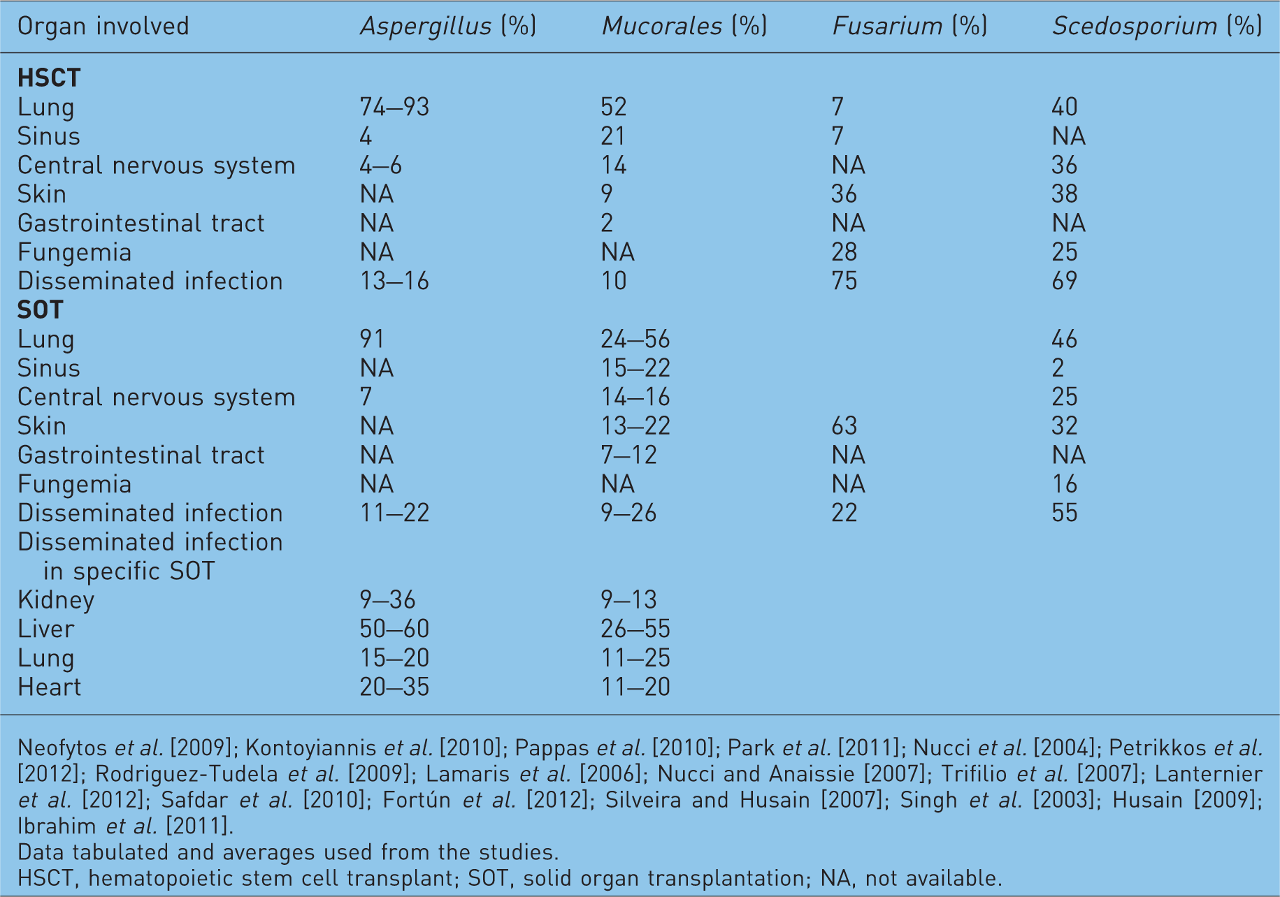
Neofytos et al. [2009]; Kontoyiannis et al. [2010]; Pappas et al. [2010]; Park et al. [2011]; Nucci et al. [2004]; Petrikkos et al. [2012]; Rodriguez-Tudela et al. [2009]; Lamaris et al. [2006]; Nucci and Anaissie [2007]; Trifilio et al. [2007]; Lanternier et al. [2012]; Safdar et al. [2010]; Fortún et al. [2012]; Silveira and Husain [2007]; Singh et al. [2003]; Husain [2009]; Ibrahim et al. [2011].
Data tabulated and averages used from the studies.
HSCT, hematopoietic stem cell transplant; SOT, solid organ transplantation; NA, not available.
Microbiologic criteria for the diagnosis of proven IFIs rely on direct tests (cytology, direct microscopy and culture) demonstrating the presence of fungal elements [De Pauw et al. 2008]. However, obtaining tissue samples or performing invasive procedures is not always feasible because of cytopenias or the poor clinical condition of these patients. Thus, the initiation of appropriate antifungal therapy is frequently delayed. The difficulty in establishing an early diagnosis is one of the primary reasons for the high mortality rates seen in IMIs [Chamilos et al. 2006; Rinaldi 1991; von Eiff et al. 1995].
Nonculture diagnostic assays such as the galactomannan (GM) and BG for the diagnosis of IFIs have been developed over the last two decades [Boudewijins et al. 2006; Mennik-Kersten and Verweij, 2006]. The advent of these indirect tests represents a major advance in the management of patients at risk for IFIs (Table 3).
Galactomannan assay
GM is an Aspergillus-specific polysaccharide residue that is incorporated into the cell wall during the initial phase of fungal growth. Eventually, GM is released into the circulation and possibly reused as a source of nutrient for further growth [Mennik-Kersten et al. 2004].
In the clinical setting, the detection of serum GM antigen has been shown to be a useful screening test for the early diagnosis of IA in patients at risk [Maertens et al. 2001; Pfeiffer et al. 2006; Sulahian et al. 2001]. Serum is the most frequently tested specimen and appears to provide the highest sensitivity (up to 95%, depending on the patient population and previous antifungal therapy) [Chamilos et al. 2006]. Galactomannan is water soluble and therefore can be detected in specimens other than serum, including bronchoalveolar lavage (BAL), cerebrospinal fluid, pleural fluid and urine [Klont et al. 2004]. Except for serum and BAL, the use of GM in other specimens remains investigational.
A recent meta-analysis study was conducted to determine the role of BAL-GM in the diagnosis of IA. In this study, BAL-GM sensitivity and specificity varied from 84% and 95%, respectively, depending on the population tested and the cut-off used [Zou et al. 2012]. BAL-GM may be used as an adjunctive tool in establishing the diagnosis of IA (see http://www.accessdata.fda.gov/cdrh_docs/pdf6/K060641.pdf and http://www.accessdata.fda.gov/cdrh_docs/pdf9/K093678.pdf). Typically, a serum GM value of at least 0.5 is considered positive. Using this suggested cutoff point, the reported sensitivity and specificity of the GM assay was 80.7% and 89.2% respectively [Chamilos et al. 2006]. Conversely, due to the lack of data, the threshold for positive BAL-GM remains under debate.
The use of serum GM is also an excellent tool for the early diagnosis of IA. Sulahian and colleagues showed that GM might be detected in serum as early as 5–8 days before the clinical manifestations of IA develop [Sulahian et al. 2001]. These results support the use of GM as a screening tool for patients at high risk of developing IA. In this setting, the detection of positive results, particularly in two consecutive serum samples, provides strong support for the diagnosis of IA [von Eiff et al. 1995; Mennick-Kersten and Verweij, 2006; Maertens et al. 2001]. Recently, some authors have also suggested that GM could serve as a surrogate marker of clinical response to treatment in patients with IA [Miceli et al. 2008; Park et al. 2011; Maertens et al. 2009; Boutboul et al. 2002]. Several studies showed that the titer of GM tends to decrease in cases that demonstrated a clinical response. Similarly, increasing GM titers were associated with poor outcomes [Park et al. 2011; Maertens et al. 2009; Boutboul et al. 2002; Woods et al. 2007; Segal et al. 2008]. False-positive reactions have also been reported in 1–18% of the tested samples and may be due to cross reactivity or false-positive GM (Table 3).
Radiographic imaging
Radiographs of the chest and sinuses have been used as a primary means of diagnostic assessment. However, they are frequently inadequate to establish a diagnosis. Initial chest computed tomography (CT) scan findings in IA are dominated by the nodule and its associated ‘halo sign’. The main finding in IA is generally a pulmonary nodule greater than 1 cm in diameter, that is, the mancronodule. It is defined as a localized, space-occupying, ovoid, soft-tissue opacity that displaces rather than conforms to the shape of the preexisting aerated lung [Georgiadou et al. 2011]. More than 90% of patients with mycologically proven IA have at least one pulmonary nodule. The halo sign apparent on the CT scan is a modifier of the macronodule. It is defined as a perimeter of ground-glass lung opacity surrounding a pulmonary nodule. On initial CT scan, a study of patients with mycologically proven IA, about 33% have one or more macronodules with a halo sign. The ‘air crescent sign’ generally follows the halo sign approximately 1 week later (Figure 2).
Computed tomography scan showing necrotic nodular infiltrate in a hematopoietic stem cell transplant recipient with pulmonary aspergillosis.
Management
Despite the advances in the field and the advent of newer technologies, identification of fungal pathogens continues to be difficult and early diagnosis is not always possible [Georgiadou et al. 2011; Miceli and Lee, 2011; Revankar and Sutton, 2010]. Because early treatment is crucial in the management of these patients, initiation of empiric antifungal therapy is not uncommon when IFI is suspected [Walsh et al. 2008].
Strategies for the management of mold infections
Current strategies for the management of IFIs include prophylaxis, empiric, preemptive, and targeted therapy (Figure 3) [Ruhnke et al. 2012; Freifeld et al. 2011]. Antifungal prophylaxis involves the administration of an antifungal drug to high-risk patient populations before the onset of signs or symptoms of infection. In addition to neutropenia during the pre-engraftment period in HSCT recipients, these patients are at high-risk for mold infection as a consequence of severe cell-mediated immunodeficiency due to GVHD and its therapy (Table 8). Similarly, certain SOT recipients are also at high risk for mold infections (Table 8). Prophylaxis with anti-mold agents has been recommended in these select patient groups [Tomblyn et al. 2009; Singh et al. 2013]. In this setting, the antifungal agent is started despite the fact that adequate microbiological diagnosis of IFIs is unavailable [Freifeld et al. 2011]. Preemptive therapy is often initiated when nonspecific radiographic signs are present or laboratory tests are suggestive of IMIs, in the absence of microbiological or histopathological confirmation of IFIs. Although preemptive antifungal therapy has been used successfully in patients with neutropenia who are febrile, there are no standard recommendations that fully support its use [Pasqualotto and Colombo, 2010; Kontoyiannis and Lewis, 2011; Lortholary et al. 2010]. Targeted therapy relies on treating microbiologically and histologically documented cases of IFIs [Ruhnke et al. 2012; Freifeld et al. 2011].
Strategies for the Management of Neutropenic Patients at High Risk for Invasive Fungal Infections.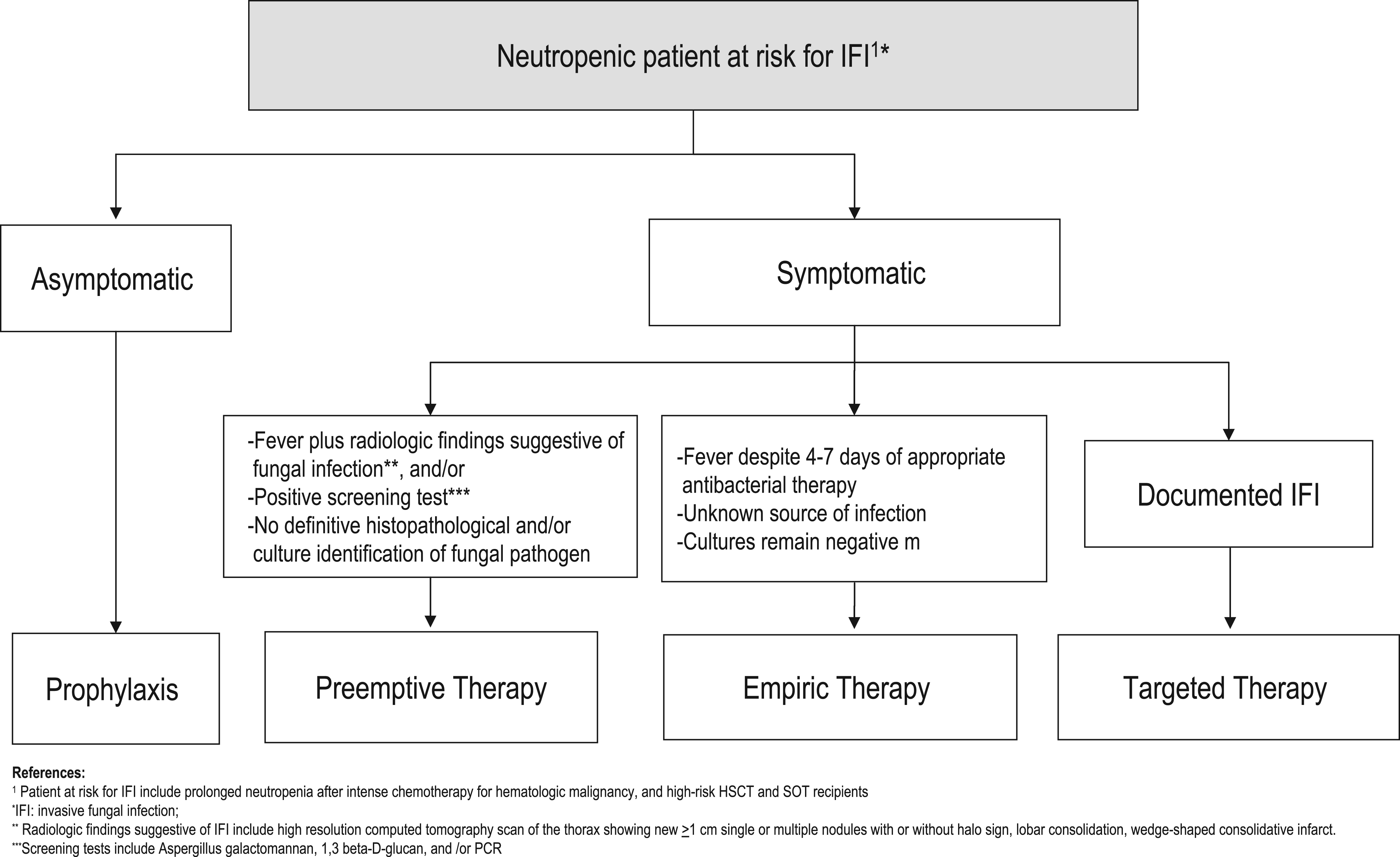
Specific management issues
Specific antifungals used for the treatment of IFI are summarized in Table 4. The management and prognosis of IA depends on the specific form of disease and the degree of immunosuppression. For over 50 years, AmB-d was the mainstay of antifungal therapy. Guidelines for the management of IA have been published by the Infectious Diseases Society of America [Walsh et al. 2008]. The current mainstay of therapy for IA is considered to be voriconazole. A randomized, multicenter study compared AMB-d with voriconazole as initial therapy for IA. This pivotal study demonstrated that initial therapy with voriconazole led to better responses and improved survival with fewer serious side effects, such as renal insufficiency and infusion-related toxicity [Pasqualotto and Colombo, 2010]. The appropriate dose of voriconazole is 6 mg/kg twice daily for 1 day, followed by 4 mg/kg twice daily.
A crucial factor in optimizing therapy in any patient with IA is the decrease or elimination of the immunosuppressant whenever possible. The recent literature suggests that if patients are diagnosed and treated early with appropriate antifungal therapy, the response rates may reach 50% or greater [Ruhnke et al. 2012].
Successful treatment of mucormycosis requires a high index of clinical suspicion for an early diagnosis [Kontoyiannis and Lewis, 2006; Ibrahim et al. 2011; Freifeld et al. 2011]. Mortality rates as high as 85% have been documented. Treatment requires reversal of the underlying condition, when possible; wide and extensive surgical removal of the affected tissue; and early antifungal therapy. Unfortunately, prospective randomized clinical trials have not been performed. Current recommendations include high-dose LFAmB at doses of 7–10 mg/kg/day [Kontoyiannis and Lewis, 2006]. The optimal duration of therapy is unknown, but a total dose of 2–6 g has been used in some cases.
In addition, posaconazole has demonstrated in vitro activity against many agents of mucormycosis and may be used as step-down therapy in patients who have responded to initial therapy with LFAmB. Voriconazole has not been shown to be active in vitro, and neither have the echinocandin group of antifungals.
The overall prognosis of the infection depends on several factors, including the site of infection, the rapidity of diagnosis, and the type and severity of immunosuppression. Although the overall mortality rate for mucormycosis is approximately 50%, the mortality rate for the rhinocerebral form is approximately 85%.
Patient survival in patients with infections due to Fusarium spp. include the variable susceptibility of the different Fusarium spp. and the immunocompromised state of the patient [Ibrahim et al. 2012]. Fusarium spp. are intrinsically resistant to many of the azoles, and occasionally, AmB. Early open-label clinical trials and compassionate clinical trials have demonstrated that voriconazole shows excellent activity against all Fusarium spp. [Lortholary et al. 2010; Herbrecht et al. 2004]. Voriconazole is used at a dose of 6 mg/kg every 12 h for two doses, followed by 4 mg/kg every 12 h. Utilization of voriconazole demonstrates an increased survival of approximately 40% compared with the historical 10% success rates seen with AmB. Studies using LFAmB at higher doses have also shown some promise. A report of the successful treatment of a Fusarium infection in a severely immunocompromised child demonstrated evidence of synergistic activity between AmB and rifampin when used together with granulocyte transfusions. Posaconazole has also shown activity against Fusarium. In a study of 23 patients with fusariosis, posaconazole demonstrated an overall success rate of 48% [Herbrecht et al. 2004]. As with all IFIs, the successful treatment depends on the host’s immune response, the early diagnosis and the early initiation of appropriate antifungal therapy. If possible, immunosuppression should be stopped or reduced and by the correction of neutropenia with growth factors. Unfortunately, prognosis remains poor with disseminated disease but correlates with the resolution of neutropenia.
The successful management of invasive scedosporiosis also depends on the early diagnosis and the early initiation of appropriate antifungal therapy, as well as the correction of the host’s immune status [Musk et al. 2006; Husain et al. 2005]. If possible, immunosuppression should be either discontinued or reduced and the neutropenia reversed. Although clinical studies have not yet been performed, voriconazole is the drug of choice in the treatment of infections due to S. apiospermum [Troke et al. 2008]. Successful outcomes have also been reported in several cases of S. apiospermum infection when posaconazole was used as salvage therapy [Troke et al. 2008; Cortez et al. 2008]. Combination antifungal therapy with AmB and caspofungin or voriconazole and caspofungin has also shown promise in vitro [Musk et al. 2006]. However, prognosis remains poor with disseminated disease, but correlates with the resolution of neutropenia [Cortez et al. 2008].
Conclusion
Invasive fungal diseases have become an infection of increasing importance in the transplant recipient. Recent advances in antifungal therapy, such as the echinocandins, voriconazole and posaconazole have made a significant impact on the selection of antifungals due to their broader spectrum of activity, their excellent safety profile, and their ease of use in these critically ill, severely immunosuppressed patients. Additionally, the earlier recognition of the high-risk patient and the known difficulty in establishing a definitive diagnosis warrant the use of early antifungal therapy in an attempt to decrease the exceedingly high morbidity and mortality associated with these infections.
Footnotes
Funding
This research received no specific grant from any funding agency in the public, commercial, or not-for-profit sectors.
Conflict of interest statement
The authors declare no conflicts of interest in preparing this article.