
Abstract
We report a case of a young woman with an aberrant right internal carotid artery (ICA) presenting as a retrotympanic reddish mass. This variant of the ICA represents the collateral pathway that is formed as a result of an embryological agenesis of the cervical segment of the ICA. The embryonic inferior tympanic artery is recruited to bypass the absent carotid segment. This hypertrophied vessel may be seen otoscopically and wrongfully considered to be a vascular middle ear tumor. Informing the otorhinolaryngologist of this important vascular variant not only obviates biopsy but also helps in careful preoperative planning of eventual middle ear procedures.
Introduction
Underdevelopment of the internal carotid artery (ICA) can occur in different degrees. In order to maintain sufficient blood flow to the brain the body will recruit collateral arteries. The collateral route may vary depending on the extent and the location of the agenesis.
In case of partial absence of the cervical segment (C1) of the ICA a bypass via the embryonic arteries that run through the temporal bone will develop, causing a pseudo-tumoral vascular mass lesion in the middle ear. Computed tomography (CT) or cone beam CT (CBCT) can easily demonstrate the vascular anomaly and provide the otolaryngologist this valuable information. Magnetic resonance angiography (MRA) can be used to confirm the diagnosis of this vascular variant.
Case report
A 21-year-old woman, with no relevant medical history, consulted the
otorhinolaryngologist with complaints of right-sided hearing loss. Audiometry showed
slight conductive hearing loss and neurological examination was normal. Otoscopy of
the right ear revealed a reddish structure behind the tympanic membrane. A tentative
diagnosis of a vascular middle ear tumor was made. Computed tomography (CT) scan of
the temporal bone was subsequently performed, showing a soft tissue lesion against
the promontory. There was, however, also a remarkable difference in size between the
vertical segment of the vertical petrous part of both internal carotid arteries
(ICA), the right being much smaller (Fig. 1a and b). The latter was also found to
run more laterally, bulging into the tympanic cavity – causing the soft tissue mass
against the promontory – before joining the horizontal petrous segment of the ICA
(Fig. 1c and d). A
right-sided partial agenesis of the ICA with collateral flow through the embryonic
inferior tympanic artery was suspected. An MR imaging (MRI) examination with MRA
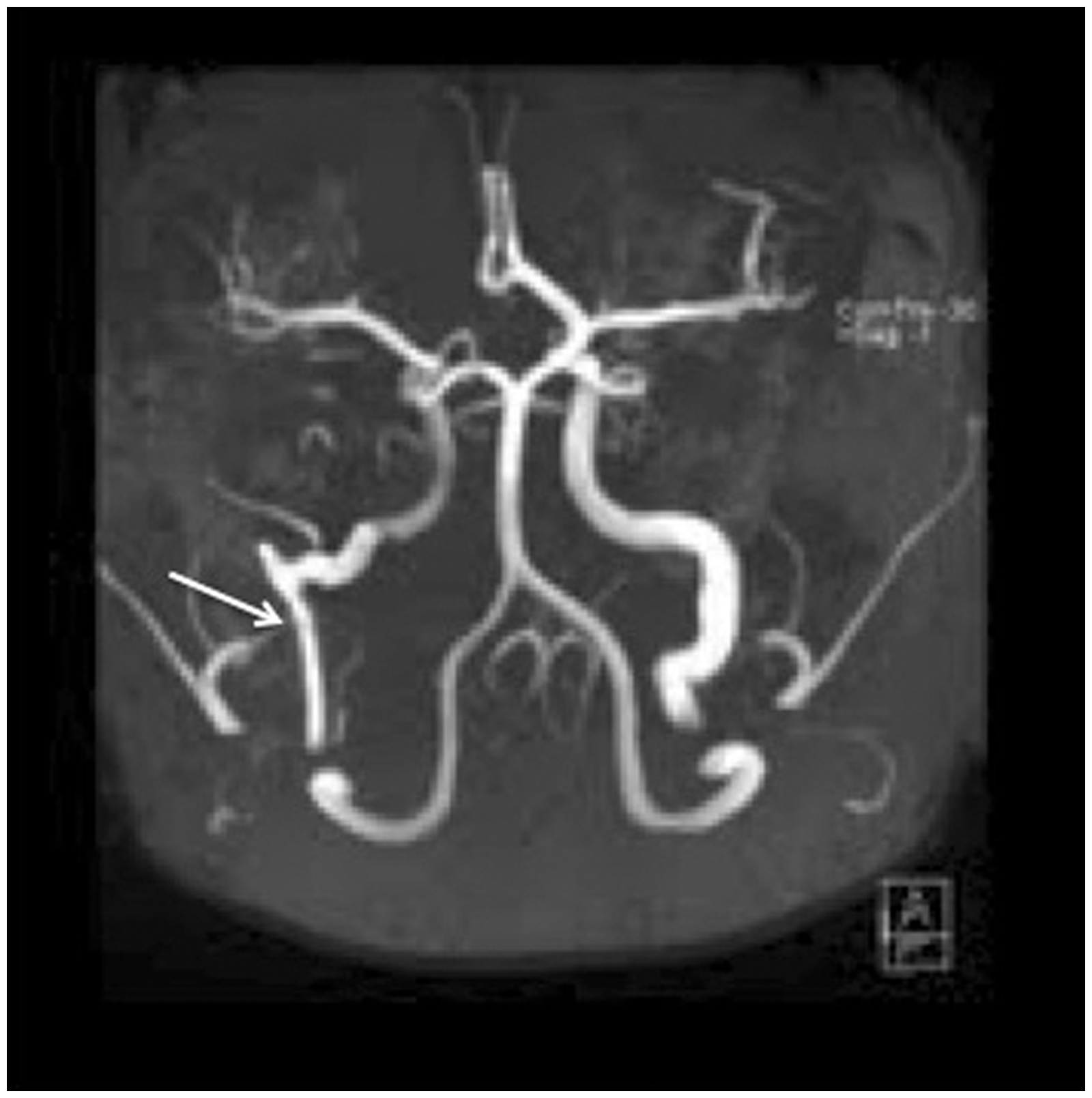
sequences was performed to confirm this diagnosis. On unenhanced three-dimensional
(3D) time of flight (TOF) MRA the difference between the normal-sized left ICA and
the thin, more laterally running right ICA can be nicely depicted (Fig. 2). Axial non-enhanced CT images at the level of the
condylar process of the mandible (a, b) and at the level of the
hypotympanon (c, d). A reduced-caliber vertical segment of the petrous
portion of the right ICA is noted (a) in comparison to the normal sized
left ICA (b). (c) The right ICA also has a more lateral course, bulging
out in the hypotympanon giving rise to a soft tissue mass lesion against
the promontory (arrow) before reaching the horizontal segment of the
petrous ICA (asterisk). (d) No such artery is found connecting to the
left horizontal segment of the petrous ICA
(asterisk). Unenhanced 3D TOF MRA of the intra-cranial
arteries nicely shows the smaller C1 segment of the right ICA running
more laterally in comparison to the normal-sized left ICA, which follows
a more medial course. Note also the coincident finding of an absence of
the A1 segment of the right anterior cerebral
artery.
Based on the imaging findings diagnosis of a partial agenesis of the right ICA with collateral circulation through the inferior tympanic artery, known as aberrant ICA, was made.
Discussion
Aberrant ICA is a rare congenital condition, with a debated pathogenesis. In the embryological stadium the third of the so-called aortic arches forms the proximal cervical portion of the ICA, while the dorsal aorta constitutes the distal portion. In aberrant ICA, also known as partial agenesis of the ICA, the presumed pathogenesis is regression or underdevelopment of the cervical part of the ICA at the skull base (1). In order to maintain adequate blood flow to the brain, an attempt to bypass this underdeveloped segment by using a collateral route is done. In most cases of underdevelopment of the ICA the circle of Willis is used to accommodate collateral flow. However, flow can also be provided by persistent embryonic vessels or branches originating from the external carotid artery (ECA). When there is agenesis of the cervical part of the ICA (C1), as in this case, the small arteries of the middle ear will be recruited to reach the horizontal petrous ICA segment. These vessels are the inferior tympanic artery, an embryonic branch of the ECA, and the caroticotympanic artery, a branch of the horizontal petrous part of the ICA. The enlarged inferior tympanic artery enters the tympanic cavity through the inferior tympanic canaliculus, passes lateral to the cochlear promontory bulging into the middle ear cavity, eventually anastomosing with the caroticotympanic artery to reach the horizontal petrous ICA segment (2,3). The intratympanic course of the aberrant ICA may suggest a vascular mass on otoscopic examination. The glomus tympanicum is the most frequent hypervascular middle ear tumor with a typical location against the promontory (4). However, in case of a glomus tympanicum, the size and aspect of the vertical and horizontal intrapetrous segment of the carotid artery is completely normal. There is also no communication between the mass lesion and the horizontal intrapetrous carotis segment in case of a glomus tumor.
Though in most cases asymptomatic, symptoms such as pulsatile tinnitus or conductive hearing loss may sometimes be seen in aberrant ICA but these are very non-specific. Aberrant ICA can therefore not reliably be discerned from vascular tumors or other vascular malformations like an aneurysm or a hemangioma (5), based on clinical findings alone (3). A biopsy or surgical exploration of a presumed hypervascular mass can be complicated by life-threatening hemorrhage or neurological deficit. Evaluation of a possible vascular mass in the middle ear should therefore be done by imaging. Even more, to avoid surgical injury, CT scan of the temporal bone should ideally be performed before any middle ear surgery. Several features suggestive of an aberrant ICA can be seen on CT or CBCT. These include – as demonstrated – the absence of the vertical segment of the ICA, distention of the inferior tympanic canaliculus, an aberrant ICA passing laterally through the tympanic space and dehiscence of the bone layer covering the tympanic segment of the ICA (6). On angiography a lateral and superior location of the carotid genu in the middle ear on the antero-posterior projection is typical (7). The ICA also runs laterally to the so-called vestibular line, a line drawn vertical from the lateral border of the vestibule (8). Since MRA is non-invasive and provides excellent visualization of the intracranial and extracranial circulation, this technique is to be preferred over conventional angiography (9).
In conclusion, an aberrant ICA is a rare vascular abnormality that may mimic a hypervascular middle ear mass lesion. Biopsy or surgical exploration of this pseudolesion can have devastating consequences. Therefore evaluation of a possible vascular middle ear mass should always be done by temporal bone CT. MRA can be used to confirm the diagnosis.
Footnotes
Conflict of interest
None declared.