
Abstract
The aim of this study was to examine the bone regeneration properties of beta-tricalcium phosphate hydrothermally converted from foraminifera carbonate exoskeleton in the repair of rat calvarial defect. These natural materials possess unique interconnected porous network with uniform pore size distribution, which can be potentially advantageous. In total, 20 adult male Wistar rats received full-thickness calvarial defect with a diameter of 5 mm. The rate of newly formed bone was measured radiologically by X-ray and micro-computed tomography and by histologic examination. After 2 weeks, the beta-tricalcium phosphate group exhibited full closure of the defect site, while control group remained unrestored at the end of the 6-week experimentation. It was observed that the newly regenerated bone thickened over the course of the experiment in the beta-tricalcium phosphate group. No soft tissue reaction was observed around the beta-tricalcium phosphate implant and the rats remained healthy. These results showed that repair of the calvarial defect can be achieved by biomimetic beta-tricalcium phosphate macrospheres, which hold potential for application as bone grafts for bone augmentation surgeries.
Introduction
In the field of biomaterial advancements, scientists are witnessing a golden era in the developments of novel and innovative materials for bone repair and regeneration. With a global population on the rise and significant increase in the number of middle-class citizens capable of spending, people are becoming more aware of available restorative surgeries that can increase an individual’s quality of life. Among these, one of the most common and practical surgeries is the use of dental implants. Depending on the patients’ condition, generally for a dental implant to be successfully integrated with the jawbone, first there must be enough bone to support the implant. In instances where there is tooth loss caused by either periodontal disease, dental caries and infections, injury, or trauma, bone augmentation procedures are required to generate more bone to the jaw before implants can be placed. This requires surgical implantation of bone graft materials, such as tricalcium phosphates (TCPs), to allow new bone to grow, a process that can take between 4 and 9 months. 1 Under the right condition, if accelerating the restoration of bone can decrease the recovery time, this will be a more ideal scenario for both the patient and health-care provider. The bone regeneration process is complex and is the subject of intense focus of research. From a material development perspective, TCPs have been extensively researched and used with success and safety in the clinical environment.2,3 Focus has shifted to making these materials more bioactive by incorporating pharmaceutics (simvastatin, bisphosphonates),4–6 proteins (bone morphogenetic proteins (BMPs)),7–9 and bioinorganics (strontium, magnesium, fluoride).10–13 While these stimulants do promote bone regeneration, the morphological features of the graft material are equally important, as bone formation requires the graft material to be porous to allow ingrowth of new bone and blood vessels. In addition, a spherical structured material would potentially be more advantageous as it has a higher contact surface area and can conform easier to the defect site. In previous studies, our group has identified a class of foraminifera carbonate exoskeleton possessing a natural spherical structure consisting of interconnected and uniform porous network that can be hydrothermally converted to beta-tricalcium phosphate (β-TCP) while preserving the original architecture. 12 This study will hence evaluate the bone regeneration ability of biomimetic β-TCP derived from foraminifera in a rat calvarial defect model.
Materials and methods
Synthesis of coralline β-TCP macrospheres
Foraminifera samples (Okinawa Business Support, Okinawa, Japan) were cleansed in sodium hypochlorite for 30 min to remove any residual contaminants and washed thrice in a sonicated water bath. Hydrothermal conversion was based on previously established protocol. 14 In brief, the samples were mixed with aqueous diammonium hydrogen phosphate (Wako Chemical Co., Tokyo, Japan) in a pressurized vessel where the Ca/P molar ratio was adjusted to 1.5 to produce β-TCP. The samples were heated to 220°C for 48 h and subsequently washed and dried.
Physicochemical characterization
The crystallographic structure of the sample was evaluated by powder X-ray diffraction (XRD) analysis (RINT-Ultima-III; Rigaku Co., Tokyo, Japan; CuKα radiation, 40 kV, 40 mA) and peak pattern matched with JCPDS database. The structural morphology of the material was observed under a scanning electron microscope (SEM) (JEOL JSM-7600F field emission SEM, 10 kV, Tokyo, Japan) at various magnifications. The in vitro degradation of the material and corresponding cellular response with bone marrow stromal cells and osteoblasts were examined in previous studies. 12 The ionic composition of the sample material was evaluated by inductively coupled plasma-mass spectroscopy (ICP-MS). An Agilent Technologies (Sydney, Australia) 7500ce series ICP-MS was used with sample introduction via a MicroMist concentric nebulizer (Glass Expansion) and a Scott-type double-pass spray chamber cooled to 2°C. The sample solution and the spray chamber waste were carried with the aid of a peristaltic pump. The ICP operating parameters and the lens conditions were selected to maximize the sensitivity of a 1% HNO3: 1% HCl solution containing 1 ng/mL of Li, Co, Y, Ce, and Tl. Helium was added into the octopole reaction cell to reduce interference. Calibration curves were constructed and the results were analyzed using Agilent Technologies MassHunter software. Approximately 0.005 g of sample was digested with 0.25 mL of 1% HNO3. Once the digestion was completed, the sample volume was made up with 5 mL water. The samples underwent a further 1:100 dilution with a 1% nitric acid solution before ICP-MS analysis. Samples were diluted further in nitric acid as required.
Animals, surgical procedure, and experimental design
The Animal Ethics Committee at Musashino University approved the animal experiments described herein. In total, 20 male Wistar rats (15 weeks old) were randomly divided into two groups: (1) control (empty defect) and (2) β-TCP. The animals were anesthetized by a combination of ketamine (40 mg/kg) and xylazine (5 mg/kg) injected intramuscularly. A cutaneous flap was created by making a mid-coronal incision through the skin, which was raised from the forehead. The periosteum was incised and elevated to expose the calvarial bone on both sides of the midline. One symmetrical, full-thickness bone defects with outer diameter of 5 mm were created with a bone trephine bur under continuous saline irrigation. Defects were either left empty or filled with five β-TCP samples. X-ray scans were taken every 2 weeks, and the animals were sacrificed at 6 weeks post surgery and evaluated histologically and radiologically.
Radiologic assessment of bone formation
The changes in the bone ingrowth area around the graft material, bone mineral density (BMD), and bone mineral content (BMC) were assessed radiologically by X-ray computed tomography (CT) scans (LaTheta LCT-200; Hitachi Aloka Medical, Ltd., Tokyo, Japan) taken at 0, 2, 4, and 6 weeks after grafting. Qualitative images of the defect were observed by 3D reconstructed images based on commercial 3D software (VGStudio MAX 2.0; Tokyo, Japan).
Histological assessment of defect
Histological analysis was performed at 6 weeks after grafting. The retrieved specimens of the rat calvaria were initially stripped of soft tissues and then fixed in 10% formalin solution (Wako Chemical Co. Ltd, Tokyo, Japan) for 2 weeks. The samples were then dehydrated in graded series of alcohol solution (60%–100%). Finally, the samples were embedded in paraffin where the blocks were sectioned using a micro-grinding machine. A series of 5 µm thick section were obtained and stained with hematoxylin and eosin (H&E). Histomorphometric analyses were performed using transmission light microscopy.
Statistical analysis
Statistical analysis was performed by using SPSS Ver. 11.5 for Windows. Analysis of variance (ANOVA) with Scheffe test was used for comparing the significance among the groups at each time point and was expressed as mean ± standard deviations. In all cases, a p value of less than 0.05 was considered statistically significant.
Results
Characterization of the material
The change in the crystallographic structure from carbonate foraminifera to β-TCP was verified by JCPDS database–matched peak patterns as illustrated in Figure 1. The spectra showed that the majority of the matched peaks are associated with β-TCP, with minor amount of calcium carbonate (aragonite: [3,0,0]) remaining in the crystal structure. In Figure 2, SEM micrographs show the morphological structure of both foraminifera and β-TCP material to possess uniform distribution of pores on the surface of the material and most importantly the preservation of these features after the conversion process. High-magnification image (4000×) of β-TCP showed the pore size of the material to be approximately 1 µm in diameter. In a previously published study (Chou et al., 2013), it was found that the surface pores were interconnected internally via porous chambers that are connected to a central porous hub.

XRD peak patterns showing matched peak of the sample after hydrothermal conversion to β-TCP.

Scanning electron micrographs showing the preservation of the material after hydrothermal conversion. High-magnification images at 400× show the uniform pore distribution through the surface of the material and at 4000× the uniformity of the pores.
Animal welfare and care
The surgical process of creating a calvarial defect can induce significant stress and trauma to the animal, and as such, all of the rats were kept in individual cages throughout the experimental period to allow space to recover individually. Figure 3 presents the CT images showing the location of the created defect site in the control (Figure 3(a)) and β-TCP (Figure 3(b)) groups. The rats were monitored on a weekly basis for the duration of the experimental period, and no histopathologic features of graft-versus-host disease or immune rejection were observed in any of the treatment and control groups. Figure 3(c) shows a healthy and consistent body weight growth during the experimental period in both experimental groups.
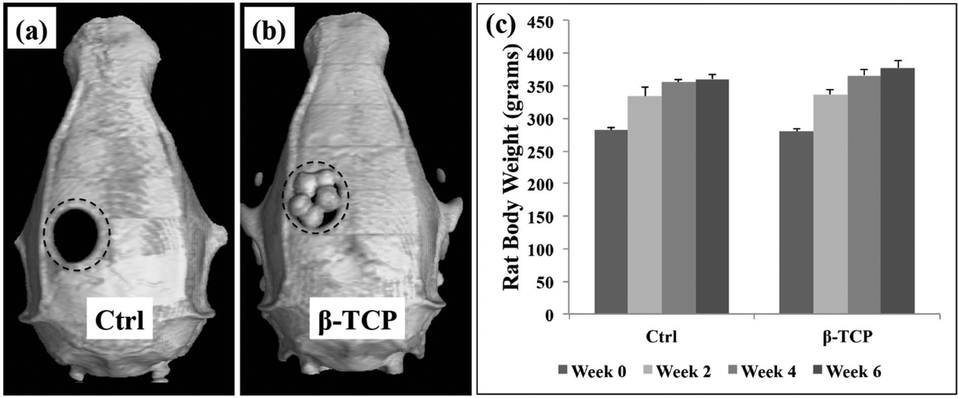
Software reconstructed image showing (a) empty defect in the control group, (b) implantation of β-TCP specimens, and (c) the preservation of the rat body weight during the study.
Radiological assessment of bone formation
The BMC, which corresponds to the amount of new bone formed within the calvarial defect (Figure 4(a)), showed that by 2 weeks post surgery, the β-TCP group induced statistically significant bone growth compared with 0 week. This significant rate of bone formation was maintained for the duration of 6-week experimental period. Compared with the empty control group which only displays a statistically significant bone formation at 4-week which at 6-week did not show further bone growth.
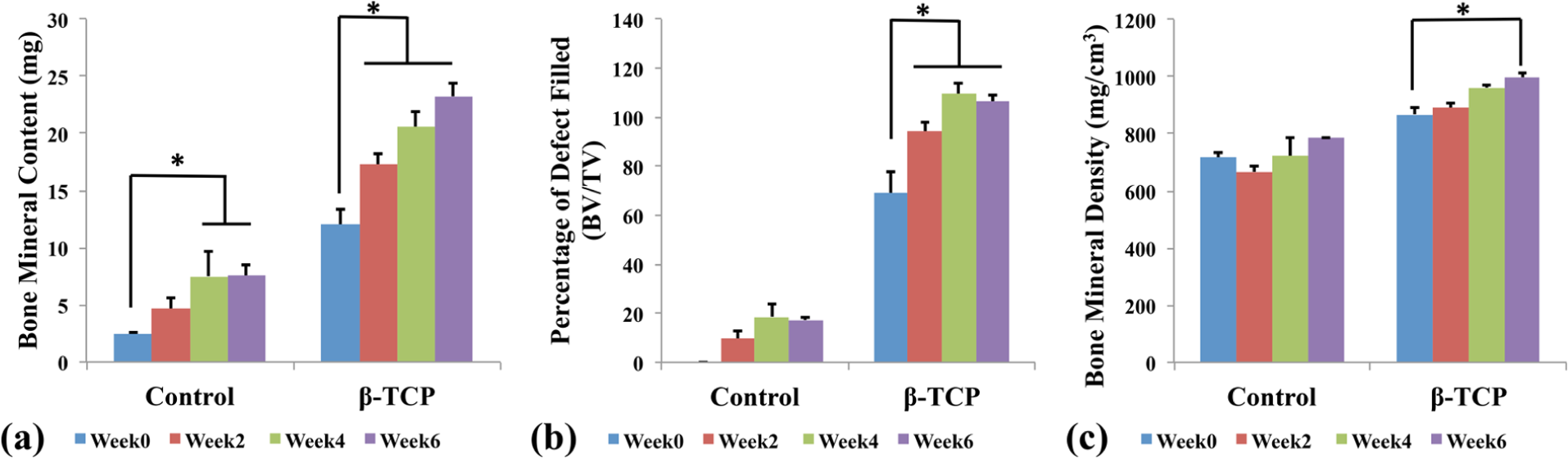
Analysis of the bone defect showing (a) the bone mineral content to be significantly higher in β-TCP group starting from 2 weeks compared with control, (b) the percentage area of the defect filled with new bone (BV/TV) showing the defect in β-TCP group to be 94% filled with new bone and samples by 2 weeks, and (c) the bone mineral density data showing β-TCP to display significant levels at 6 weeks compared with control.
The data presented in Figure 4(b) illustrate the bone volume/total volume (BV/TV) at the defect site that corresponds to the percentage of the defect filled with bone. It should be noted that for the β-TCP sample, at 0 week, the percentage filled was 70%, corresponding to the volume of the sample rather than bone. At 2 weeks, the control group’s BV/TV was at 17%, which did not change significantly till the end of the experiment. At 0 week, the β-TCP samples covered ~70% of the defect, which quickly increased to 94% by 2 weeks, which is in agreement with the BMC results. By 4 weeks, the BV/TV for β-TCP was 109% and remained the same until 6 weeks. This suggests that the defect was completely filled with bone by 4 weeks along with the embedded β-TCP. It should be noted that when the β-TCP was implanted, the samples were slightly bigger than the defect area and therefore the final calculated BV/TV was over 100%. This can be further visualized in later figures.
The BMD results (Figure 4(b)) show the control group to maintain a non-significant level of BMD (~700 mg/cm3), which remained at similar levels throughout the experimental period of 6 weeks. β-TCP showed significant increase in BMD between 0 (867 mg/cm3) and 6 weeks (997 mg/cm3).
Figure 5 shows progressive bone restoration at the defect site viewed from the top, and Figure 6 shows coronal view of the progressive bone restoration at the defect site. The restoration of bone at the defect site in the control group plateaued around 4 weeks with no significant changes observed at 6 weeks. This again is in agreement with the presented BMC and BV/TV data. It can be clearly seen from the images that in the β-TCP group, at 2 weeks, the empty space in the defect has been covered, and at 4 weeks, the bone restored was able to cover the entire defect, which again correlates with the corresponding BMC and BV/TV data.
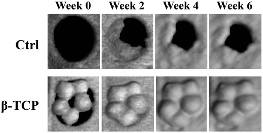
CT images showing the overall recovery of the calvarial defect. Control group showed minimal recovery, while β-TCP showed bone recovery from 2 weeks.
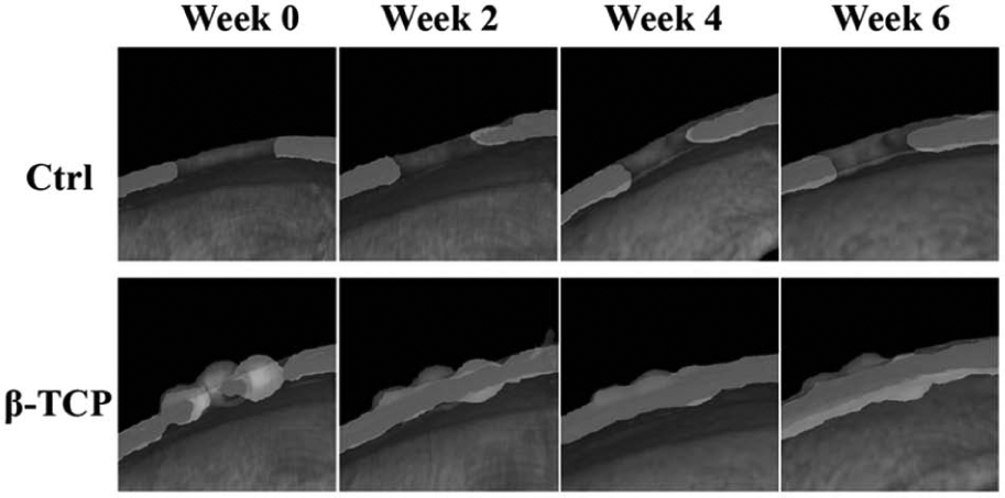
Software reconstructed coronal image of the center of the defect shows the empty control to achieve only minor bone restoration, while β-TCP showed defect closure by 2 weeks and thickening of the bone continuing till 6 weeks.
Histological findings
Figure 7 shows the H&E stained samples of the empty control and β-TCP groups at 6 weeks. The images show similar bone restoration of the defect site as observed in the previous CT images. In the control group (Figure 7(a)), minimal bone restoration was observed at the center of the defect. The β-TCP samples (Figure 7(b)) showed that the defect was completely restored with new bone to the same level of the original bone thickness. On closer observation of the β-TCP samples, it can be seen that the new bone infiltrated throughout the material suggesting a good integration between β-TCP and the surrounding tissue. Furthermore, the internal porous chamber structure can be observed from the image.

H&E stained slice showing minimal bone restoration in the empty control group, while β-TCP showed complete recovery of the defect with bone infiltrating the interior chambers of the material.
Discussion
The need for a biologically active bone graft substitute is an ongoing challenge for both biomaterial scientist and clinicians, and while suitable materials are available on the market, areas for improvements are still required. Calcareous material originating from the marine environment has shown to possess unique structural properties15–17 that can be adapted for biomedical purposes. The use of hydrothermal treatment as a process for converting calcareous materials to biocompatible calcium phosphates has been well documented18–20 and has been shown to preserve the fidelity of the original structure. Crystallographic evaluation by XRD showed the main crystalline phase of the foraminifera material to be calcium carbonate in the form of aragonite [JCPDS-00-0586] and TCP [JCPDS-00900169] after the hydrothermal treatment. While the spectra did show minor retention of the carbonate structure, it should be noted that the inorganic phase of the human bone is carbonated allowing the calcium phosphate material to be more osteoconductive and more resorbable than stoichiometric hydroxyapatite. 21
SEM images showed that the structure of the materials was preserved during the conversion process and most importantly the pores were uniform in size and well distributed throughout the surface of the material. This is a crucial factor as uniform pore size can potentially allow better infiltration of bone cells resulting in faster bone integration between the material and the surrounding environment. The interconnectivity of these porous chambers is one of the key attractiveness in the potential use of these materials as bone remodeling cells are able to adhere and proliferate allowing bone formation and infiltration of blood vessels. 22
The aim of this study was to evaluate the bone regenerative properties of using biomimetic foraminifera calcareous exoskeleton as a scaffold for bone restoration in a rat calvarial defect model. Following implantation of the β-TCP samples into the defect, the conditions of the rats were carefully monitored and showed no sign of inflammation or material rejection during the 6-week implant period. In the defect site, the BMC showed significant bone growth in the presence of the β-TCP starting from 2 weeks post operation. This shows a much higher rate of bone healing of the defect compared with the empty control group. We hypothesize that this observation is the result of the β-TCP material acting as a scaffold capable of stimulating the localized environment through the natural release of calcium and phosphate ions as the material degrades and reabsorbed by osteoclast cells. This can potentially accelerate the bone remodeling process thereby promoting bone regeneration of the rat calvarial defect. The uniformity and the interconnectivity of the pores from the β-TCP may also allow for increased cell infiltration into the material, which again contributes to the overall bone restorative process.
While the promotion of new bone mineral is crucial, the change in BMD is equally important. The BMD levels in the control group showed no significant changes, which can be explained by the lack of bone restoration at the defect site. β-TCP on the other hand displayed gradual increase in BMD during the experimental period, and at 6 weeks, significant BMD levels were observed. These results suggest that the β-TCP material was able to initiate an accelerated bone formation at the defect site during 2–4 weeks, and between 4 and 6 weeks, the bone mineral became denser as a result of continuous bone formation. This is also reflected in the BV/TV results showing that 94% of the defect was filled at 2 weeks, and by 4 weeks, the defect has completely recovered.
An overview of the defect site detailing the bone restoration shown in the CT images in Figure 5 reinforces the discussed BMC, BMD, and BV/TV results that the β-TCP displayed complete closure of the defect by 2 weeks. The empty control group, without any material to stimulate the bone remodeling process, showed a much slower rate of healing with a maximum of ~17% of the defect being restored during 6 weeks. Given more time, the defect may eventually be restored.
As previously discussed, H&E stained samples showed similar observation as those from the CT images. The newly formed bone in the β-TCP group was potentially thicker than the original calvarial bone and is regularly arranged. Furthermore, new bone can be seen infiltrating the β-TCP material completely displaying the porous chambers of the material at the same time. This suggests a good tissue–material integration and the biocompatibility of the β-TCP samples.
The presented results are consistent with the regenerative properties of synthetically produced β-TCP material in the restoration of bone defects.23,24 This study demonstrates that a material derived from the natural marine environment can possess the same stimulatory properties on bone formation. Furthermore, traditional production of β-TCP material requires high-temperature sintering between 800°C and 1000°C compared with the 200°C required for hydrothermal treatment.11,25 Current and future studies will require investigating the cellular mechanism and pathways involved in the actions of the β-TCP material observed in this study and comparing it with clinically used β-TCP samples.
The results presented in this study suggest the therapeutic potentials of using marine-derived β-TCP from foraminifera in the restoration of bone in a rat calvarial defect. While it is not envisaged that the material will replace traditional production of β-TCP grafts, the aim of this study is intended to provide an alternative source of improved biomaterial for bone restoration applications.
Conclusion
This study showed that foraminifera calcareous exoskeleton can be converted to biocompatible β-TCP while preserving the interconnected porous architecture. Evaluating the bone regeneration abilities of the material in a rat calvarial defect model, the results illustrated a faster bone regeneration rate compared with empty control group while maintaining a steady inclination toward BMD. With these, it is envisioned that this material can be potentially advantageous for use as a bone graft for bone augmentation surgeries or maxillofacial and craniofacial applications.
Footnotes
Acknowledgements
The authors gratefully acknowledge the support from Japanese Society for the Promotion of Science, Musashino University, University of Technology, Sydney, and Tokyo Medical and Dental University.
Data accessibility. Upon acceptance of this article, the data presented will be submitted to open access Data Dryad for public accessibility
Declaration of conflicting interests
The authors declare that there is no conflict of interest.
Funding
This study received financial support from Japanese Society for the Promotion of Science, Musashino University, University of Technology, Sydney, and Tokyo Medical and Dental University.