
Abstract
Introduction
A spinning top is a traditional game in some countries. The top is often made of wood or mud and the bottom is made of a sharp metal pin. Penetrating injuries can occur if the spinning top is incorrectly thrown.
Case report
We report a spinning top penetrating injury on the nose of an 8-year-old girl. Clinical examination revealed a spinning top that had penetrated her nasal bridge. Plain computed tomography (CT) of paranasal sinuses revealed a spinning top with its sharp pointed nail penetrating through the left nasal bone into the left nasal cavity. Examination under general anesthesia and rigid nasoendoscopy showed the tip of spinning top situated in between middle turbinate and nasal septum. The spinning top was then removed following the direction of entry.
Discussion
While air gun pellet facial injuries in pediatric patients have been reported, other playthings as culprits of facial penetrating injuries is still unheard of. Projectile objects can easily penetrate pediatric facial skeleton due to thinner facial bones. It is vital to exclude life- and sightthreatening injuries first, followed by imaging to assist decision-making and surgical planning. Computed tomography (CT) imaging is the gold standard for the radiographic evaluation of facial injuries. It assists in evaluating trauma extent, trajectory, depth, and course of foreign body. Following that, examination under anaesthesia including rigid nasal endoscopy is warranted.
Conclusion
We would like to highlight the importance of parental supervision and the value of pre-operative imaging. Neglecting seemingly harmless children’s toys can potentially cause fatal injuries.
Introduction
A spinning top is a traditional game in some countries. The top is often made of wood or mud and the bottom is made of a sharp metal pin. Significant penetrating injuries can occur if a spinning top is thrown incorrectly and lands on a vital body part. This case report presents the first reported incident of an injury caused by a spinning top. It emphasizes the potential for significant injuries despite their rarity.
Case report
An 8-year-old girl was brought to the emergency department with a spinning top stuck on her nose. The incident occurred when one of her friends accidentally threw the spinning top at her face, resulting in the penetration of her nose.
Clinical examination revealed a spinning top that had penetrated her nasal bridge, specifically 0.5 cm below the nasal root at the left paramedian region (Figure 1). Anterior rhinoscopy showed a blood clot in the left nostril without active bleeding from the wound. Her skull X-ray in anterior-posterior view showed a sharp radiopaque object penetrating the left nasal cavity at the left paramedian region of the nasal bridge, while lateral view showed the object lodged between the nasal root and the maxillary crest. Clinical examination revealed spinning top penetrating patient’s nasal bridge 0.5 cm below the nasal root.
Further imaging with plain computed tomography (CT) of the brain and paranasal sinuses revealed that the spinning top’s sharp pointed nail had penetrated the left nasal bone and entered the left nasal cavity, precisely between the left middle turbinate and nasal septum. Soft tissue density opacity was observed in the left anterior and posterior ethmoidal sinuses, indicating blood accumulation. Cerebrospinal fluid leakage was unlikely as the injury site was away from the skull base in the CT assessment. Additionally, the nasal septum was displaced to the right (Figures 2 and 3). Axial view of plain computed tomography (CT) of the brain and paranasal sinuses revealed a spinning top with its sharp pointed nail penetrating through the left nasal bone into the left nasal cavity. Coronal view of plain computed tomography (CT) of the brain and paranasal sinuses revealed a spinning top with its sharp pointed nail penetrating through the left nasal bone into the left nasal cavity between the left middle turbinate and nasal septum.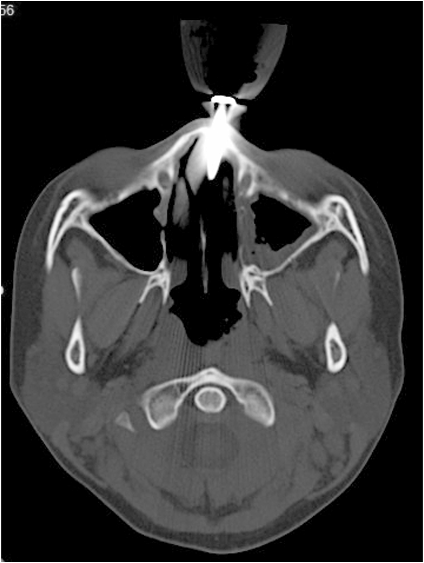

The patient underwent examination under general anesthesia to extract the spinning top from the nasal bridge. Intraoperatively, rigid nasoendoscopy revealed the tip of spinning top to be situated between the middle turbinate and nasal septum, with minimal blood clot and abrasion at the nasal septum and middle turbinate. The spinning top was removed with caution following the direction of entry. After removal, there was a communicating wound between the point of entry and the point of exit in the left nasal cavity. A small piece of bone at the exit point was also extracted. The external penetrating wound was then repaired by subcutaneous and skin closure. Following the operation, she was discharged home well with oral antibiotic and analgesia. During subsequent follow-up visits, she remained well and her nasal wound had healed with minimal scarring.
Discussion
While intranasal foreign bodies and transnasal penetrating injuries are commonly encountered in the pediatric population, it is uncommon to encounter a penetrating injury directly through the nasal bone, which barely missed the orbit. A penetrating injury happens when an object penetrates the tissues and remains lodged within the structures. Following that, the object is considered a foreign body. 1 Ordinarily, such injuries occur on organs with larger surface areas, such as the trunk and extremities. They are uncommon in the smaller facial region as the face is either moved away from the incoming object or the object is deflected away via natural protective reflexes. 2
Craniofacial penetrating injuries in adults often involve objects such as knives, metal rods, firearms, pipes, nail guns, machetes, and spear guns. 2 On the contrary, penetrating injuries in pediatric cases often include objects like sticks, chopsticks, writing instruments, toothbrushes, cylindrical toys, and straws. However, these injuries usually occur through the oral cavity, causing damage to the palate, posterior oropharynx, tonsillar region, cheek, tongue, and mouth. 3 Cases of air gun pellet facial injuries in pediatric patients leading to foreign body retention in the paranasal sinuses have been reported. 4 However, literature reporting other playthings as culprits of facial penetrating injuries is still unheard of to our knowledge.
Pediatric facial penetrating injuries require caution as their facial bones and orbital walls are thinner compared to those in adults, making it easier for projectile objects to penetrate their facial skeleton. 2 This explains how a seemingly harmless object such as a spinning top can cause injury, as a low-velocity penetrating foreign body can fracture pediatric facial skeleton with ease. 5 In cases of facial traumas with severe or multiple injuries, life- and sight-threatening injuries should be excluded first. Once the patient is stabilized, imaging techniques such as plain radiography, CT, and magnetic resonance imaging (MRI) can assist in decision making and surgical planning. 6
CT imaging is considered the gold standard for radiographic evaluation of facial injuries. In addition to assessing the extent of trauma, it assists in evaluating the trajectory, depth, and course of a foreign body. 3 Special attention should be paid to wooden foreign bodies, such as twig remnants or pencils, as they can be difficult to recognize due to the attenuation of air initially. As these foreign bodies start to absorb moisture, they become inseparable from the surrounding soft tissues. 3 Apart from that, evaluation of the most distal tip of the penetrating object is also crucial. 2 Facial penetrating traumas can cause injuries to various areas, including the base of skull, nasal cavity, orbit, cranial nerves, and even vascular structures such as the internal carotid artery. 7 Therefore, it is vital to exclude injuries to the anatomical structures in close proximity. Notably, Antonio reported a screwdriver injury that pierced the maxilla, subsequently penetrating the skull base. 8 In another article, El-Anwar described a left orbital penetrating injury in a five-year-old child, where the object pierced the left lacrimal bone and traversed the nasal cavity by perforating the nasal septum. The reported object then penetrated the right orbital lamina papyracea, stopping just right before the right optic nerve. 5 These examples emphasize that while foreign body penetrating injuries are usually obvious when the objects are attached to the entry wounds, evaluation of the depth and course prior surgical intervention is also vital. In instances where vascular injury is suspected, the utilization of contrast-enhanced CT or CT angiography may be deemed appropriate, given the limitation of non-contrast CT in assessing extravasation. Nonetheless, in our particular case, non-contrast CT was expediently chosen owing to the absence of fasting requirement for the patient and the absence of clinical indications suggestive of vascular injury.
In penetrating injuries, examination under anesthesia in a controlled environment is important as no removal of objects should be attempted in the field or in the emergency room. Apart from causing pain especially in an uncooperative pediatric patient, any bleeding that occurs following bedside removal will be difficult to manage. 2 After external wound examination, internal examination via rigid nasal endoscopy with various degrees of visualization allows us to navigate the affected nasal cavity. Anatomical structures of interest include the sinuses, turbinates, nasal septum, orbital floor and base of skull. Vascular injury should also be ruled out, as the anterior ethmoidal artery might course lower than usual in the nasal cavity. Furthermore, endoscopic examination can identify any remaining foreign body fragments. Although uncommon, retention of foreign body fragments in the nasal cavity or paranasal sinuses has to be considered, as it can lead to complications, such as sinocutaneous fistula, intermittent nasal hemorrhage, secondary infections, and sinusitis. Transnasal endoscopy can aid in the management of these cases. 9 Cetinkaya et al. described the importance of transnasal endoscopy in not only guiding foreign body removal, but also in revealing the skull base defect caused by the penetrating object. Subsequently, the cribriform plate defect was repaired endoscopically. 10 Following the removal of the penetrating object and examination under anesthesia, any hard or soft tissue injuries should be addressed simultaneously. Only then we can restore the compromised facial functions and aesthetics. 2 Fortunately, apart from the nasal bone fracture and abrasion in the left nasal cavity, our patient did not sustain other injuries.
Conclusions
While penetrating injuries are more common in the adult population, often resulting from accidents or intentional acts, pediatric penetrating injuries usually occur due to lack of parental supervision. This case report emphasizes the importance of parental supervision and caution in the pediatric population. Additionally, we would also like to stress the significance of pre-operative imaging, given the variability of injury it can reveal. Lastly, neglecting seemingly harmless children’s toys can potentially lead to severe and even fatal injuries.
Footnotes
Author contributions
J.H.K. wrote the first draft of the manuscript. B.C.T. reviewed and revised the manuscript. I.S.S. reviewed the manuscript. All authors read and approved the final version of the manuscript.
Declaration of conflicting interests
The author(s) declared no potential conflicts of interest with respect to the research, authorship, and/or publication of this article.
Funding
The author(s) received no financial support for the research, authorship, and/or publication of this article.
Ethics statement
Data availability statement
Data sharing is not applicable to this article as no datasets were generated or analysed during this study.