
Abstract
Background
Stature estimation is important in medical investigations to identify victims and is most accurate using anatomical methods utilizing the entire skeleton. However, in cases of mass fatalities, it has been calculated using regression analysis formulas of different body parts. Radiology especially post-mortem computed tomography (PMCT) is increasingly being used in victim identification as it is non- invasive and allows for multi planar reconstruction.
Methods
This study aims to generate stature estimation formulae using regression analysis of the vertebral column incorporating population that form the Malaysian ancestry i.e. Malay, Chinese and Indian using PMCT. This was a retrospective study involving 115 subjects who had a PMCT. Subjects consisted of Malaysian males and females (92 males and 23 females) aged 18–70 years. Cervical length (CL), thoracic length (TL), lumbar length and the whole spine length (WSL) incorporating cervical, thoracic and lumbar length were compared against cadaveric length.
Results
Whole spine length had the highest correlation coefficient of 0.570 and showed highest correlation with cadaveric length among female subjects compared to males. Whole spine length amongst the Malay was the highest followed by the Chinese and Indian ethnic groups (r = 0.671, 0.629, 0.402).
Conclusion
Our study concluded that the use of WSL from PMCT images can be used for body height estimation amongst Malaysians. This study has also successfully generated population-specific database for Malaysian experts to perform stature estimation using measurements of the vertebral column.
Keywords
Introduction
Mass fatality incidents (MFI) which may be due to environmental (e.g. earthquakes, tsunamis, hurricanes), medical (e.g. famine, disease), vehicle (e.g. aircraft, car, train, ship), industrial (e.g. explosions, fires) or terrorist (e.g. chemical, biological, radiological, nuclear or explosive attacks) are currently on the rise worldwide. 1 In these situations, not only is it important to identify the manner and cause of death but also rapidly and accurately identify victims for juridical reasons as well as to provide closure to the families.2-4
Stature estimation together with sex, age and ancestry are important in medical investigations to identify victims and is most accurate using anatomical methods utilizing the entire skeleton.5,6 However, in most cases of MFI where the body is severely mutilated and the entire skeleton is not available, stature estimation has been calculated using mathematical methods with the help of regression analysis formulas from measurements of the skull, sternum, vertebral column, scapula, long bones of the upper and lower limbs, hands and feet with the help of imaging or the hybrid method which is a combination of anatomical and mathematical methods.3,5,7–23
Radiology has been used for human identification since 1927 and today with the use of post-mortem computed tomography (PMCT) imaging in most if not all forensic institutes worldwide, it is being used with increased frequency in human identification. post-mortem computed tomography has the added advantage over the conventional method of stature estimation in that it is non- invasive avoiding maceration processes, quick to perform and allows for reconstruction as well as conservation of images.1,4,8,15,17,24–26
There have been studies done in Europe and Asia using imaging of the spine to determine stature looking at two or three vertebrae of a spinal segment.3,9,11 Some studies however used segments of the spine i.e. either cervical or lumbar spine in determination of stature.12,27
In Malaysia, there is lack or scarcity of data on stature estimation amongst the three main ethnic groups using PMCT. Thus, this study aims to generate stature estimation formulae for Malaysians using regression analysis of the whole spine incorporating population that form the Malaysian ancestry i.e. Malay, Chinese and Indian as previous studies of stature estimation amongst Malaysians were done using radiographs of upper and lower limb lengths. It is particularly important to have stature estimation data catering for the Asian population as most stature estimation in the past used regressions based on the European and American population, which may not be applicable to the Asian population. In addition, this study will also compare different segments of the spine i.e. cervical, thoracic and lumbar to determine which segment of the spine is most accurate in stature estimation.
Methods
Study subjects
This was a retrospective study conducted within local guidelines with approval by the local ethics committee of both the university and the Ministry of Health Malaysia (NMRR 36308). Subjects included 115 medico-legal autopsy cases (Police 61 Form, thus a waiver for consent) brought to the National Institute of Forensic Medicine, Hospital Kuala Lumpur, Malaysia who had a whole body PMCT done in accordance with the Institute’s protocol. Subjects were made up of Malaysian males and females (92 males and 23 females) with ages ranging from 18 to 70 years incorporating population that form the Malaysian ancestry i.e. Malays, Chinese and Indians. Subjects with fractures, congenital conditions or neoplasms of the spine, sacralization or lumbarization of the spine and decomposed bodies were excluded from the study. Cadaveric length was measured from head to toe during post mortem examination using sliding calipers and age of the subjects were obtained from identification documents based on date of births.
Post-mortem CT scan imaging
A native PMCT scan was performed within 6 h of arrival of the deceased to the Institute. Post-mortem computed tomography was performed prior to any manipulation of the body using a 64-slice (Toshiba Aquilion 64 TSX-101A, Japan) multi-detector computed tomography (MDCT) scanner, a machine wholly dedicated for autopsy use. Examinations were performed in a cranio-caudal direction from head to toe using 1 mm slice thickness for the head region and 2 mm slice thickness for the thorax, abdomen and pelvis down to the toes. Scans were performed using pre-defined scanning protocols: 120 kVp, Auto set mAs (Caredose), FOV 500 (LL), 1.0 × 32 raw detector collimation and 0.844/standard pitch. Curved Multiplanar reformatted (MPR) images of vertebral body height and intervertebral disc spaces, as well as the three-dimensional (3D) image reconstruction of images involving the entire vertebral column was done and viewed using bone window on the Osirix workstation, OsiriX MD V.8.5 installed in a 27-inch iMac computer.
Actual Height (AH) of all cases were obtained from cadaveric length measurements. An ideal mid-sagittal image through the odontoid process of the second cervical vertebra (C2) down to the lower border of the fifth lumbar vertebra (L5) was used to outline the vertebral curvature using Osirix software taking into account the natural lordosis of the cervical and lumbar spine as well as the natural kyphosis of the thoracic spine. Slight manipulation of the vertebral column was required to ease the Whole Spine Length (WSL) and to acquire the best image of the spine in coronal plane (Figure 1). Whole Spine Length was measured from the tip of the odontoid process of C2 vertebra to the lower border of L5 vertebra. For the measurement of specific spinal segment groups; the Cervical Length (CL) was measured from the tip of the odontoid process of C2 to the lower border of the seventh cervical vertebral body (C7), the Thoracic Length (TL) was measured from the lower border of C7 body to the lower border of the 12th thoracic vertebral body (T12) while the Lumbar Length (LL) was measured from the lower border of T12 body to the lower border of L5 (Figure 1). All measurements mentioned were independently measured and recorded by 4 observers and the average reading was computed to compensate inter-observer error. Curved Multiplanar reformatted (MPR) and three-dimensional (3D) image reconstructionof the spine. WSL = whole spine lengthmeasurement, CL = cervical spine length measurement, TL = thoracic spine length measurement and LL= lumbar spine length measurement.
Statistical analysis
Statistical analysis was performed using IBM Statistical Package for Social Sciences (SPSS) version 22.0. Mean, standard deviation (SD), and standard error (SE) for Age, AH, WSL, CL, TL and LL were calculated for all subjects. Normality test was performed on the measurement parameters using Kolmogorov-Smirnov statistic with p > .05. The significance and confidence interval p-value of SPSS analysis throughout the study was p < .05. Taking into account several previous studies, Kevin Norton’s and Tim Old’s methodology was applied by computing the technical error of measurements (TEM) as well as the coefficient of reliability.
The correlation between AH and each of the four measurements including WSL, CL, TL and LL was elucidated using Pearson’s product-moment correlation coefficients (r). Simple linear regression analysis was performed for each of the four measurements to derive regression formulae for stature estimation. The standard error of estimation (SEE) and adjusted coefficient of determination was then calculated to assess the significance of the regression equation.
Further validation analysis of the regression formulae of stature was performed using 15% holdout subjects (18 males and 5 females) from the total subjects. The remaining 85% of the total subjects were used to generate new regression formulae to be tested using the holdout subjects according to sex and ethnicity. The formulae were then used to estimate the stature for each individual and compare it to the actual stature of the holdout subjects.
Results
A total of 115 cases were included in this study, of which 92 were males and 23 were females. The ratio of males to females and the Malay: Chinese: Indian ratio was 9:2 and 5:4:2 respectively reflecting the proportion of postmortem cases by sex and ethnicity. The mean age ± SD was 43.39 ± 12.57 years whilst the mean AH ± SD was 161.61 ± 6.27 cm.
Descriptive statistics for age, actual height, WSL, CL, TL and LL. (measurement in centimeters).

The relative TEM for all the measurement parameters between observers were lower than the standard 2% and reliability at higher than 0.8 except for the lumbar segment. However, there was no significant difference for inter-observer error analysis based on the paired sample t-test at p > .05 for lumbar segment. Hence, this showed that the measurements taken by the observers in this study were acceptable for further statistical analysis.
Linear regression formulae in terms of ethnicity.

Linear regression formulae in terms of sex.
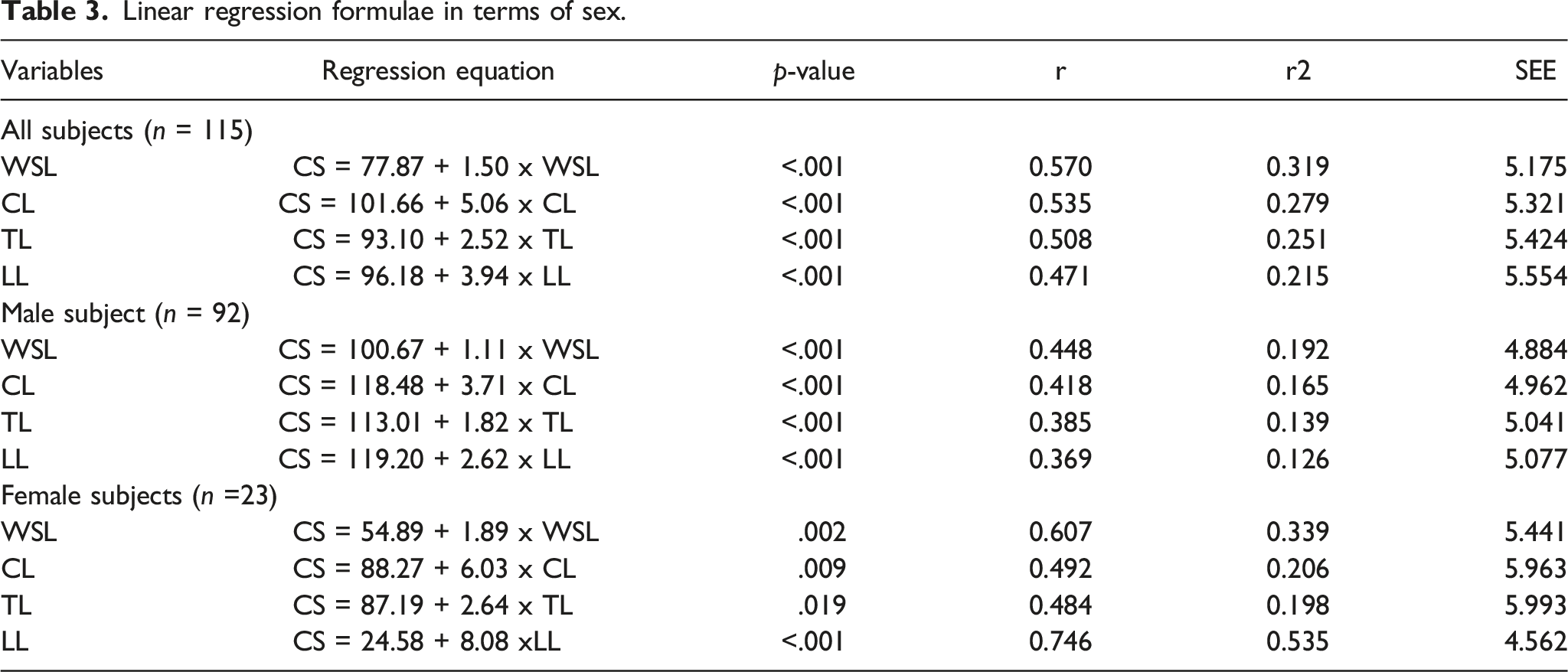
In Table 3; WSL had the highest correlation with AH among female subjects compared to males. It was also similarly applicable to the cervical, thoracic and lumbar segments of the vertebra column. WSL has also the highest correlation with stature in combined sex at r = 0.570. However, lumbar segment showed extra-ordinary higher correlation at r2 = 0.535 with height in female subjects compared to the other segments.
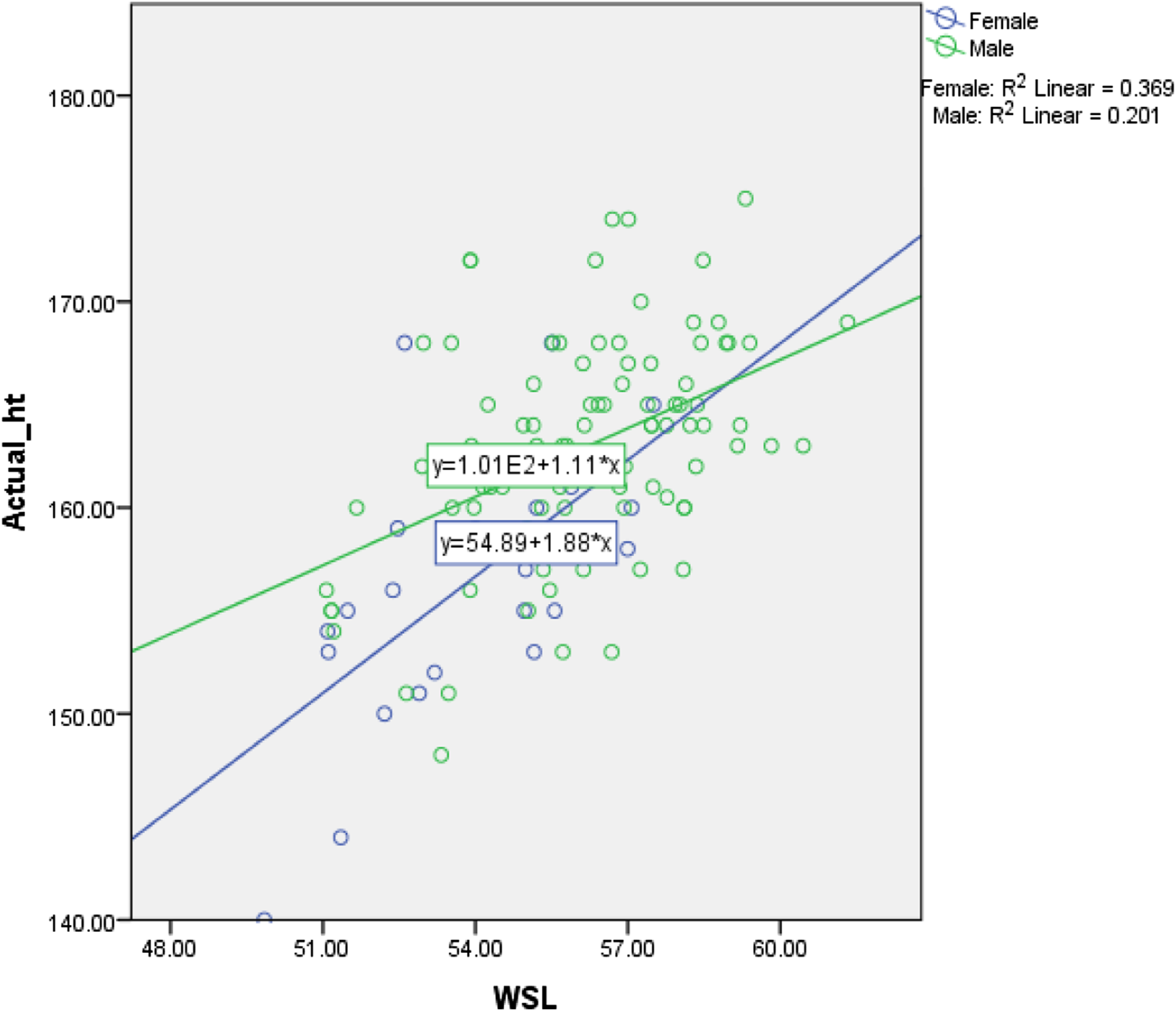
Scatterplot graphs representing the AH versus WSL based on ethnicity (Figure 2), AH versus WSL based on sex (Figure 3), AH versus each parameter (WSL, CL, TL and LL) for overall sample size (Figure 4) all showed positive slope with varying spread. Scatterplot of actual height versus WSL based on ethnicity. Scatterplot of Actual height versus WSL based on sex. Scatterplots of actual height versus each parameters (WSL, CL, TL and LL) for overall sample size.

Validation analysis of the regression formulae in terms of sex and ethnicity.

Discussion
Accurate estimation of stature represents an important goal in forensic evaluation and over the years many methods have been developed to obtain such details using different parts of the body to establish a relationship between stature and body segments. Body segments used in the past include the skull, sternum, spine, upper and lower limbs imaged using different radiological imaging methods which included the use of X-rays, computed tomography (CT) and magnetic resonance imaging (MRI).3,5,7–23
Studies have proven that though osteometric measurements using dry bones were considered the gold standard by most anthropologists, the use of imaging especially PMCT is equally acceptable when compared to measurements obtained from dry bones with the advantage of being faster and reproducible. 28
Though most studies have shown that the lower extremity has the greatest association with body stature, there are often missing or badly fragmented body parts found at the scene of death. For this reason, in this present study, we used the spine as it could be often found intact as a whole or as part of the body trunk. Our study also decided to undertake only the Malaysian population, as most studies done previously showed that genetic and environmental components have a strong influence on skeletal development with equations derived from certain populations not being suitable for another originating from a different nation, or even a different continent. 29
Our study as shown in Table 2; indicated that WSL has the highest correlation with stature consistent with data from a previous study.13,27,30 In terms of segments of the spine, our study determined that cervical segment of the spine performed better when both sexes were grouped together as well as in males. However, the lumbar segment of spine was most accurate when females alone were considered which was similar to the previous studies which stated that the lumbar spine was better.13,30 A study however stated that the TL of spine correlated most closely to the AH. 27
Two major meta-analyses also found that ethnicity-specific and sex-specific methods provide negligible or no improvement in accuracy when estimating stature, while at the same time increased the complexity of applying these methods due to arbitrary parameters of group membership and the difficulty of assigning an unknown to those groups.31,32 This study has generated a formula that can be used by both sexes and the three main ethnic groups in Malaysia, which is especially important for stature estimation in unknown cases. However, different population data is beneficial as it will reduce the variation effect due to population-specific genetic pool. In fact, hormonal changes in different sexes could also affect the stature which makes sex-specific formulae more relevant in known sex cases.
Comparison of correlation coefficient (r) and standard error of estimation (SEE) of different studies.

The limitation of this study included the scarcity of available cases in the existing database. This was because initially a total of 412 cases were extracted, however 297 cases had to be excluded due to several issues such as incomplete scan images, images scanned using the old protocol, homicide cases, sacralization etc. leaving us with a remaining 115 cases to work on. Given some additional time and sources, a search for another 14 cases amongst Indian samples would have provided the minimum number to run a proper regression analysis. Having said that, contrary to a previous study by Jason D. which stated that the thoracic spine length showed the highest correlation with AH, our study found that the cervical group had the highest correlation to AH. 27 This theory however needs to be re-evaluated by further studies which specifically aim to compare the correlation of individual vertebral spine segments to AH.
Conclusion
It can be concluded that the use of WSL or if not available, cervical spine length from PMCT images can be used for body height estimation amongst Malaysians. However, the regressions could be improved in future with a larger sample size to better represent diversity of the population. Furthermore, from the clinical perspective, although full body CT scans are not routinely done, we believe that to some extent stature estimation of completely bedridden and gravely ill patients can be done by measuring the spine length using CT slices of the thorax-abdomen-pelvis.
Footnotes
Acknowledgement
The authors would like to thank Afrina Aqilah Binti Abd Shukor, Nur Najla Aqilah Binti Jamil, Mas Aina Binti Mohd Nasir and Muhammad Azrul Bin Talib, year 3 medical students from the Faculty of Medicine UiTM who performed this study as part of their elective program project as well as the Director General of Health Malaysia for his permission to publish this article.
Authors’ contributions
Availability of data and materials
The datasets generated and/or analysed during the current study are available from the National Institute of Forensic Medicine, Hospital Kuala Lumpur.
Ethical approval
Ethical approval for this study was obtained from the Research Ethics Committee, Universiti Teknologi MARA and the National Medical Research Registry, Ministry of Health Malaysia (NMRR- 36308).
Informed consent
Informed consent was not required from the next of kin in this study as it involved retrospective PMCT data which is a pre requisite to an autopsy under the police (P61).
Declaration of conflicting interests
The author(s) declared no potential conflicts of interest with respect to the research, authorship, and/or publication of this article.
Funding
The author(s) received no financial support for the research, authorship, and/or publication of this article.