
Abstract
Purpose
Imaging changes in the pituitary volume during pregnancy remains scantly researched. This study set out to assess the differences in total, anterior, and posterior pituitary volume in pregnant women compared to nulliparous and post-partum women.
Materials and Methods
A retrospective review was completed of women that had undergone MRI imaging of the brain. Patients were divided into three cohorts: pregnant, nulliparous, and post-partum (defined as being within 12 months of delivery). Anterior and posterior pituitary volumes were manually measured.
Results
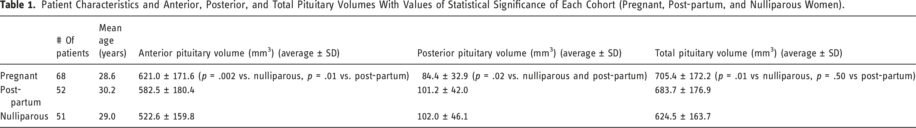
171 patients were included, of which 68 were pregnant, 52 were post-partum, and 51 were nulliparous. The average anterior (621.0 ± 171.6 mm3) and total (705.4 ± 172.2 mm3) pituitary volumes were significantly larger in pregnant patients than nulliparous women (522.6 ± 159.8 mm3 and 624.5 ± 163.7 mm3, respectively) (p = .002 and p = .01, respectively). The posterior pituitary volume was significantly smaller in pregnant women (84.4 ± 32.9 mm3) compared to both post-partum (101.2 ± 42.0 mm3) and nulliparous (102.0 ± 46.1 mm3) women (p = .02 for both).
Conclusions
The anterior and total pituitary volumes are significantly larger during pregnancy persisting into the post-partum period. The posterior pituitary volume, conversely, decreases during pregnancy, and returns to its normal size in the post-partum period.
Introduction
The hormones created from the pituitary are several, but well-known. The anterior lobe releases adrenocorticotropic hormone (ACTH) which stimulates the adrenals, follicle-stimulating hormone (FSH) and luteinizing hormone (LH) which act on the ovaries and testes, growth hormone (GH) which stimulates bone and muscle mass growth, prolactin which stimulates breast milk production, and thyroid-stimulating hormone (TSH) which acts on the thyroid gland. The smaller posterior lobe produces anti-diuretic hormone (ADH) which regulates renal water absorption and oxytocin which contracts the uterus during childbirth and aids in lactation. 1
Pregnancy induces substantial physiological changes in many organs during pregnancy, and the pituitary is no exception.2,3 The gland increases in volume by over a third by the end of gestation. 4 The anterior pituitary becomes 2–3 times larger, mostly due to the hyperplasia and hypertrophy of lactotroph cells which synthesize and release prolactin.5,6 Other types of anterior pituitary cells remain unchanged or decrease in size. Specifically, both gonadotrophs (which regulate LH and FSH) and somatotrophs (which regulate GH) decrease in number during pregnancy. 6 There is evidence that somatotrophs are transformed into lactotrophs during pregnancy, contributing to increased prolactin production. 7
Pregnancy and the post-partum period are times of elevated risk for pituitary disorders, including lymphocytic hypophysitis and Sheehan syndrome with pituitary infarction. 8 As such, it is crucial to be familiar with the expected changes of the pituitary gland in pregnant and post-partum patients. This study sought to assess the changes in the pituitary volumes on MRI in cohorts of women that were pregnant, post-partum, and nulliparous.
Materials and Methods
Patient Population
This study was performed following approval by the local institutional review board. A retrospective review was completed of women that had undergone imaging of the brain with MRI between 1/1/2000 and 12/31/2020. Included patients were divided into three cohorts: pregnant at the time of imaging, nulliparous (no prior births), and post-partum (defined as being within 1 year of the most recent birth). Patients were found using a retrospective review of available MRIs of the brain, with review of the electronic medical record used to confirm pregnancy status. According to the inclusion criteria, no further patients were collected after all cohorts had over 50 patients. The pregnancy status and demographic information of each patient was assessed using a review of electronic medical records. The breastfeeding status of post-partum patients was also recorded, when available. According to the criteria for this study, any MRI examinations that were limited by artifact (e.g., motion) or poor quality were set to be excluded, though no such exams were observed.
MRI Imaging
Pituitary measurements were performed using either sagittal T1 MPRAGE images (TR = 2500 ms, TE = 1.88 ms, slice thickness = 1 mm, FOV = 240 x 240 mm) or sagittal T1 FLAIR images (TR = 11,000 ms, TE = 155.8 ms, slice thickness = 4 mm, FOV = 220 x 220 mm). Volumes were measured using the institutional PACS system (Visage, Visage Imaging Inc., San Diego, CA). A single radiology resident used slice-by-slice regions of interest to measure both the anterior and posterior pituitary volumes in each patient.
Statistical Analyses
Means and standard deviations were calculated for all continuous variables. All statistical calculations were performed using BlueSky Statistics software (Bluesky Statistics LLC, Chicago, IL, USA). Student’s t-test was used to assess for statistically significant difference between continuous variables. For all calculations, the threshold for statistical significance was set to p = .05.
Results
A total of 171 patients were included in this study, all of which were women. 68 were pregnant (average age: 28.7 ± 4.7), 52 were post-partum (average age: 29.8 ± 6.0), and 51 were nulliparous (average age: 27.6 ± 8.0). There was no significant difference in age between the pregnant and post-partum (p = .31) or nulliparous (p = .37) cohorts. Of the 52 post-partum women, 37 (71.2%) were breastfeeding at the time of imaging. The indications for brain MR imaging varied among patients, with headache, syncope, and vertigo being the most common. None of the patients were imaged specifically for pituitary pathology.
Patient Characteristics and Anterior, Posterior, and Total Pituitary Volumes With Values of Statistical Significance of Each Cohort (Pregnant, Post-partum, and Nulliparous Women).
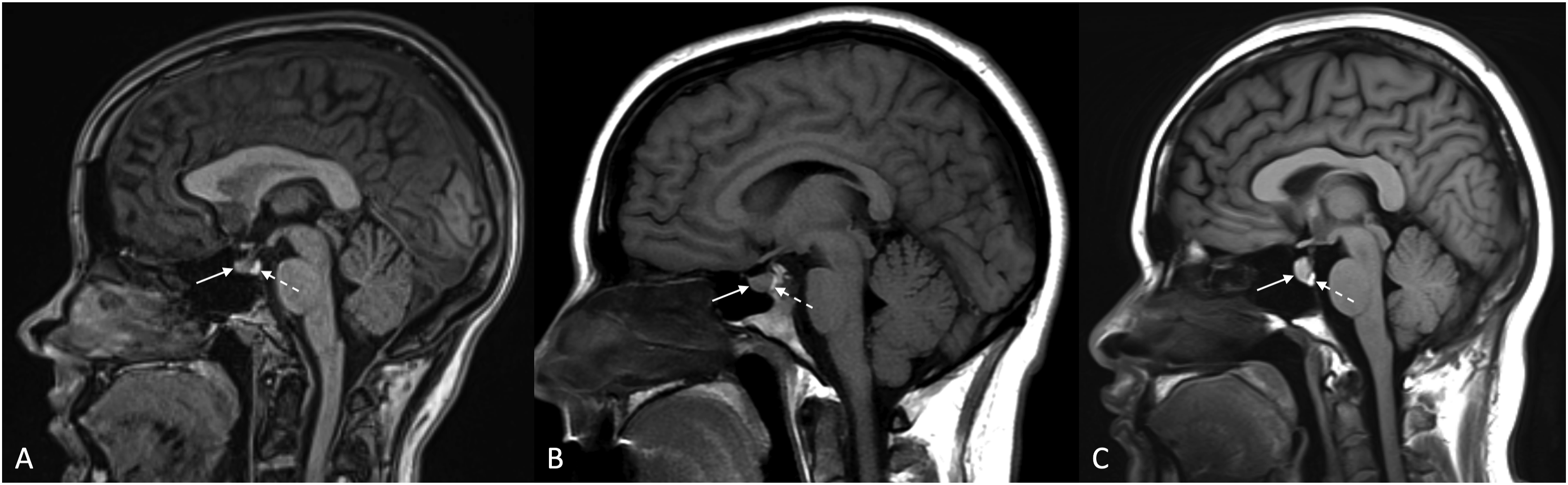
Examples of anterior (solid arrows) and posterior (dashed arrows) on sagittal images in nulliparous (a), pregnant (b), and post-partum (c) women.
Among post-partum women, anterior pituitary volumes were not significantly different in those that were breastfeeding (611.0 ± 180.0 mm3) compared to those that were not (533.1 ± 175.1 mm3) (p = .49). Posterior pituitary volumes were also not significantly different in those that were breastfeeding (95.5 ± 40.1 mm3) compared to those that were not (111.0 ± 44.5 mm3) (p = .36).
Discussion
To our knowledge, this is the first study to specifically compare anterior and posterior pituitary volumes in pregnant, nulliparous, and post-partum women on MRI. The results indicate that anterior and total pituitary volumes significantly increase during pregnancy and remain larger in the first post-partum year. The posterior pituitary volume, conversely, is decreased during pregnancy compared to both nulliparous and post-partum women.
The observation of anterior pituitary hypertrophy during pregnancy is not surprising. As stated above, lactotrophs in the anterior pituitary – responsible for the synthesis of prolactin – are known to increase in quantity and size during pregnancy. 6 The increased total pituitary volume during pregnancy is related to the hypertrophy of the anterior lobe. The observations of the posterior pituitary, meanwhile, are somewhat less clear. It is possible that mass effect related to the anterior pituitary expansion leads to relative compression of the posterior pituitary against the rigid osseous dorsum sella. The posterior pituitary returned to its normal size in the post-partum period (101.2 cm3 compared to 102.0 cm3 in nulliparous women). This could reflect decreased mass effect from the slightly (though not significantly) smaller anterior lobe, or slight growth due to the increased production of oxytocin during breastfeeding.
Our findings are clinically relevant. Enlargement of the pituitary gland during pregnancy, though benign, could potentially be mistaken for a variety of pathologic conditions. For example, spontaneous intracranial hypotension characteristically causes engorgement of the pituitary gland. Furthermore, some pathologic conditions may become symptomatic during pregnancy, in part due to transient enlargement of the pituitary gland. One recent study, for instance, described a series of patients with tuberculum sella meningiomas that caused progressive visual decline during pregnancy, prompting urgent surgical management. 9 Understanding the normal changes of the pituitary gland during pregnancy is crucial both from a diagnosting standpoint, and with regard to appropriate management of pathology.
Initial studies on pituitary size in pregnancy were based on autopsy results.10,11 Scheithauer et al., for example, found that the most notable change during pregnancy was the increase in cells responsible for prolactin synthesis. Hyperplasia of these cells was seen as early as 1 month of conception and gradually disappeared after months following abortion or delivery. 9
Prior MRI studies, using smaller patient cohorts, have consistently shown pituitary hypertrophy during pregnancy. Gonzalez et al., for example, found that there was a significant increase in pituitary volume during pregnancy (around 130%), though the anterior and posterior pituitary components were not individually measured. 4 Dinç et al. similarly showed a 120% increase in total pituitary volume, but did not specifically assess the two pituitary lobes. 12 The observed increase in size peaked at 3 days after delivery.
This study has limitations shared by retrospective reviews. For example, assessment of breastfeeding status was based on review of the electronic medical record. The cohort was not large enough to subdivide patients into various stages of post-partum time intervals. Similarly, we did not assess the gestational age at the time of imaging during pregnancy. Finally, this study did not assess the effect of prior pregnancies on the observed findings. Future studies, using larger cohorts, may be useful to create a more comprehensive timeline of expected pituitary changes during subset intervals of the pregnancy and post-partum periods.
Conclusions
The anterior and total pituitary volumes are larger during pregnancy, which continues into the post-partum period. The posterior pituitary is smaller during pregnancy, and returns to its normal size in the post-partum period.
Footnotes
Declaration of conflicting interests
The author(s) declared no potential conflicts of interest with respect to the research, authorship, and/or publication of this article.
Funding
The author(s) received no financial support for the research, authorship, and/or publication of this article.