
Abstract
Objective
Osteoarthritis (OA) is more prevalent in females. We hypothesized that changes in articular cartilage (AC) constituents with aging may cause differences. Herein, we aimed to compare the changes in AC constituents with aging in male and female normal rats.
Design
The glycosaminoglycan (GAG) and collagen (COL) contents of the AC in knee, hip, and shoulder joints of male and female rats were quantified and compared between age groups and sexes.
Results
The amount of GAG was decreased in multiple joints in both males and females with aging. In females, it had a significant decrease in all joints measured. The decrease in GAG with aging was more severe in females than in males. Even in young rats, the amount of knee joint GAG was significantly less in females than in males. The amount of COL in the AC was unchanged with aging in both sexes.
Conclusions
The drastic GAG decrease with aging in female normal rats may explain the higher prevalence and more severe OA in females.
Introduction
The regeneration disability of the articular cartilage (AC) commonly causes the widespread of osteoarthritis (OA) among elderly people. Patients with OA are increasing globally with aging and are estimated to occur in >10 million by research on osteoarthritis/osteoporosis against disability (ROAD) study in Japan. 1 The OA onset is earlier in females than in males according to the National Institute for Longevity Sciences-Longitudinal Study of Aging (NILS-LSA) study, 2 and the incidence steeply increases in females aged >50 years.3,4 From a meta-analysis of OA prevalence and incidence, the severity is higher in females than in males.5,6 The OA prevalence is also higher in females in various countries,7-9 indicating that its incidence is higher in females than in males regardless of the lifestyle. Therefore, we supposed that the constitutional changes in AC with aging have intrinsic differences between sexes.
The AC is hyaline cartilage and one of the richest extracellular matrix (ECM) tissues with fewer chondrocytes. Chondrocytes are embedded in the abundant ECM with 60%-80% of water content. 10 The ECM is mainly composed of glycosaminoglycans (GAGs) and collagen (COL) fibers. The AC constituents are continuously changing with aging. The difference in amount and ratio of constituents corresponds with the difference in mechanical properties.11-13 In normal AC with aging, the amount of GAG is significantly decreasing, while the amount of COL is stable in human knee 14 and horse joints. 15 As GAG decreasing is seen as one of the OA characteristics, the decrease in GAG with aging in normal AC facilitates the development of OA in elderly people. 16 That is, sex differences in the prevalence and severity of OA may be due to differences in the decrease in GAG with aging between the sexes. However, there are few comparison data between the sexes. We hypothesized that the amount of GAG is more decreasing in females than in males with aging even in normal animals. In this study, we compared the amounts of GAG and hydroxyproline (Hyp) as an indicator of COL between young and old normal male and female rats. We also compared these amounts between sexes and between the protrusion side and the fossa side in the knee, hip, and shoulder joints.
Methods
Animals
Wistar/ST rats were purchased from Shimizu Laboratory Supplies Co. (Kyoto, Japan) and dissected at young (7-9 weeks; male, 4; female, 5), and old (>1 year; male, 7; female, 7), respectively. They were anesthetized using CO2. Dissection was started after the termination of respiration was confirmed. ACs from the femur (femur) and tibia (tibia) in the knee, from the femoral head (head) and lunate articular of the acetabular fossa (acetabulum) in the hip, and from the humerus head (humerus) and the glenoid fossa of the scapula (scapula) in the shoulder joints were harvested carefully.
Ethical Approval
This study was performed with permission from the Animal Research Committee at Shimane University (IZ24-98 and IZ27-125).
Solubilization of AC
The harvested tissues were freeze-dried and solubilized with thermolysin from Bacillus thermoproteolyticus (Code No. 3504; Lot. No. 650201; Peptide Institute, Inc, Osaka, Japan; 900 U) at 70 °C for 24 hours in 200 mM ammonium acetate (pH 8.0), 6 mM CaCl2, 20 mM sodium acetate, and 0.88 mg/mL bovine serum albumin in the total volume up to 100 μL per 1 mg dry tissue weight which was modified from the previous methods. 17 Then, the samples were centrifuged at 20,000g for 15 minutes. The supernatant was collected as the analytes.
Measurement of GAG
A part of the analytes was added to 18 times volume of chilled ethanol, was kept overnight at 4 °C, and centrifuged at 20,000g for 15 minutes. Precipitation was reconstituted with distilled water. The total GAG of the sample was quantified with 1, 9-dimethyl methylene blue using the Blyscan GAG assay kit (Biocolor, Carrickfergus, UK).
Measurement of Hyp
The proportion of Hyp in COL fibers is about 13%-14% in various mammal tissues. 18 The modification of Pro to Hyp quickly occurred after translation, and the location of Hyp was almost determined.19,20 Five microliters of the analyte was added to 45 μL of 10 N HCl. The solution was heated at 100 °C overnight for COL hydrolysis. The Hyp content of the solution was quantified with chloramine T and dimethylamino benzaldehyde, following Woessner. 21
Statistical Analysis
The data were analyzed statistically using R ver.3.6.3 free software. The amount of GAG and Hyp and the GAG/Hyp ratio, at each AC between the sexes and between young and old, was compared using the Mann-Whitney test. The data were also analyzed using Friedman’s test to clarify the variation of ACs in the same individual rat.
Results
The amounts of GAG between the young and old rats were compared (Fig. 1A and
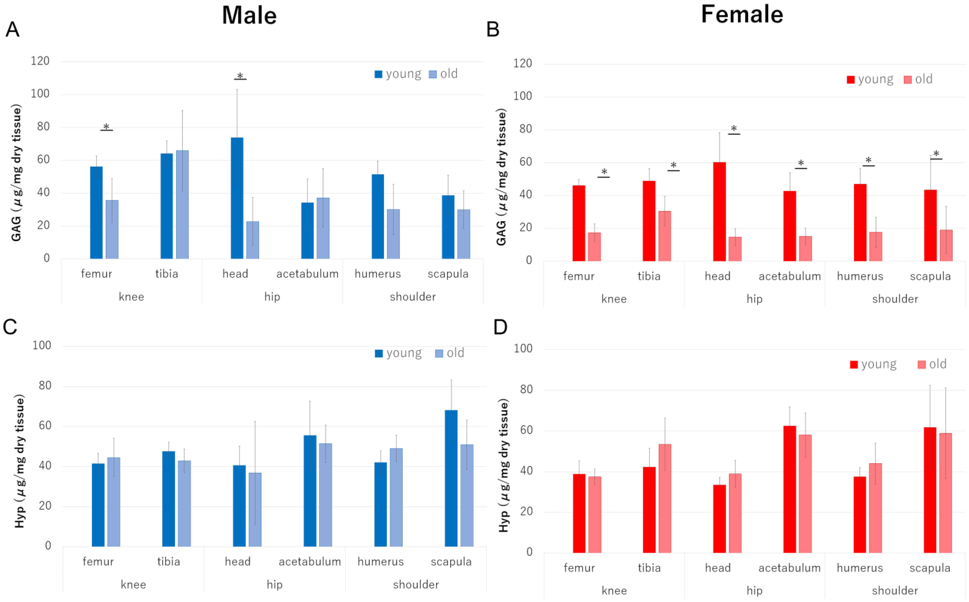
Comparison of multiple articular cartilage constituents (GAG and Hyp) between young and old rats. (
The decrease in GAG content with aging in all ACs was more pronounced in females than in males (Fig. 2A). In males, the amount of GAG did not decrease in the tibia and acetabulum, and it decreased by approximately 40% in the femur and humerus. In females, the decrease was approximately 60% in the femur, tibia, humerus, and scapula. The decrease at the head was approximately 75% and 70% in females and males, respectively. In contrast, the amount of Hyp did not decrease by more than 35% in males and females (Fig. 2B).
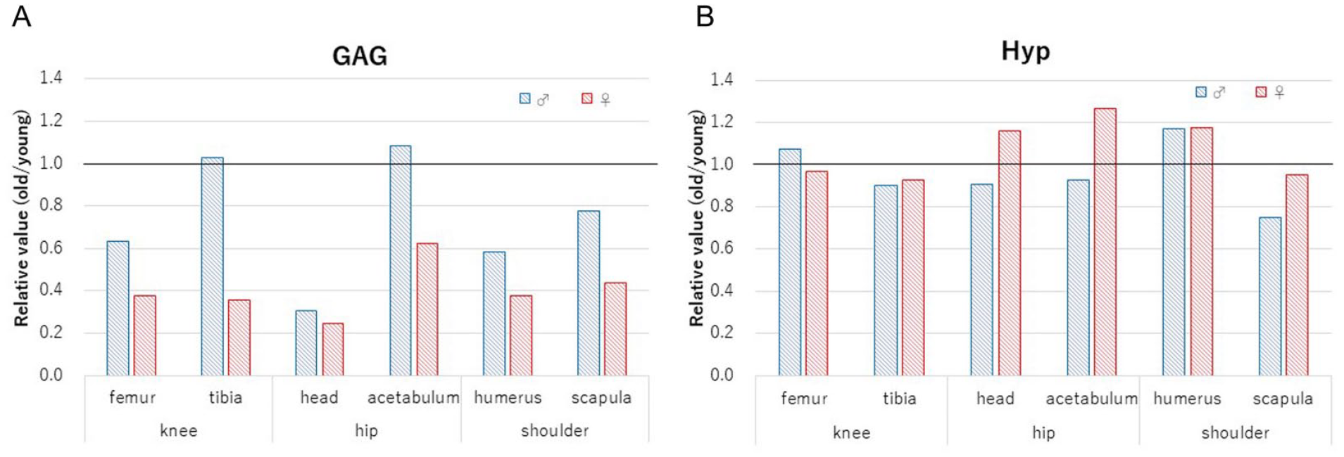
Relative value of the amount in the old based on the amount in the young. The relative value (old/young) was shown in both males and females. (
The amounts of GAG and Hyp were compared between the sexes (Fig. 3). In young knee joints, the amounts of GAG were significantly less in females than in males (femur P = 0.024, tibia P = 0.024) (Fig. 3A). In other young joints, the amounts of GAG were not significantly different between the sexes (head P = 0.53, acetabulum P = 0.42, humerus P = 0.53, and scapula P = 0.93) (Fig. 3A). In the old joints, significant differences were observed at the femur, tibia, and acetabular ACs (P = 0.010, 0.0051, and 0.0025, respectively) (Fig. 3B). In the other old ACs, no significant differences were observed (head P = 0.34, humerus P = 0.11, scapula P = 0.11) (Fig. 3B). In all the ACs, no significant differences in the amount of Hyp between sexes were observed in the femur (young, P = 0.78; old, P = 0.27), tibia (young, P = 0.32; old, P = 0.15), head (young, P = 0.32; old, P = 0.11), acetabulum (young P = 0.53, old P = 0.64), humerus (young, P = 0.16; old, P = 0.64), and scapula (young, P = 0.41; old, P = 0.64) (Fig. 3C and
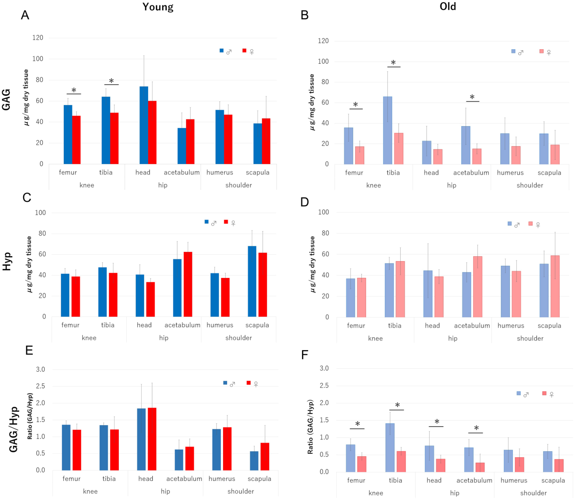
Comparison of multiple articular cartilage constituents (GAG and Hyp) between male and female rats. (
The amounts of GAG and Hyp in the ACs of the shoulder, hip, and knee joints of the young and old rats are shown in Table 1. Among the ACs from the protrusion and fossa sides in these joints of the same individual, the amounts of GAG and Hyp, as well as the GAG/Hyp ratio, were compared. Even in the young group, the amount of GAG varied among the ACs in the same male, and the amount of Hyp varied among the ACs in the same female. The GAG/Hyp ratio also varied significantly among the ACs in both young males and females.
The Amounts of GAG and Hyp, and the Ratio of GAG/Hyp in Young and Old, and Male and Female Rats.
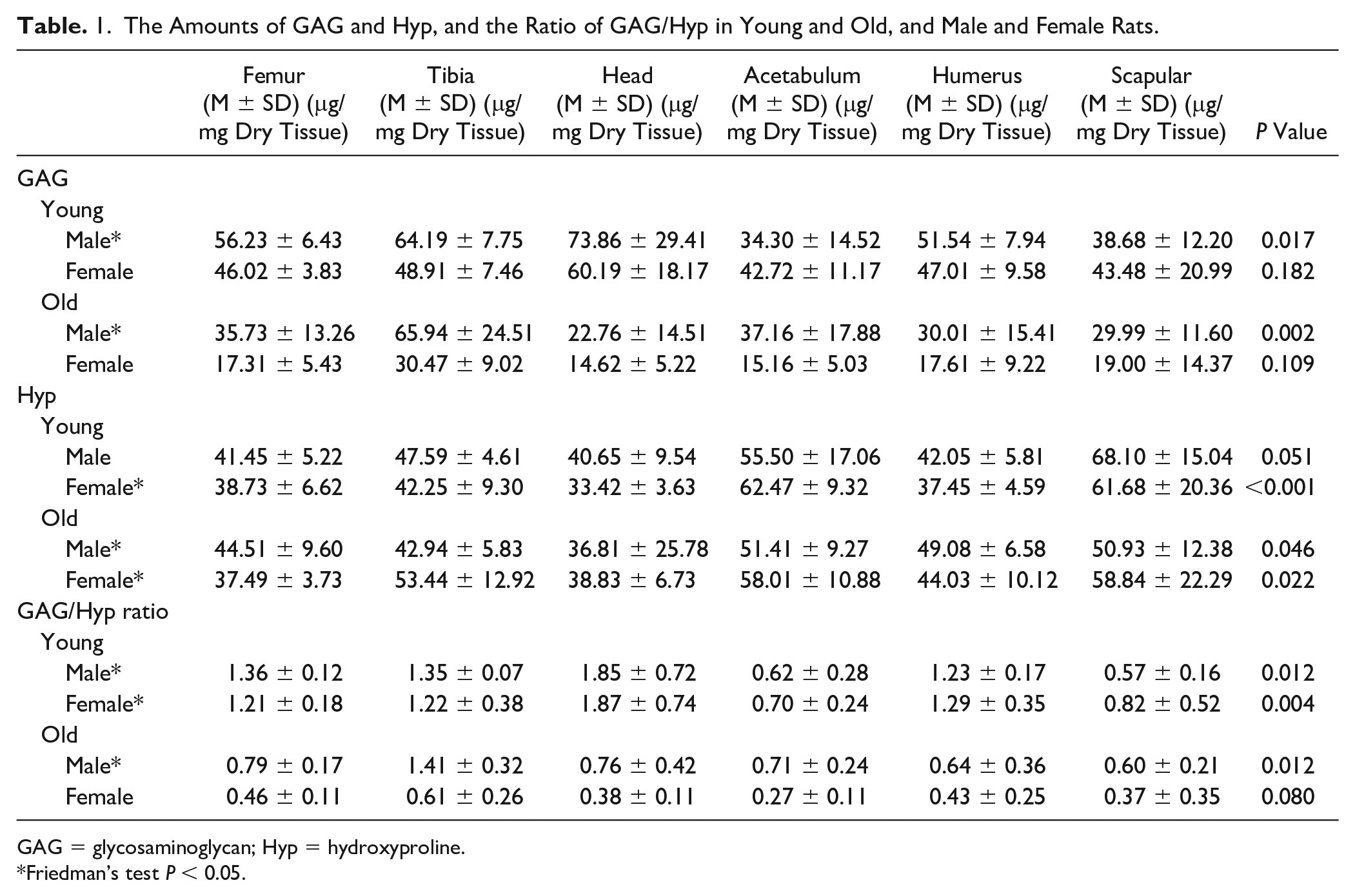
GAG = glycosaminoglycan; Hyp = hydroxyproline.
Friedman’s test P < 0.05.
Discussion
We indicated the decrease in GAG at multiple ACs with aging (Figs. 1A and
Generally, the effect of sex hormones is assumed to be the most likely cause of sex difference. As the estrogen treatment after menopause decreases the OA incidence, 25 estrogens may have a protective effect on AC to the progression to OA in elderly people. In contrast, a clear relationship between OA and estrogen use was not found in the Framingham knee OA study 26 and in a systematic analysis from an epidemiological database. 4 Thus far, it is unclear whether estrogen and OA incidence have a relationship. Therefore, the relationship between hormones and the decrease in GAG with aging is unclear.
The GAG decrease with aging occurred in various ACs (Fig. 1A and
As the OA prevalence is also different in each joint,28-30 the constituents (GAG and COL) in the young group between the sexes were compared (Fig. 3A and
In this study, we indicated that the decrease in GAG with aging in normal rats was greater in females than in males at all joints measured (Figs. 1A and
Footnotes
Acknowledgments and Funding
We would like to express special gratitude to Prof. M. Tsuchiya for useful advice to complete this study. We would also like to acknowledge Prof. T. Miura of Kyusyu University for technical advice, Enago (![]() ) for the English language review, and the Interdisciplinary Center for Science Research, Organization for Research and Academic Information, Shimane University, for technical expertise. This work was supported by Grant-in-Aid for Scientists (C) of Scientific Research (KAKEN), Japan (grant nos 25462371, 16K01821); Grant-in-Aid for Encouragement of Scientists of Scientific Research (KAKEN), Japan (grant nos 25930009, 26930007, 15H00583); and “Shimane University Grants for Joint Research Project led by Female Researchers” under the MEXT “Initiative for Realizing Diversity in the Research Environment (Collaboration Type).”
) for the English language review, and the Interdisciplinary Center for Science Research, Organization for Research and Academic Information, Shimane University, for technical expertise. This work was supported by Grant-in-Aid for Scientists (C) of Scientific Research (KAKEN), Japan (grant nos 25462371, 16K01821); Grant-in-Aid for Encouragement of Scientists of Scientific Research (KAKEN), Japan (grant nos 25930009, 26930007, 15H00583); and “Shimane University Grants for Joint Research Project led by Female Researchers” under the MEXT “Initiative for Realizing Diversity in the Research Environment (Collaboration Type).”
Declaration of Conflicting Interests
The author(s) declared no potential conflicts of interest with respect to the research, authorship, and/or publication of this article.
Ethical Approval
This study was performed with permission from the Animal Research Committee at Shimane University (IZ24-98 and IZ27-125).