
Abstract
Objective
Osteoarthritis is a painful, chronic joint disease affecting man and animals with no known curative therapies. Palliative nonsteroidal anti-inflammatory drugs (NSAIDs) are commonly used but they cause adverse side effects prompting the search for safer alternatives. To address this need, we evaluated the anti-inflammatory activity of avocado/soybean unsaponifiables (ASU), glucosamine (GLU), and chondroitin sulfate (CS) with or without the NSAID carprofen.
Design
Canine chondrocytes were propagated in microcarrier spinner culture and incubated with (1) control medium, (2) ASU (8.3 µg/mL) + GLU (11 µg/mL) + CS (20 µg/mL) combination for 24 hours; and/or carprofen (40 ng/mL). Cultures were next incubated with control medium alone or IL-1β (10 ng/mL) for another 24 hours. Production of PGE2, IL-6, IL-8, and MCP-1 (also known as CCL-2) were measured by ELISA.
Results
Chondrocytes proliferated in microcarrier spinner culture and produced type II collagen and aggrecan. Stimulation with IL-1β induced significant increases in PGE2, IL-6, IL-8, and MCP-1 production. The increases in production were suppressed by carprofen as well as [ASU+GLU+CS]. The combination of carprofen and [ASU+GLU+CS] reduced PGE2 production significantly more than either preparation alone. The inhibitory effect of carprofen on IL-6, IL-8, and MCP-1 production was significantly less than that of [ASU+GLU+CS], whereas the combination did not reduce the production of these molecules significantly more than [ASU+GLU+CS] alone.
Conclusions
The potentiating effect of [ASU+GLU+CS] on low-dose carprofen was identified in chondrocyte microcarrier spinner cultures. Our results suggest that the combination of low-dose NSAIDs like carprofen with [ASU+GLU+CS] could offer a safe, effective management for joint pain.
Introduction
Osteoarthritis (OA) is a debilitating joint disease affecting humans and animals. 1 In dogs, OA is a common orthopedic problem particularly in large breeds. 2 The pain and immobility associated with OA have been attributed to an imbalance of anabolic and catabolic processes resulting in the destruction of articular cartilage and subchondral bone remodeling. A prominent biological feature of OA is the overproduction of pro-inflammatory molecules including PGE2, cytokines, chemokines, nitric oxide, and reactive oxygen species, all of which are produced by joint tissue cells in cartilage, synovium, subchondral bone, tendon, and ligament.3-11 PGE2 is known to activate degradative enzymes capable of breaking down cartilage. 5 It has also been shown to sensitize pain afferents innervating the joint.8-12 In addition to PGE2, inflammatory molecules IL-6, IL-8, and MCP-1 have also been reported to play a role in the pathogenesis of OA by inducing pain and activating matrix metalloproteinase (MMP) enzymes that degrade cartilage.6-19 Inhibition of PGE2 synthesis is the conventional treatment regimen for controlling inflammation and pain in OA.20-23 The efficacy of nonsteroidal anti-inflammatory drugs (NSAIDs) in the treatment of OA strongly correlates with their ability to inhibit PGE2 production. However, long-term use of NSAIDs at higher doses is associated with serious gastrointestinal, renal, and hepatic side effects.24,25 There has been much speculation of whether the toxicity of NSAIDs can be reduced without affecting their therapeutic efficacy through a combination with other agents with known anti-inflammatory properties.26,27 The possibility of reducing the side effects of NSAIDs through combination therapies has been explored.28-30 In a clinical study in dogs with OA, Fritsch et al. found that the effect of carprofen can be enhanced when combined with dietary supplement omega-3 fatty acids. 26 Their finding supports the notion that a combination therapy may offer an advantage over the use of carprofen alone.
Cell-based screening assays are particularly useful to identify agents that may allow reduction in NSAID dosage. We and others have previously characterized a chondrocyte microcarrier spinner culture system that simulates features of the cartilage environment in the joint.31-36 Chondrocytes propagated in microcarrier spinner cultures continued the production of type II collagen, high-molecular-weight aggrecan, and exhibited behavior similar to that of their counterpart cartilage. The present study used chondrocyte microcarrier cultures stimulated with cytokine IL-1β to induce production of inflammatory biomarkers: PGE2, IL-6, IL-8, and MCP-1. These key inflammatory molecules have been documented to play significant roles in the pathogenesis of OA. The inhibitory activity on their production by carprofen and [ASU+GLU+ CS] (avocado/soybean unsaponifiables [ASU], glucosamine [GLU], and chondroitin sulfate [CS]) were then measured. The aim of this study is to determine the anti-inflammatory effect of an NSAID carprofen used in the conventional treatment regimen, and a nonpharmacologic [ASU+GLU+CS] for controlling inflammation and pain in OA.
Using our dynamic cell-based assay, we evaluated the anti-inflammatory activity of carprofen and [ASU+GLU+CS]. Carprofen is an NSAID most commonly used in the treatment of osteoarthritic dogs.20-23 The compounds ASU, GLU, and CS, separately or in combination, have been documented to reduce inflammation in vitro and in vivo in man and animals.37-59 ASU, GLU, and CS have also been reported to be beneficial in the management of OA without adverse side effects.48-59
Methods
Propagation and Characterization of Canine Monolayer and Microcarrier Cultures
Chondrocytes were isolated from healthy looking articular cartilage of 4 beagle dogs (Celsis Laboratory Group, Baltimore, MD) as previously described. 45 Cells were first propagated in monolayer for up to 3 passages in chondrocyte medium (Cell Applications Inc., San Diego, CA) and were subsequently seeded (4 × 10⁶ cells/flask) into cross-linked microcarriers derived from bovine type I (Advanced Biomatrix, San Diego, CA). The microcarrier spinner cultures were spun at intermittent spin cycle (30 minutes active and 2 hours static, 30 rpm, 37°C, 5% CO2) for 24 hours. Flasks were filled up to 120 mL volume and then spun at 60 rpm for 7 days. Chondrocyte seeded microcarriers were analyzed by phase-contrast microscopy, scanning electron microscopy (SEM), and transmission electron microscopy (TEM) (Johns Hopkins School of Medicine Cell Imaging Facility, Baltimore. MD). Viability was determined using Trypan blue dye uptake. 31 Cells used in experiments had 100% viability.
Phenotype Analyses of Collagen and Aggrecan: Immunofluorescence and Western Blot
Chondrocytes (1 × 10⁴ cells/well) seeded onto 8-well chambered slides were fixed with 10% v/v formalin and were double stained for type I and type II collagen using a cocktail containing a goat anti-type I antibody (1:500, Southern Biotechnology Associates, Birmingham, AL) and a mouse monoclonal anti-type II antibody (1:50, Calbiochem, La Jolla, CA) as detailed by Heinecke et al. 45 Aggrecan was immunostained with mouse monoclonal anti-aggrecan as previously described (1:20, U.S. Biological, Swampscott, MA). 45 Cell nuclei were counterstained with DAPI for 30 minutes. Staining was analyzed using epifluorescent microscopy and digital images were captured with a camera (Epifluorescent TE200 microscope, Spot Camera, Nikon, Melville, NY).
For Western Blot analysis, culture supernatants were (20 µg protein) run on 4 to 15 minigels for 1 hour at 200 V in 1× Tris/glycine/sodium dodecyl sulfate buffer and transferred to polyvinylidene fluoride in buffer containing 2.5% v/v methanol, for 30 minutes 100 V. Nonspecific binding was minimized using a commercial blocking solution before incubation overnight with either mouse anti-type II or goat anti-type I collagen. Membranes were washed and then incubated with either horseradish peroxidase-conjugated donkey anti-mouse, or rabbit anti-goat IgG antibody (1:50,000) for 1 hour. Membranes were soaked in chemiluminescent detection reagent and then exposed to the film in the dark for 2 minutes.
Titration of Carprofen Anti-Inflammatory Activity Using Chondrocyte Monolayer Cultures
The dose titration was performed using monolayer cultures. This in vitro method is routinely used for dose titrations of various compounds due to its ease of use. The goal of the titration studies was to identify a concentration of carprofen that yields only partial inhibition of PGE2 production, preferably less than 50% inhibition. This allows detection of further inhibitory effects with combinations products. Chondrocytes (1 × 10 cells 5 ) were plated in monolayer culture for 24 hours at 37°C, 5% CO2, and then stimulated with IL-1β (10 ng/mL) for another 24 hours. PGE2 was quantitated in supernatant fluids as described below.
Treatment Procedures
Chondrocyte seeded microcarrier spinner cultures (10 mL) were pre-incubated with control media alone (chondrocyte media, Cell Applications Inc., San Diego, CA) [ASU (8.3 µg/mL, NMX1000, Nutramax Laboratories, Inc., Edgewood, MD, USA) + GLU (11 µg/mL (FCHG49, Nutramax Laboratories, Inc.) + CS (20 µg/mL, TRH122, Nutramax Laboratories, Inc.], carprofen (40 ng/mL, Pfizer, Groton), or the combination of [ASU+GLU+CS] and carprofen for 24 hours at 37°C, 5% CO2. The cultures were next incubated with control media alone, or stimulated with IL-1β (10 ng/mL) for another 24 hours. The concentrations of ASU, CS, and GLU were selected based on their reported anti-inflammatory effect in vitro and on clinically relevant doses used in human and canine studies.37-59 The clinically relevant doses used in osteoarthritic dogs and man were reported to relieve pain and discomfort and within the range of what could be available in the joint.37-59 Clinically relevant concentrations of carprofen used were based on previous in vivo studies.20 -23
PGE2, IL-6, IL-8, and MCP-1 (CCL-2) Quantitation Using ELISA
PGE2, IL-6, IL-8, and MCP-1 from culture supernatant were quantified using immunoassay kits (R&D System, Minneapolis, MN), and optical density was measured using the SpectraMAX 340 microplate reader (Molecular Devices, Sunnyvale, CA, USA) at 450 nm with wavelength correction at 540 nm.
Statistical Analysis
Pairwise multiple comparisons were carried out using one-way ANOVA, Tukey post hoc using Sigma Stat, Windows Version 3.11 (Systat Software Inc., Chicago IL), where P < 0.05 was considered statistically significant.
Results
Characterization of Canine Chondrocytes
Chondrocytes cultures in monolayer used for dose titrations studies were 100% viable and had doubling times ranging from 3 to 7 days. They displayed heterogeneous shapes with spheroid to elongated morphology. Immunofluorescence analysis of passage 3 monolayer cultures showed a mixed population with some chondrocytes stained solely for type II collagen, some only stained for type I collagen, while others stained for both type I and type II collagen ( Fig. 1A ). Chondrocytes also stained positive for aggrecan ( Fig. 1B ). These monolayer cultures were used for ease and reliable identification of carprofen concentration to be used in combination studies.
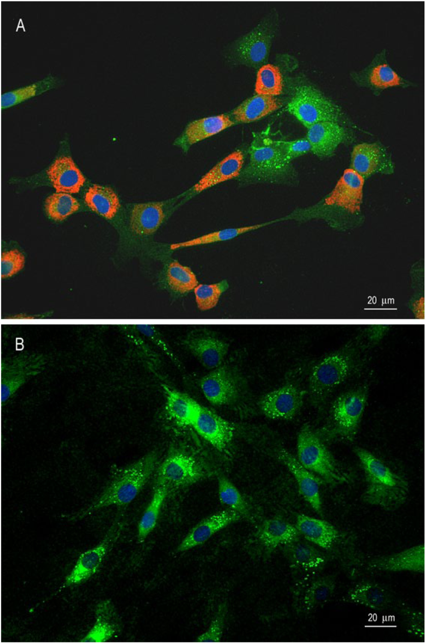
Immunofluorescent staining of monolayer chondrocyte cultures (passage 2) for type I collagen, type II collagen, and aggrecan. (
Chondrocytes in microcarrier cultures were 100% viable and proliferated with doubling times of ~3 to 4 days ( Fig. 2 ). Chondrocyte-seeded microcarriers produced extracellular matrix (ECM) materials and formed aggregates over a period of 7 days indicating metabolic activity ( Fig. 2A and B ). SEM showed refractile viable chondrocytes with well-defined intact structures on the surface of microcarrier ( Fig. 2C ). TEM showed a chondrocyte with intricate cytoplasmic extensions infiltrating the microcarrier surfaces ( Fig. 2D ). Western blot analysis of culture supernatants retrieved from 7 day microcarrier cultures confirmed production of type II collagen protein in all 4 chondrocyte lines ( Fig. 3A-D ). There was negligible production of type I collagen in these microcarrier spinner cultures. As we have reported earlier, aggrecan could not be analyzed by Western blot due to its very high molecular weight. 32 The expected goal of maintaining phenotypic integrity with continued production of type II collagen was achieved in chondrocyte microcarrier cultures.31-36
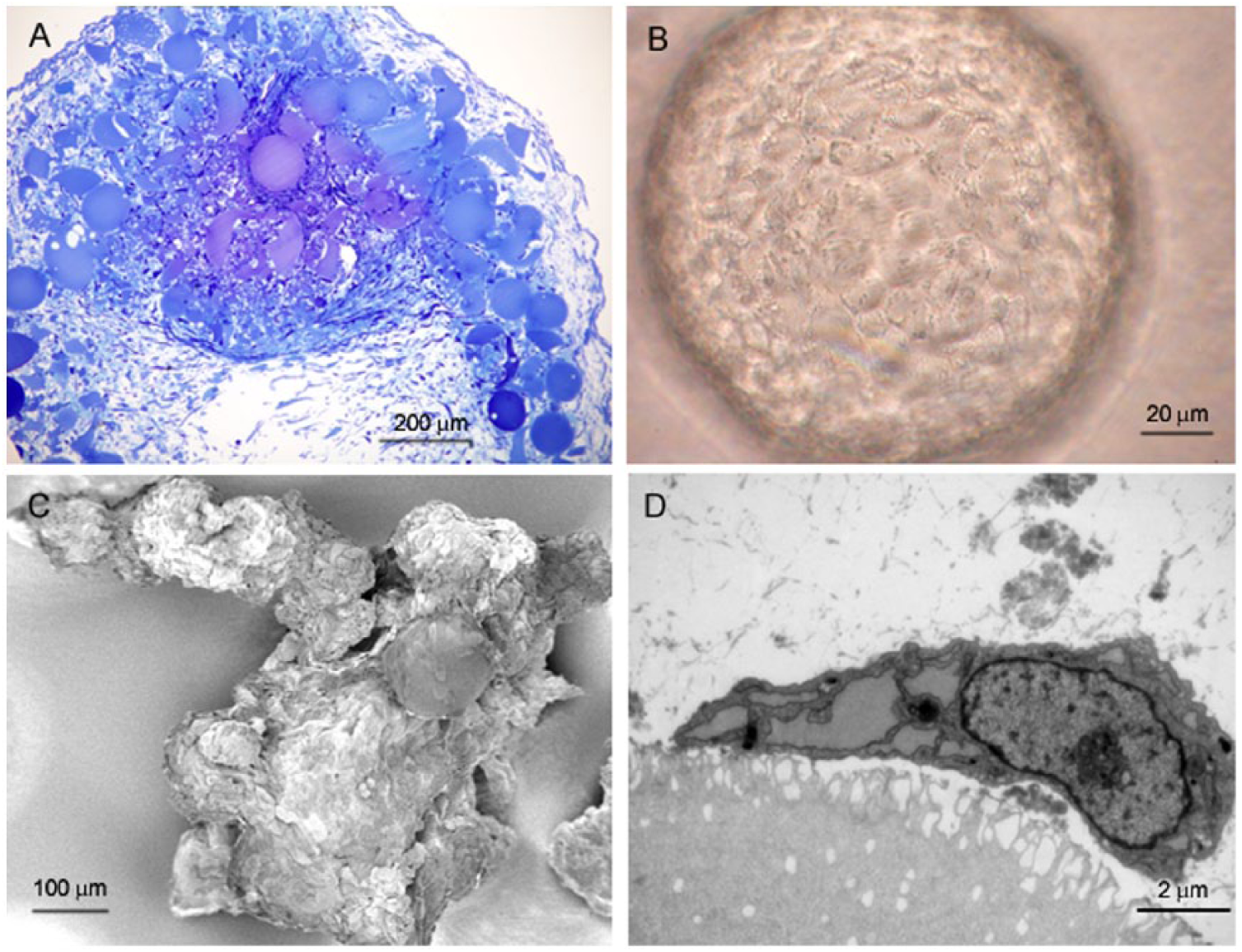
Morphological analyses of chondrocyte-seeded microcarrier 7 day cultures. (
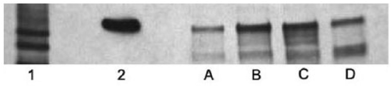
Western blot of type II collagen from chondrocyte microcarrier spinner culture supernatant. Lane
IL-1β Stimulation of PGE2 Production in Chondrocyte Monolayer and Microcarrier Cultures
Chondrocytes stimulated with IL-1β significantly increased PGE2 compared to nonstimulated control cells in monolayer and microcarrier cultures (P < 0.001; Figs. 4 and 5 ). IL-1β stimulated chondrocytes in microcarrier spinner cultures produced up to 200-fold greater amounts of PGE2 compared to nonstimulated controls incubated in media alone ( Fig. 5 ).
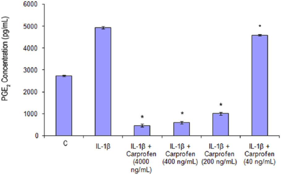
Dose-response effect of carprofen (4,000-40 ng/mL) on PGE2 production in IL-1β-stimulated chondrocyte monolayer cultures. PGE2 levels were measured in cell culture supernatant by ELISA. Results are presented as mean ±1 SD, n = 3. *Denotes significant (P < 0.001) differences between the treatments IL-1β alone and combination of IL-1β + carprofen.
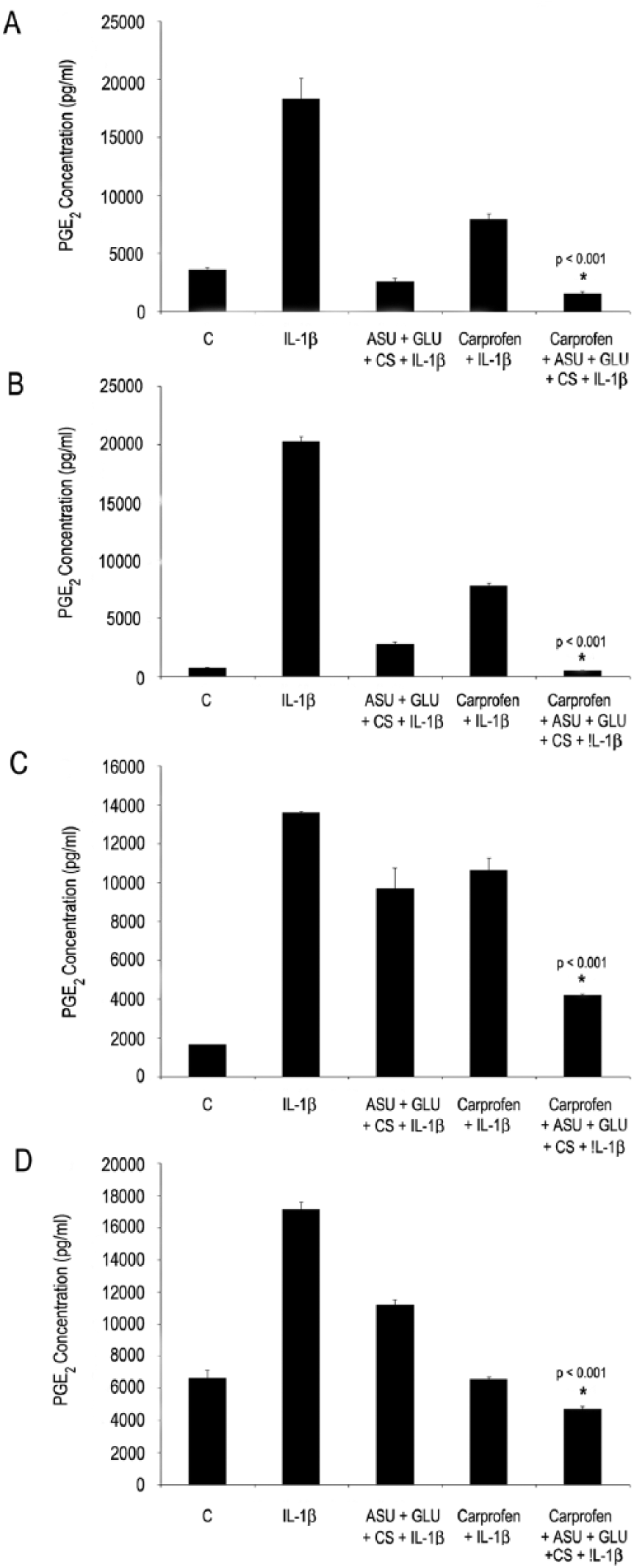
Effect of [ASU+GLU+CS] and carprofen on PGE2 production in IL-1β-stimulated chondrocyte microcarrier spinner cultures. PGE2 levels were measured in cell culture supernatant by ELISA. Results are presented as mean ± 1SD, n = 3. *Denotes significant (P < 0.001) differences between the combination treatment (IL-1β + carprofen + [ASU+GLU+CS]) and (IL-1B + carprofen alone) or (IL-1β + [ASU+GLU+CS] alone). (
Dose Titration of Carprofen in IL-1β-Stimulated Chondrocyte Monolayer Cultures
To determine concentrations of carprofen that would partially but not completely suppress PGE2 production for combination studies, a dose-response titration was performed including levels of carprofen detectable in blood following a therapeutic dose. The inhibition of IL-1β-induced PGE2 production by carprofen was concentration dependent ( Fig. 4 ). Clinically relevant concentrations of carprofen (4,000 to 200 ng/mL) significantly reduced PGE2 production down to baseline levels. These concentration ranges were detectable after oral carprofen dosing of 2 to 4 mg/kg in dogs.12,20-22 The finding that IL-1β less than doubled PGE2 production suggests that chondrocytes may have been previously stimulated possibly in vivo and were no longer as responsive to further stimulation. That carprofen at 200, 400, and 4,000 ng/mL significantly reduced PGE2 levels lower than the baseline negative control reinforces the therapeutic efficacy of this range of concentrations. The low concentration of NSAID at 40 ng/mL had a small inhibitory effect on PGE2 production and was selected for subsequent combination studies in microcarrier spinner culture.
Inhibition of IL-1β-Induced PGE2, IL-6, IL-8, and MCP-1 Production in Chondrocyte Microcarrier Cultures
[ASU+GLU+CS] treatment significantly reduced IL-1β-induced PGE2 production in all 4 chondrocyte lines (P < 0.001; Fig. 5A-D ). PGE2 production with [ASU+GLU+CS] treatment decreased down to ~15% to 61% of IL-1β-induced PGE2 levels ( Fig. 5A-D ). Carprofen (40 ng/mL) treatment also significantly reduced IL-1β-induced PGE2 production in all 4 chondrocyte lines (P < 0.001; Fig. 5A-D ). In comparison PGE2 production with carprofen treatment decreased down to 31% to 78% of IL-1β-induced PGE2 levels. The [ASU+GLU+CS] and carprofen combination significantly inhibited PGE2 production compared to [ASU+GLU+CS] (P < 0.001) alone, or carprofen alone (P < 0.001). Chondrocytes cell lines used were from different dogs, at different cell passages with possibly different PGE2 production rates, which could contribute to the observed variability.
Nonstimulated chondrocyte cultures produced detectable amounts of IL-8 and MCP-1 but not IL-6 ( Fig. 6A-C ). IL-1β stimulation significantly increased production of IL-6, IL-8, and MCP-1 ( Fig. 6A-C ; P < 0.001). [ASU+GLU+CS] treatment inhibited production of IL-6 down to ~6%, IL-8 down to ~24%, and MCP-1 down to ~17% of IL-1β induced levels (P < 0.001; Fig. 6A-D ). Carprofen at 40 ng/mL treatment reduced production of IL-6 down to ~25%, IL-8 down to ~62%, and MCP-1 down to ~39% IL-1β induced levels (P < 0.001; Fig. 6A-D ). The combination of [ASU+GLU+CS] and carprofen significantly reduced the production of IL-6, IL-8, and MCP-1 greater than that of carprofen alone (P < 0.001). However, the inhibitory effect of the combination was not greater than the reduction from treatment with [ASU+GLU+CS], P > 0.05.
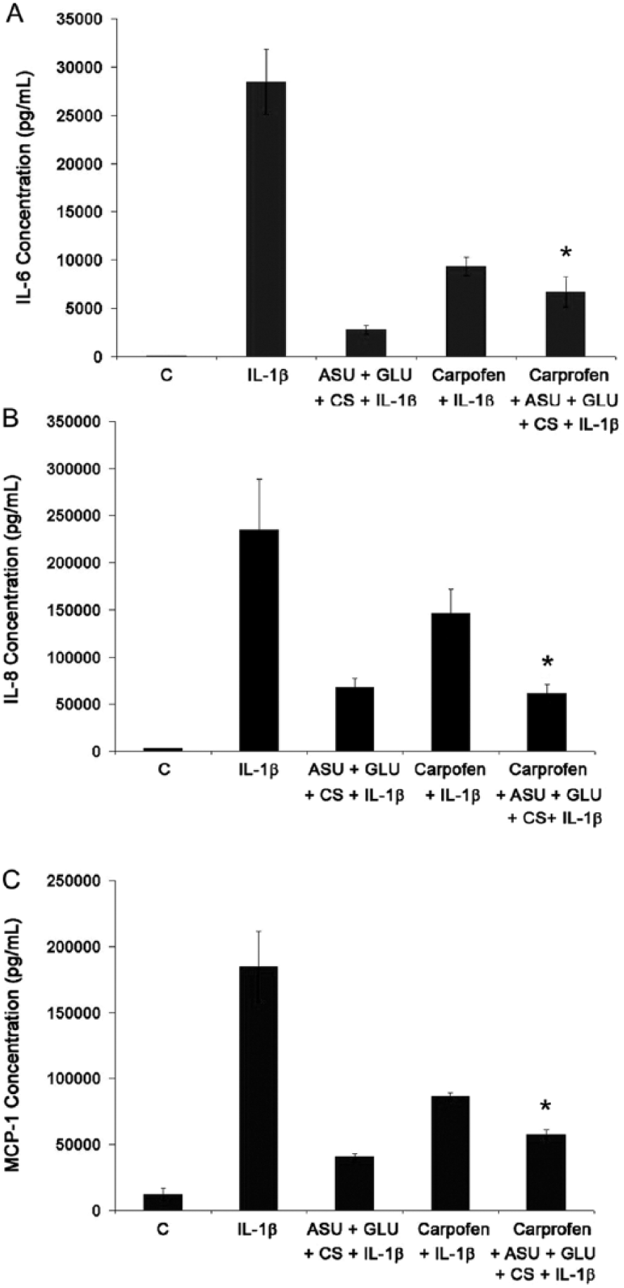
Effect of [ASU+GLU+CS] and carprofen on IL-6, IL-8, and MCP-1 production in IL-1β-stimulated chondrocyte microcarrier spinner cultures. IL-6, IL-8, and MCP-1 levels were measured in cell culture supernatant by ELISA. Results are presented as mean ± 1 SD, n = 3. *Denotes significant (P < 0.001) differences between the combination treatment (IL-1β + carprofen + [ASU+GLU+CS]) and (IL-1 β + carprofen alone). (
Discussion
The present study demonstrates that the microcarrier spinner culture can be used to propagate canine chondrocytes with ease as we and others have shown previously for human chondrocytes.31-36 Chondrocytes are exposed to conditions in microcarrier spinner cultures that resemble more closely the biomechanical conditions present in joints.31-36 Similar to earlier observations, chondrocytes propagated for several passages in monolayer culture can be subsequently grown in microcarrier spinner culture, remain viable, and continue to produce type II collagen as well as aggrecan ( Figs. 1 - 3 ).31-36 In contrast, chondrocytes propagated in monolayer culture undergo phenotypic changes and shift to producing type I collagen.60,61 Their responsiveness to stimuli is reflected in increased PGE2, IL-6, IL-8, and MCP-1 (CCL-2) production following exposure to IL-1β ( Figs. 4 - 6 ). Overproduction of these molecules in osteoarthritic joints is associated with inflammation and pain.3-11
As a disease-relevant cell-based assay, chondrocyte microcarrier spinner cultures facilitate testing anti-inflammatory drugs and nonpharmacologic products alone and in combination. The principal finding of this study is that carprofen or [ASU+GLU+CS] significantly inhibited PGE2 production while the inhibitory effect of the combination was significantly more than either agent alone ( Fig. 5 ). In contrast, the inhibitory effect of carprofen on IL-6, IL-8, and MCP-1 production is significantly less than [ASU+GLU+CS], while the combination did not significantly reduce production of these molecules more than either agent alone ( Fig. 6 ). Inhibiting IL-6, IL-8, and MCP-1 production has important clinical implication as their accumulated levels in the joint have been correlated to radiographic grading and pain-related scores in OA.13-16 PGE2, IL-6, IL-8, and MCP-1 pro-inflammatory molecules have been documented to mediate joint pain through interaction with nociceptors.6-19 IL-6 is suspected to contribute to OA pathology through increasing the number of inflammatory cells in synovial tissue and through amplifying the effects of IL-1β on MMP synthesis. Increased synovial fluid levels of IL-6 has been proposed as a predictor of knee OA.13-16 Chemokines IL-8 and MCP-1 have been identified as signaling molecules in addition to their well-known chemotactic function.6-8,17-19 Upregulated cell receptors for IL-6, IL-8, and MCP-1 in joints are thought to trigger production and activation of cartilage degradative enzymes and are thought to play a role in the pathogenesis of OA.6-19,62 Reduction of PGE2, IL-6, IL-8, and MCP-1 production in the joint is considered critically important in attenuating the pathogenesis of OA.6-19,62
Earlier studies showed that the anti-inflammatory effect of ASU alone or in combination with other compounds is associated with inhibition of COX-2, the molecular target of carprofen.38,45 It is well documented that NSAIDs like carprofen inhibit PGE2 synthesis and is the conventional treatment regimen for controlling inflammation and pain in OA.38,45 The production of PGE2 is regulated by COX-2, the molecular target of NSAIDs like carprofen.
Assuming that reductions in these markers are achieved via the same mechanisms, it is possible that a maximum decrease in production levels of IL-6, IL-8, and MCP-1 had already been achieved, similar to a floor effect. Alternatively, the combined effect of carprofen and [ASU+GLU+CS] suggests the involvement of targets different from those affected by carprofen alone ( Fig. 6 ). This may also help explain the observation that the combination significantly potentiated suppression of PGE2 production but not that of IL-6, IL-8, and MCP-1.
In a recent study, a therapeutic concentration of another NSAID, phenylbutazone, significantly reduced PGE2 but not IL-6, IL-8, and MCP-1 production in stimulated chondrocytes. 63 In contrast, [ASU+GLU+CS] significantly reduced production of these inflammatory molecules suggesting mechanisms different from phenylbutazone. 63
The observation that the combination of carprofen and [ASU+GLU+CS] reduced PGE2 production significantly more than either treatment alone suggests that the same clinical efficacy may be achieved in a clinical setting with a lower dose of carprofen, below therapeutic doses. It is also possible that the low-dose carprofen has a potentiating effect on [ASU+GLU+CS] in inhibiting PGE2 production. The results are in line with the observed enhanced effect of carprofen when combined with omega-3 fatty acid in alleviating OA severity in dogs. 26 Considering the multiple effects of omega-3 fatty acids on cell functions, it is difficult to provide a mechanistic explanation for the observed effect. 64 Fritsch suggested that combination with omega-3 fatty acids supplementation could allow reduced carprofen dosage. 26 Similarly, our finding that [ASU+GLU+CS] potentiated the anti-inflammatory effect of a low concentration of carprofen suggests the benefit of a combination approach in the management of OA. In addition, the anti-inflammatory effect of NSAIDs is relatively quick compared to slower acting [ASU+GLU+CS], thus providing the benefit of covering the time gap for activity.
In conclusion, this study provides evidence that the combination of low-dose carprofen and [ASU+GLU+CS] can inhibit PGE2, IL-6, IL-8, and MCP-1 production. A lower dose of NSAID together with nonpharmacologic [ASU+GLU+CS] may promote palliative efficacy while minimizing adverse side effects. Attenuating multiple pathways leading to inflammation and joint destruction may facilitate a safe, effective management of OA. The transferability of our in vitro data using the canine chondrocyte OA model to the in vivo settings needs further evaluation in vivo.
Footnotes
Acknowledgments and Funding
We would like to thank Dr. Reinhard Grzanna for designing the immunohistochemistry and Western blot experiments as well as for constructive review of the article.
Declaration of Conflicting Interests
The author(s) declared the following potential conflicts of interest with respect to the research, authorship, and/or publication of this article: Mark W. Grzanna, Lowella V. Fortuno, Angela Y. Au, and Carmelita G. Frondoza are former employees of Nutramax Laboratories, Inc., but do not hold stocks or royalties. Erica J. Secor has no conflict of interest.
Ethics Approval
Ethical approval was not sought for the present study because the study used canine tissues purchased from Celsis Laboratory Group, Baltimore, MD.
Animal Welfare
Guidelines for humane animal treatment did not apply to the present study because the study used canine tissues purchased from Celsis Laboratory Group, Baltimore, MD.