
Abstract
Objective
The effects of hydrostatic pressure (HP) on the matrix synthesis by human articular chondrocytes have been reported elsewhere. In order to optimize the production of extracellular matrix, we aimed to clarify the effects of repetitive HP on metabolic function by human articular chondrocytes.
Design
The human articular chondrocytes were expanded and embedded within a collagen gel/sponge scaffold. We incubated these constructs with and without HP followed by atmospheric pressure (AP) and repeated the second HP followed by AP over 14 days. Genomic, biochemical, and histological evaluation were performed to compare the effects of each regimen on the constructs.
Results
The gene expressions of collagen type II and aggrecan core protein were significantly upregulated with repetitive HP regimens compared with a single HP or AP by 14 days (P < 0.01 or 0.05). Matrix metalloptoteinase-13 (MMP-13) in AP was upregulated significantly compared to other HP regimens at day 14 (P < 0.01). No significant difference was observed in tissue inhibitor of metalloproteinases-II. Immunohistology demonstrated that application of HP (both repetitive and single) promoted the accumulation of specific extracellular matrix and reduced a MMP-13. A single regimen of HP followed by AP significantly increased the amount of sulfated glycosaminoglycan than that of the AP, whereas repetitive HP remained similar level of that of the AP.
Conclusions
Repetitive HP had a greater effect on anabolic activity by chondrocytes than a single HP regimen, which will be advantageous for producing a matrix-rich cell construct.
Introduction
A cell-based therapy has been used to promote cartilage repair in articular cartilage or in osteochondral defects. 1 In particular, autologous chondrocyte implantation (ACI) has been commercially available for more than 2 decades. More than 20,000 patients worldwide have been treated with ACI as the first tissue-engineering product.1-3 However, ACI requires a complex surgical procedure that would be improved with a cell construct, for example, matrix-assisted chondrocyte implantation, 4 chondrocyte collagen gel construct, 5 and mesenchymal stem cells. 6
We have developed a platform technology to manufacture the autologous chondrocyte/gel/sponge construct with a treatment of cyclic hydrostatic pressure (HP) to promote the accumulation of cartilage extracellular matrix (ECM) by human articular chondrocytes (hACs).7,8 This cell construct is composed of a 3-dimensional (3D) uniform cell distribution and a substantial amount of newly synthesized ECM. Thus, the HP-loading was expected to instruct cultured hACs to recover their chondrocyte characteristics at the weightbearing site in articular cartilage.9,10
We previously demonstrated that HP-loading followed by atmospheric pressure (HP-AP) was an effective regimen for promoting the expression of anabolic genes and the accumulation of ECM by middle zone–derived bovine articular chondrocytes (bACs) seeded in collagen gel. 11 Recently, various cell culture technologies using mechanical stimuli have been developed to engineer chondrocyte constructs and are well-documented elsewhere.12,13 However, the effects of HP have been studied at various magnitudes, frequencies, regimen, and durations by other investigators using their custom-designed cell culture systems (bioreactors), and expedient monolayer, construct, and explant model.13-18 Particularly, we thought that rationale of the regimen of HP was unclear. It was very important information to manipulate metabolic turn over by chondrocytes with HP for engineering cartilage. Previous studies demonstrated that intermittent HP-loading (1-10 MPa, 0.5-1 Hz, 4 hours per day for 4 days) increased Col-2 and Agg expressions by monolayer cultures of bACs or hACs compared with a single HP-loading.17-20 A different study reported a significant increase of 35-sulfate incorporation into glycosaminoglycan in bovine cartilage explants due to cyclic HP at 5 MPa, 0.5 Hz for 1.5 hours, whereas significant inhibition was detected in monolayer chondrocytes. 21 Those results were not easily compared because of the various different culture conditions: shorter (1.5 hours) or longer (4 days) regimen of HP, intermittent or cyclic HP, and so on. We demonstrated that a 3-day loading regimen was appropriate to detect metabolic genes. 22 If this regimen was effective to alter anabolic gene expression, we hypothesized that a repetitive HP-AP regimen would have greater potential to promote chondrogenesis than a single HP-AP. We evaluated this regimen using our well-validated culture system23,24 with gene expressions for the anabolic, catabolic, and chondroprotective functions and their corresponding antibodies and accumulations of cartilage ECM by hACs isolated from full depth articular cartilage in middle-aged subjects.
Methods
Isolation and Expansion of Human Articular Chondrocytes
Discarded articular cartilage from 3 patients (40-62 years old) who underwent total knee replacement was obtained with the approval of an Institutional Review Board protocol (2013D002589) of Brigham and Women’s Hospital, Boston, Massachusetts. Pieces of the macroscopically intact cartilage (International Cartilage Repair Society grade 0) were harvested from the femoral condyle using a scalpel (No. 22, BD, Franklin Lakes, NJ), minced, and rinsed in phosphate-buffered saline (PBS, Life Technology, Carlsbad, CA). The cartilage pieces were then digested with 0.15% collagenase (CLS-1, Worthington, Lakewood, NJ) dissolved in Ham’s F12 medium (F-12) including 100 units/mL penicillin and 100 µg/mL streptomycin (Life Technology) at 37°C for 18 hours with gentle rotation. The isolated hACs were seeded to 100 mm cell culture dishes and subcultured in Dulbecco’s modified Eagle’s medium (DMEM)/Ham’s F12 medium with osmolarity ranging from 300 to 320 mOsm (1:1, Life Technology) including 1% insulin-transferrin-selenium (ITS, Life Technology), 10% fetal bovine serum, 100 units/mL penicillin and 100 µg/mL streptomycin at 37°C, 3% O2, and 5% CO2 in air. After 2 passages, the cells were harvested with 0.15% collagenase for experiments. Five hundred thousand cells per 40 µL were suspended in a cold neutralized 0.3% collagen type I solution (PureCol, Advanced BioMatrix, Carlsbad, CA). The cell suspension was placed on a low-adherent polystyrene dish (Fisher Scientific, Pittsburgh, PA). The cell suspension was contained within a desired area on the hydrophobic dish surface, subsequently absorbed by placing a honeycomb collagen sponge (6 mm in diameter, 2 mm thick; CSH-96, Koken, Tokyo, Japan) onto the suspension, and solidified at 37°C for 1 hour in an incubator (
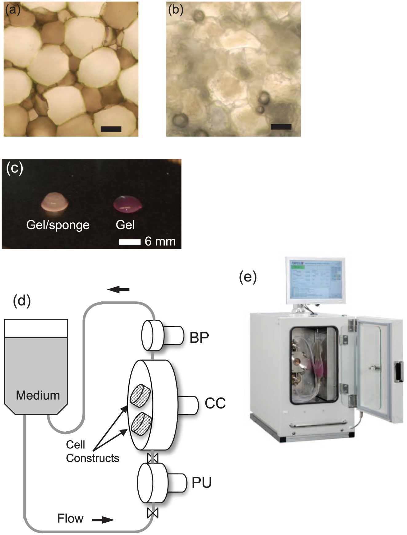
Human articular chondrocyte (hAC)/sponge construct and cell culture system. (
HP Loading Regimen
Sixteen cell constructs were divided into 4 groups: (1) Group HP-AP; HP-loading for the first 4 days with a treatment of cyclic HP at 0 to 0.5 MPa, 0.5 Hz followed by AP for 10 days; (2) Group AP-HP; AP for 7 days followed by HP-loading for 4 days and a second AP for the last 3 days; (3) Group repetitive HP-AP (Rep HP-AP); HP-loading for 4 days followed by AP for 3 days and the second HP-loading for 4 days followed by AP for the last 3 days; and (4) Group AP; AP for 14 days throughout the experiment (
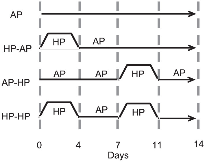
The loading regimen of application of HP and AP. HP, hydrostatic pressure; AP, atmospheric pressure.
Gene Expression Profiles with Quantitative Polymerase Chain Reaction
To produce gene expression profiles, collected samples (one construct in each group at each time point) were washed with D-PBS, and total RNA was extracted with TRIzol Reagent (Life Technology) following the manufacturer’s instructions. Briefly, the samples were homogenized using a pellet pestle (Kimble-Chase, Thermo-Fisher) and mixed with 200 µL chloroform and were shaken for 15 seconds. They were then centrifuged at 14,000 rpm for 15 minutes at 4°C. After centrifugation, approximately 400 µL of the clear aqueous phase was transferred to a new microfuge tube, and 400 µL of 100% isopropanol was added to precipitate RNA. The solution was left to settle down for 10 minutes and was then centrifuged at 14,000 rpm for 15 minutes at 4°C. After removing the supernatant, the RNA pellet was washed with 1 mL of cold 75% ethanol and centrifuged at 12,000 rpm for 5 minutes at 4°C. After removing the new supernatant, the pellet was incubated at 55°C to allow it to loosen and was then suspended in 30 μL of RNA free water (Invitrogen). Total RNA was measured with a fluorescent-tag and microfluidic capillary electrophoresis (Bioanalyzer, Agilent Technologies, Santa Clara, CA). The samples and fluorescent-labeled specific primers (TaqMan, Life Technology) were mixed for use in quantitative-PCR (7900HT, Applied Biosystems, Foster City, CA) using TaqMan primers: Col-2: Hs00264051_m1 (all TaqMan primers are from Life Technology), aggrecan core protein (Agg): Hs00153936_m1, MMP-13: Hs00233992_m1, TIMP-2: Hs00234278_m1, and GAPDH: Hs03929097_g1. Expression Suite Software v1.0.4 was used to analyze the data.
Biochemical Evaluation
We measured amounts of sulfated glycosaminoglycan (S-GAG) and DNA within a cell construct at day 14. The cell constructs were collected and digested with 125 µg/mL papain solution (Sigma-Aldrich) dissolved in 5 mM L-cystein-HCl (Sigma-Aldrich), 0.1 M sodium phosphate (Fisher), and 5 mM Na2-EDTA (Fisher) at pH 6.0 for 18 hours at 60°C. The samples were mixed with 1,9 dimethyl-methylene blue (DMMB, Sigma-Aldrich) dissolved in ethanol, sodium formate, and formic acid (Fisher) at pH 3.5 adjusted with 0.1N NaOH. Shark chondroitin sulfate was used for the standard S-GAG. Immediately after mixing the sample and dye, we measured the optical density of the sample at 570 nm with a microplate reader (Model550, Bio-Rad, Hercules, CA). We also measured the DNA concentration of the sample mixed with a fluorescent dye (Hoechst 33258, Thermo Fisher) dissolved in a 10 mM Tris-NaCl-EDTA buffer at pH 7.2 using a mini-fluorometer (TBS-380, Turner Biosystems, Sunnyvale, CA). Calf thymus DNA was used for the standard DNA.
Histological Evaluation
One cell construct from each group at day 14 was fixed in a 2% paraformaldehyde/0.1 M cacodylate buffer (pH 7.4) at 4°C, embedded in paraffin, and cut into 10-µm sections for immunohistological staining. The dewaxed sections were rinsed with PBS, pretreated with 0.3% hydrogen peroxide, and blocked with 3% normal horse serum (Vectastain ABC, Vector Laboratory, Burlingame, CA) in a humidified chamber for 20 minutes at room temperature, followed by a primary antibody against collagen type II (Col-2, 1:100, Abcam, Cambridge, MA) and matrix metalloproteinase-13 (MMP-13, 1:100, LifeSpan BioSciences, Seattle, WA). The sections were then rinsed 3 times and incubated with a biotinylated second antibody according to the manufacturer’s instructions (Vector Laboratory). For the keratan sulfate (KS) antibody, the dewaxed sections were incubated with 0.1 U/mL chondroitinase ABC (Seikagaku America, Falmouth, MA) to expose epitopes at 37°C for 30 minutes. The sections were blocked with 3% normal serum followed by a monoclonal antibody against KS (1:200, Seikagaku America) for 1 hour at room temperature and stained with a biotinylated second antibody according to the manufacturer’s instructions (Vector Laboratory). Color was developed with 3,3′-diaminobenzidine (Vector Laboratory). Counterstaining was performed with Harris’s hematoxylin (Sigma-Aldrich) for Col-2 and KS, and Contrast RED (KPL, Laboratories, Gaithersburg, MD) for MMP-13.
Data Analysis of Gene Expression with qPCR
The cycle threshold (ΔCT) value of each sample was calculated according to the difference between the CT value of Col-2, Agg, TIMP-2, MMP-13 and the housekeeping gene (glyceraldehyde 3-phosphate dehydrogenase: GAPDH). The average of four technical replicates for each sample was used. Relative quantities (RQ) of the expression of each gene (ΔΔCT) were calculated according to the difference between the ΔCT of AP control at day 4 (value, 1.0) and the ΔCT of each sample at each day. RQ of the expression of each gene (ΔΔCT) were analyzed with one-way analysis of variance to compare all culture conditions at each day followed by Bonferroni-corrected t tests, using P < 0.05 as statistically significant (Stata, version 13; Statacorp LP, College Station, TX).
Results
The experiments were conducted 3 separate times using hACs isolated from the 3 subjects. After 2 passages, 8 × 10⁶ to 10 × 106 cells were used for each experiment. The hACs maintained polygonal shape in a monolayer culture by seeding to the cell scaffolds. The hACs were homogeneously distributed within the collagen gel/sponge construct. Macroscopically, the cell construct maintained the same shape after overnight culture as the one which was seeded (
Molecular Evaluation with qPCR Assay
We obtained anabolic and catabolic gene expression profiles from 3 experiments at days 4 and 11 directly following the first and second 4-day HP-loading, respectively. In addition, the profiles after off-loading were obtained at days 7 and 14. The data were organized chronologically as days 4, 7, 11, and 14. We obtained the RQ of each gene normalized with GAPDH and subsequently compared the RQ as 1.0 at day 4 under each culture condition following time points.
The Col-2 expression in AP was 7-fold greater at day 7 than at day 4. The expression maintained a similar level by day 14 (
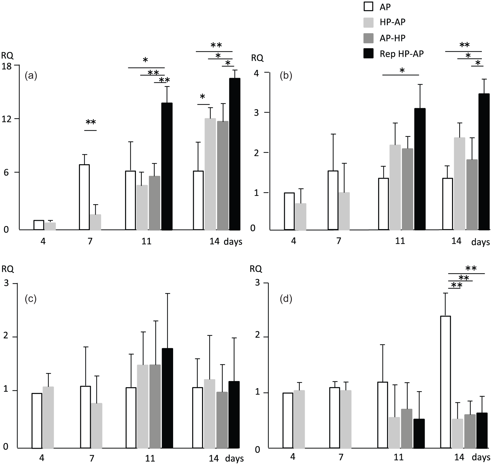
Relative quantity (RQ) of chondrocyte phenotype, chondroprotective, and degenerative markers. (
The Agg expression in AP maintained similar RQ levels as 1.0 by day 14 (
The TIMP-2 expression in AP maintained a similar level throughout the experiments (
The MMP-13 in AP maintained the similar level from day 4 to day 11 and increased 2.4-fold from days 11 to 14 (
Immunohistological Evaluation
We harvested a cell construct from each treatment group at day 14. The constructs were fragile when they were collected with forceps for immunohistological evaluation. Ten-micrometer paraffin sections were stained with antibodies against KS, Col-2, and MMP-13 (
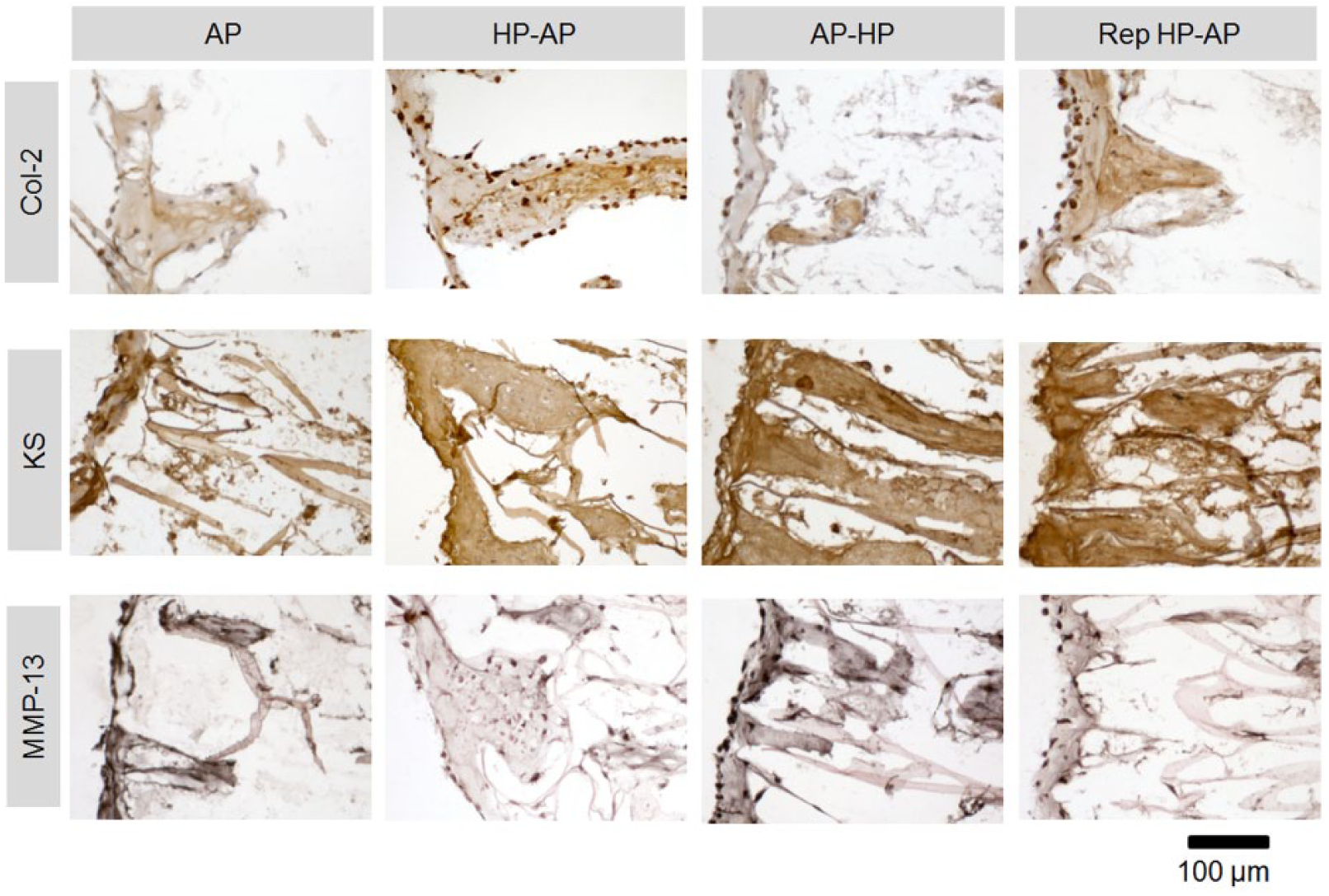
Immunohistology of human articular chondrocytes (hACs) within a collagen sponge at day 14. Col-2, collagen type II; KS, keratan sulfate; MMP-13, matrixmetalloproteinase-13.
Accumulation of Newly Synthesized S-GAG and DNA
hACs produced aggrecan, which accumulated within a construct and leaked into the culture medium. We measured S-GAG as a part of an aggrecan composite accumulated within the constructs using dimethyl methylene blue (
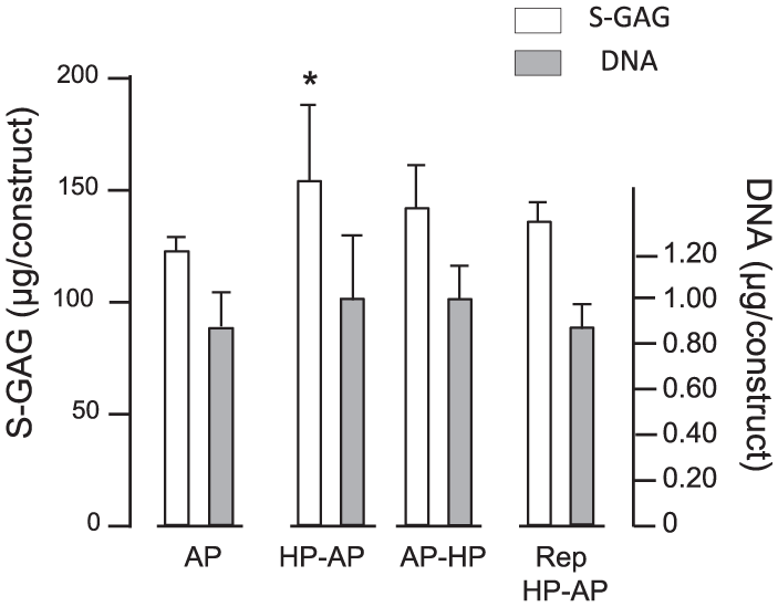
Accumulation of sulfated glycosaminoglycan (S-GAG) and DNA at day 14. AP, atmospheric pressure (no HP); HP, hydrostatic pressure (0.5 MPa, 0.5 Hz). *P < 0.05. Error bar indicates SD.
Discussion
We focused on the effects of HP-AP (HP-loading followed by AP regimen) on anabolic (Aggrecan, Col-2), catabolic (MMP-13), and chondroprotective (TIMP-2) activities by hACs seeded within a 3D collagen gel/sponge scaffold to improve accumulation of ECM within chondrocyte constructs. Our data indicated that the HP-AP upregulated anabolic function, whereas it downregulated catabolic function with hACs by day 14. These characteristics suggested that HP-AP may be beneficial to engineer a mature chondrocyte construct in vitro prior to implantation.
Duration of HP-Loading and HP-AP Regimen
Experiments to clarify the effects of HP-loading have been conducted at various durations. In those studies, chondroitin sulfate (glycosaminoglycan) and proteoglycan-core protein (aggrecan gene expression) were measured to determine major metabolic products by chondrocytes. To determine the quantity of those molecules, we should consider the synthetic mechanisms of those molecules and how they differ from one another. Thus, it seems that 35 S incorporation and its accumulation as chondroitin sulfate proteoglycan need to account for a time lag in response to HP-loading because protein synthesis may take a longer time via upregulation of gene expression than sugar production.20,21 In our previous studies, we determined the duration of the first HP-loading to be four days because the RNA expression of Col-2 was detected with a minimal 3 to 4 days’ HP-loading by bAC seeded in collagen gel.11,22,23 Although 2D versus 3D culture conditions affected metabolic functions of chondrocytes, the 4 days of HP-loading was appropriate to detect mRNA expression of ECM molecules in vitro. Moreover, our previous studies showed that the accumulation of S-GAG increased significantly by bACs or porcine articular chondrocytes within a cell construct with a treatment of HP (0.5 MPa, 0.5 Hz) for at least 4 days followed by additional AP.8,11 In our current study, upregulation of Col-2 and Agg expressions were detected within 7 days after the first HP-loading. Thus, the 7-day duration of the HP-AP was appropriate for detecting changes in gene expression.
Magnitude of HP
We conducted this present study with cyclic HP-loading at 0 to 0.5 MPa, 0.5 Hz, which was a 1/10 lower magnitude than Smith’s and Parkkinen’s experiments, and 1/100 lower than Kunitomo’s.20,21,26 However, intracellular calcium concentration in bACs increased significantly in response to continuous HP at 0.1 MPa for 5 minutes. 27 Thus, we thought that 0.5 MPa was biologically effective for the present study. The magnitude of HP for in vitro experiments was still in debate because it might be relevant to in vivo. Rational for the magnitude often cited Hodge’s hip joint data. 28 Their data indicated that the peak magnitude was 3.8 MPa as a “contact pressure” on articular surfaces in hip joints. This magnitude obviously included both pure HP and pure deviatoric stress within articular cartilage. Thus, a range in HP of between 0.1 MPa and 3.8 MPa is probably a biologically effective magnitude in vitro and in vivo.
Repetitive HP
An intermittent HP-loading regimen can be conducted in many ways. Smith’s previous data and ours indicated that 4 days of loading is minimal for detecting changes in gene expressions.11,20 Since we had an undefined rationale for the regimen in an in vitro study being relevant to physiological changes, we simply repeated the first HP-AP regimen. The second HP-loading immediately upregulated expression of anabolic molecules after the first HP-AP. Thus, the second HP-loading transduced to the cells within the accumulated ECM as seen in Parkkinen’s and Kunitomo’s studies.21,26 Moreover, their studies indicated that the resting period of AP between the first and second HP-loadings restored sensitivity to the loading via recovered signal molecules.29-31 Nevertheless, our results suggested that HP-AP was an effective regimen for producing a chondrocyte construct filled with newly synthesized ECM.
Catabolic and Chondroprotective Gene Expressions
We measured MMP-13 expression as a catabolic molecule because active MMP-13 has the potential to break down Col-1, which affects the loss of accumulated ECM within a cell construct.32-35 In our study, HP-loading (both a single time and repetitive) tended to downregulate MMP-13 expression, whereas AP upregulated its expression significantly at day 14.
These results were in line with previous studies although the previous ones used human chondrocyte in the monolayer or bovine cartilage in the alginate beads.36,37 Thus, our results confirmed the inhibitory role of HP for MMP-13 and upregulation of MMP-13 in the AP condition by the hACs in the 3D culture.
On the other hand, each treatment maintained TIMP-2 expression at a similar level throughout the experiment. Thus, these expression profiles indicated that HP was a potential factor in promoting ECM accumulation, which was a result of the stimulation of anabolic molecules and the reduction of catabolic molecules.
Newly Synthesized ECM Accumulation within a Cell Construct
In the immunohistological evaluation, denser accumulation of Col-2 was seen in the cell construct treated with HP-AP and Rep HP-AP compared with AP, which was consistent with Col-2 gene expression at day 14. On the other hand, less accumulation of MMP-13 was seen in the cell construct treated with HP-AP and Rep HP-AP compared with AP. This observation was consistent with the gene expression of MMP-13 at day 14. These observations possibly suggested that Col-2 was upregulated by day 14, but active MMP-13 broke down Col-2 (enzymatic substrate) in AP. Inconsistency between gene expression and immunohistology was found in the AP-HP treatment. Since HP-loading was conducted later in the AP-HP, it was thought that MMP-13 was activated at an earlier time prior to HP-loading. We used a KS antibody to detect an aggrecan molecule, which has abundant S-GAG produced by hAC. Although MMP-13 had a chance to break down the KS, the GAG was not a substrate of enzymatic activity by MMP-13. Thus, KS could remain within the construct showing dense matrix. Therefore, our results of gene expression and immunohistology indicates that HP-loading is useful to prevent degeneration of newly synthesized ECM and contribute to promoting the accumulation of ECM within a cell construct for engineering articular cartilage. If the MMP-13 activity would be minimized, HP-loading should be applied from an earlier culture time.
Furthermore, single HP loading promoted more S-GAG accumulation compared with AP and Rep HP-AP. These results were inconsistent with gene expressions. Efficiency of product accumulation by cells should differ from the activity of its synthesis in cells. One possibility for the difference is caused with fluid compression of culture medium due to cyclic HP, which may have removed accumulated S-GAG to the medium phase. If this was the case, we should be able to increase S-GAG accumulation within a construct enclosed with some membrane material. This interpretation can be supported by the presence of a superficial zone in the native articular cartilage. 38
As relevant to clinical applications, cell culturing for cell therapy has been recognized as an important process, not only for expanding cell numbers and maintaining phenotypes but also for promoting regeneration and integration with the host tissue. Manufacturing a cell construct using algorithm of HP-AP loading has a potential to improve clinical outcomes, for example, joint function and pain scale.39,40 In addition, cellular qualities, including Col-2 expression, CD44 expression, and cell viability, at the time of implantation could have a positive influence on clinical outcomes after ACI. 41 Therefore, the ability to enhance anabolic functions by repetitive HP before implantation may lead to better clinical outcomes.
In conclusion, repetitive HP-AP stimulated anabolic function and reduced catabolic function, which will be advantageous for engineering cartilage tissue. By manipulating physicochemical changes, for example, duration, frequency, and magnitude, the quality of the engineered chondrocyte construct can be controlled for desired purposes.
Footnotes
Acknowledgments and Funding
The authors thank Julia Mullokandova for the preparation of the manuscript. TO has received a training fellowship from Funabashi Orthopaedic Hospital (Chiba, Japan). This project has been partially supported as a sponsorship research project by PURPOSE (Shizuoka, Japan).
Declaration of Conflicting Interests
The author(s) declared no potential conflicts of interest with respect to the research, authorship, and/or publication of this article.
Ethical Approval
Ethical approval for this study was obtained from Institutional Review Board protocol of Brigham and Women’s Hospital, Boston, Massachusetts(2013D002589).
Informed Consent
Not applicable
Trial Registration
Not applicable.