
Abstract
Objective
Hyaline cartilage degenerative pathologies induce morphologic and biomechanical changes resulting in cartilage tissue damage. In pursuit of therapeutic options, electrical and mechanical stimulation have been proposed for improving tissue engineering approaches for cartilage repair. The purpose of this review was to highlight the effect of electrical stimulation and mechanical stimuli in chondrocyte behavior.
Design
Different information sources and the MEDLINE database were systematically revised to summarize the different contributions for the past 40 years.
Results
It has been shown that electric stimulation may increase cell proliferation and stimulate the synthesis of molecules associated with the extracellular matrix of the articular cartilage, such as collagen type II, aggrecan and glycosaminoglycans, while mechanical loads trigger anabolic and catabolic responses in chondrocytes.
Conclusion
The biophysical stimuli can increase cell proliferation and stimulate molecules associated with hyaline cartilage extracellular matrix maintenance.
Introduction
Hyaline cartilage is an avascular tissue characterized by having abundant extracellular matrix (ECM) that is mainly composed of collagen type II and proteoglycans (PG).1-3 This tissue is formed by a unique cellular type, the chondrocyte, which is responsible for maintaining the integrity of the ECM.4,5 Within the skeletal system, hyaline cartilage is found in two specialized structures of the long bones: the articular cartilage and the growth plate.1,5 The first acts as a smooth, lubricated, low-friction surface that receives mechanical loads and facilitates motion between opposing joints6,7; while the second, which is located between the epiphysis and diaphysis, is responsible for the longitudinal growth and shape of long bones.8,9
Histologically, both articular and growth plate cartilages are tissues stratified in different zones according to chondrocytes phenotype and spatial arrangement of the ECM. Accordingly, articular cartilage is organized in 5 zones: superficial, intermediate, medium, deep, and calcified, 10 whereas the growth plate is arranged into 3 zones: reserve, proliferative, and hypertrophic. 11 In both tissues, the molecular structure of the ECM within the hyaline cartilage provides dimensionality, elasticity and strength to the tissue.3,10
Given its location, hyaline cartilage is exposed to mechanical loads that generate different signals implicated in the physiological regulation of chondrocytes behavior and several pathological processes, considering that chondrocytes sense and respond to different mechanical loads.2,12-14 For example, mechanical loading has been shown to affect cell deformation, fluid flow, nutrient and ion concentration gradients, pH changes, and anabolic and catabolic activity of ECM components and stimulation of growth factor synthesis.15-20 In turn, overweight or strong impact loading have been proven to cause damage in the ECM and chondrocyte apoptosis, resulting in dysfunction of the tissue and subsequent damage.6,13,21,22 Several studies have shown that hyaline cartilage behavior is also affected by electrical and electromagnetic stimuli, resulting in changes in cell dynamics, such as migration, 23 differentiation,24-26 morphology,27-30 proliferation,31-36 and gene expression.37-50 This implies that mechanical, electrical, and electromagnetic stimulation may have an important role in the regulation of hyaline cartilage in both normal and pathological conditions.
Based on the above, understanding the influence of biophysical stimuli in the biology of hyaline cartilage not only provides useful information regarding their role in cartilage physiology in vivo but also supplies new tools for tissue engineering and regenerative medicine focused on the develop of new therapeutic approaches for the treatment of injuries in articular cartilage and growth plate. In this context, we performed a review of literature of the past 40 years of in vivo and in vitro studies, highlighting their results, advances, main limitations, and perspectives. The information presented summarizes the current understanding about the role of both the electrical stimulation over chondrocytes intended for articular cartilage recovery and the mechanical stimuli over growth plate and its influence on biological events during human bone development.
Electrical Stimulation for Tissue Engineering Hyaline Cartilage
Electric Fields
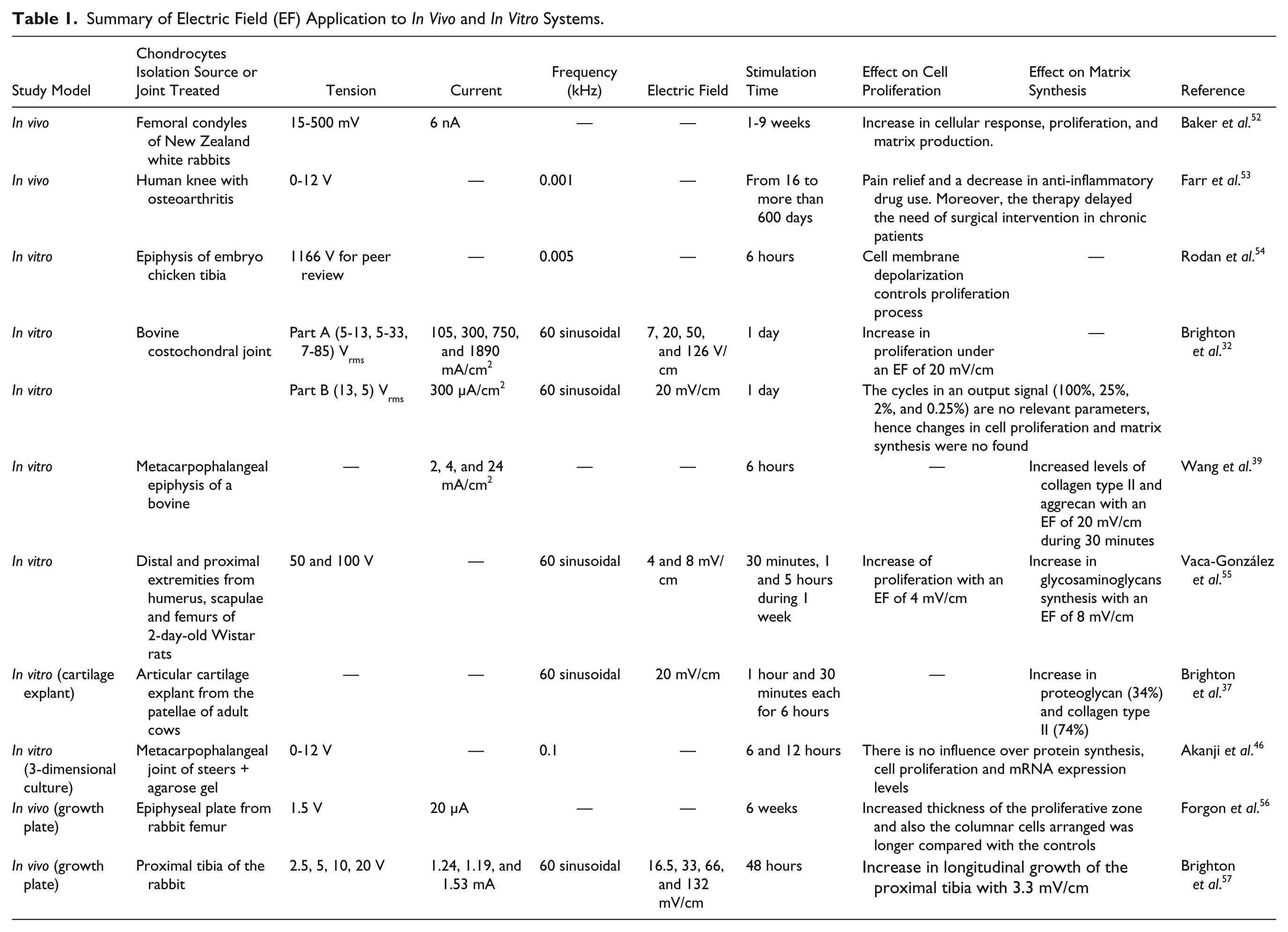
Electrical stimulation has been used to improve chondrocyte proliferation rate and the synthesis of characteristic ECM molecules. Electric fields (EFs) for cartilage regeneration have been used in in vivo and in vitro systems ( Table 1 ).31,35,38,46,51-53
Summary of Electric Field (EF) Application to In Vivo and In Vitro Systems.
In Vivo studies
The first reported use of electrical stimulation to influence cartilage was in 1974 by Baker et al. 52 who assessed the influence of a bimetallic platinum electrochemical device in the recovery of full-thickness injuries of articular cartilage. The electrochemical device was implanted in the lateral femoral condyle at the location of the injury and voltages from 15 to 500 mV were applied. Results showed a 71% of increase in the production of ECM in treated animals. 52 This finding set the basis for the application of electrical therapy in patients presenting with joint pathologies. The experience reported up to now in the literature has evidenced that the application of the EFs lead to pain improvement in patients with knee osteoarthritis. For instance, Farr et al. 53 used a transcutaneous electric stimulator in 288 patients with knee osteoarthritis. Patients who received electrical stimulation for more than 750 hours (73% of total population) reported both pain relief and a decrease in anti-inflammatory drug use (45.3%). Moreover, the therapy delays the need of a surgical intervention in chronic patients. 53
In Vitro Studies
The technique of delivering electrical stimulation to cell culture plates consists of an indirect coupling system that uses external parallel electrodes connected to a power supply ( Fig. 1A ).31,38,39,54 The custom-designed devices are noninvasive capacitive coupling systems that can be used to stimulate any cellular type, and they are based on the same principle used in medical stimulation devices.51,53,54
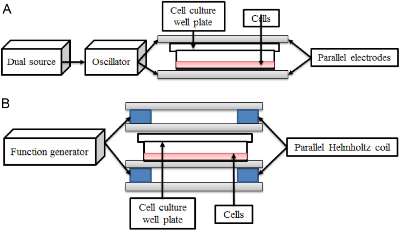
Schematic diagram of an indirect coupling system to stimulate in vitro cell cultures. (
In vitro, the application of EFs to monolayer cultures has shown to increase cell population and to stimulate synthesis of main molecules that compound cartilage such as collagen type II, glycosaminoglycans (GAGs), and aggrecan. For instance, Rodan et al. 55 demonstrated that increased chondrocyte proliferation observed after EFs stimulation was triggered by a depolarization in the cell membrane, it means that the Na+ and Ca2+ fluxes generated by the EFs trigger DNA synthesis of chondrocytes. More descriptive studies were carried out by Brighton et al., 34 in which they stimulated chondrocytes with different voltage values (10, 100, 250, and 1000 V) to a frequency of 60 kHz (sinusoidal-wave form). Their results indicated that voltages of 10 and 1000 V decreased cell proliferation; while a voltage of 250 V increased the GAGs synthesis. 34 In these studies, no data about the magnitude of the EFs were reported.
Similar studies were performed by Armstrong and Brighton where chondrocytes were stimulated with several EFs (15, 22.5, 30, and 45 mV/cm). Their results demonstrated an increase in cell population when EFs of 15, 22.5, and 30 mV/cm were applied. An increase in GAGs synthesis (66%) was obtained after stimulation with EFs of 45 mV/cm.31,33 From these studies, it was possible to conclude that an EF close to 20 mV/cm stimulates chondrocyte dynamics. Brighton et al. 32 in later studies reported that an EF of 20 mV/cm increases the cell proliferation by 50%; while Wang et al. 39 in a more detailed study elucidated that this EF of 20 mV/cm increases aggrecan and collagen type II synthesis. On the contrary, researchers such as Nakasuji et al. 35 have applied EFs of greater magnitude (50, 100, 250, and 500 mV/cm) to chondrocytes cultured in vitro. The results showed that EFs of greater magnitude also affect cell proliferation and ECM synthesis. 35 Because of the discrepancy regarding the magnitude of the EFs required to stimulate the in vitro cultures, Vaca-González et al. 54 implemented a new methodology to calculate EFs and modulate proliferation and GAGs synthesis. Results allowed to establish the dielectric constant of the cell culture medium (complex permittivity and conductivity), and also showed that EFs of 4 mV/cm applied during 30 minutes increment cell population; while EFs of 8 mV/cm applied during 5 hours maintain a stable GAGs synthesis. 54
Studies Performed in Explants and 3-Dimensional Cartilage Constructs
The EFs have also been applied to cartilage explants and tridimensional constructs. 56 Brighton et al. 37 stimulated cartilage explants with EFs of 20 mV/cm. Results showed an increase in PG (34%) and collagen type II (71%) after 14 days of stimulation. 37 Because EFs of 20 mV/cm have been shown to influence the cellular dynamics, the research group of Brighton studied the effect of the electrostimulation in osteoarthritic human cartilage explants. In this work, osteoarthritic explants stimulated for 14 days using 20 mV/cm EFs showed 1.4- and 1.5-fold increase in PG and collagen type II, respectively. 38 EFs have been also applied to 3-dimensional (3D) cartilage constructs used for cartilage repair; however, to our knowledge, the reports in literature are limited and the results have been contradictory. For instance, Szasz et al. 45 stimulated 3D chondrocyte-seeded agarose gels, obtaining increases in cell density and GAGs synthesis. In contrast, Akanji et al. 46 investigated the effects of direct current on cell proliferation and matrix synthesis, using a 3D chondrocyte-agarose model system. Their results demonstrated that EFs have no influence over protein synthesis, cell proliferation, and mRNA expression levels. 46
Effect of EFs Over the Growth Plate
Applying electrical stimulation across growth plate cartilage has little effect on composition, 57 but can alter the thickness of zones within the cartilage,57,58 and ultimately the length of the bone.57,59 However, high voltage reduces bone growth and maintains cartilage in a quiescent state. 59
Electromagnetic Fields
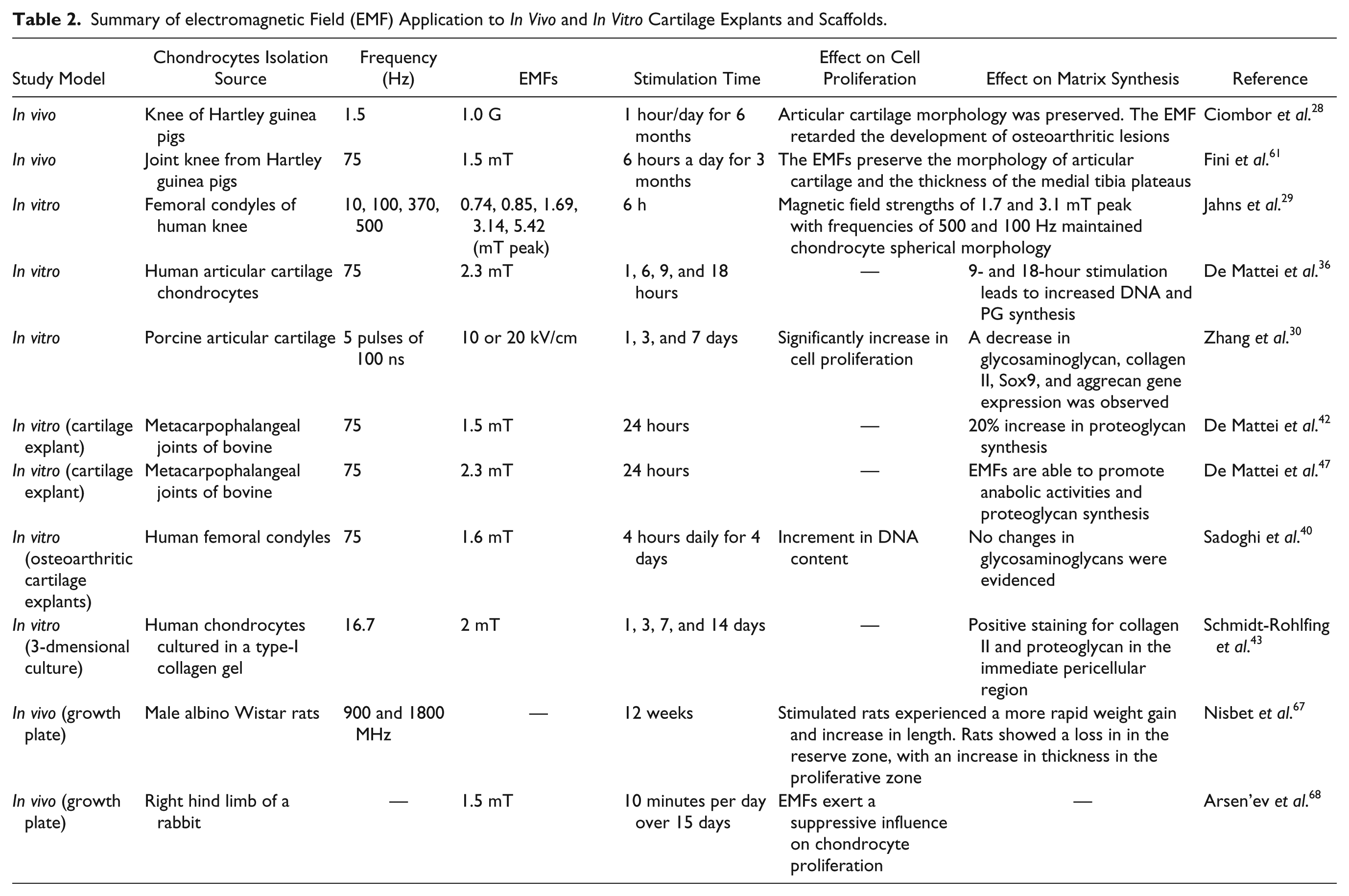
Similar to EFs, electromagnetic fields (EMFs) have been applied in in vivo and in vitro studies using cartilage and growth plate explants, and tridimensional structures ( Table 2 ).27,28,30,42,44,49,60 Similar to EFs, the technique of deliver EMFs consists of an indirect coupling system that uses external parallel coils connected to a function generator ( Fig. 1B ).28,29,47,60 This kind of noninvasive configuration has the advantage of generating small currents and potentials in the proximity of the targeted cells, and also it generates a homogeneous EMF across the cell culture. 51
Summary of electromagnetic Field (EMF) Application to In Vivo and In Vitro Cartilage Explants and Scaffolds.
In Vivo Use of EMFs to Heal Osteoarthritis
In vivo animal models have been used to study the influence of EMFs in the recovery of knee osteoarthritis. Ciombor et al. 28 showed that articular cartilage morphology was preserved and the development of osteoarthritic lesions were retarded when EMFs were applied. EMFs have also been implemented in clinical studies to treat knee osteoarthritis. Results have evidenced that electromagnetic stimulation contributes to preserve articular cartilage morphology,60,61 improvement in joint motion, tenderness, and a reduction in pain.62-65
In Vitro Studies Using Cell Cultures
The application of EMFs in monolayer cultures have shown preservation of chondrocytes morphology, 29 increased DNA synthesis, 36 and enhancement of proliferation and GAGs synthesis in human chondrocytes. 66 It has also been demonstrated that EMFs significantly increase cell population, but decrease the synthesis of characteristic molecules of hyaline cartilage such as collagen type II, aggrecan, SOX 9, and GAGs. 30
Studies Performed in Explants 3-Dimensional Cartilage Constructs
Healthy and pathological tissue explants, have also been treated with EMFs.40,42,49 The use of EMFs over cartilage explants have shown increase in PG synthesis and stimulation of anabolic activities,42,47 while the effect of EMFs in osteoarthritic explants have evidenced an improvement in osteoarthritis of grade I and III, increasing PG synthesis and counteracting catabolic activities. 49 However, it was also shown that EMFs have no effect on DNA and PG synthesis. 40 The application of EMFs has also been investigated in osteoarthritic chondrocytes cultured in 3D structures. Results showed that the PG concentration of osteoarthritic chondrocytes cultured in alginate scaffolds was stored in the cell culture medium after stimulation was finished. 27 Normal articular chondrocytes cultured in 3D constructs were also stimulated with EMFs. In studies performed by Schmidt-Rohlfing et al. 43 and Nicolin et al., 44 the chondrocytes that were cultured in a collagen membrane showed that this scaffold is a useful bioengineering construct since allowed increases in cell proliferation, collagen type II synthesis, and PG in the immediate pericellular region.
Effect of the EMFs Over the Growth Plate
Studies have been carried out to study the effects of EMFs over growth plate development. A recent study showed that male albino rats stimulated with EMFs experienced a more rapid weight gain and increase in length. Moreover, the stimulated rats showed a loss in cartilage matrix density in the reserve zone, with an increase in thickness of the reserve and proliferative zones. In addition, measurements of the growth plate thickness showed that the trabecular zone was thinnest and the reserve and proliferative zones were thickest. There were no significant differences between the groups with respect to the thickness of the hypertrophic zone. 67 A similar study evidenced that EMFs exert a suppressive influence on chondrocyte proliferation in the growth plates and promote expansion of the differentiate fraction. 68 The epiphyseal plate has been also stimulated through in vitro cultures; for instance, the costochondral junction growth plate of a rat was exposed to different EMFs and changes of temperature. Results indicated that longitudinal growth of the costochondral junction was only observed when EMFs were applied in an environment where the temperature increased. 69
Mechanical Stimulation for Tissue Engineering Hyaline Cartilage
Compressive Loads
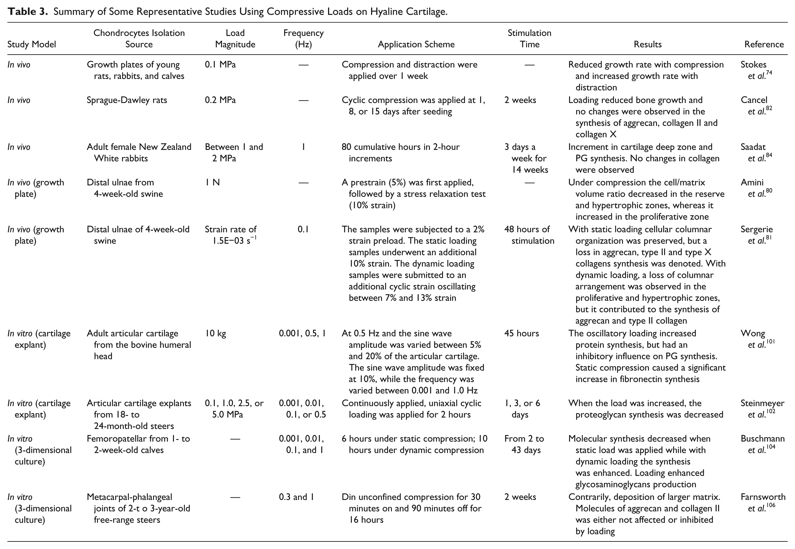
Because of the fact that hyaline cartilage, especially the one found in joints and growth plate, is mainly subjected to compressive mechanical stimuli,22,70,71 several in vivo and in vitro studies have been performed in order to identify the effects of mechanical compression in cell dynamics (proliferation, hypertrophy, and apoptosis),1,72-79 cell height,72,74,80 and the biosynthetic behavior of chondrocytes, assessed mainly in terms of expression of molecules of the ECM (collagen types II and X and aggrecan) and some enzymes (MMP 13 and ADAMTS-4/5).81,82 In addition, changes in the expression of metalloproteinase inhibitors (TIMP-1 and TIMP-2), growth factors (VEGF), and other molecules are influenced by load ( Table 3 ).16,79,83-85 Up to now the specific mechanisms involved in mechanotransduction are not well understood; however, 2 kinds of proteins have been involved in these processes: integral membrane proteins associated in ECM interactions (e.g., integrins) and mechanosensitive ion channels.86-89 Furthermore, signaling mediated by the primary cilium and MAPK pathway have also been implicated.90-95 Regarding the devices used to apply compressive loads, the technique of deliver mechanical loads consist of a system that uses loading actuators that exert an axial force ( Fig. 2A ).7,92,94 This devices present an advantage of maintain the cell cultures in atmospheric conditions of 37°C and 5% CO2 while mechanical loads are been applied. 96
Summary of Some Representative Studies Using Compressive Loads on Hyaline Cartilage.
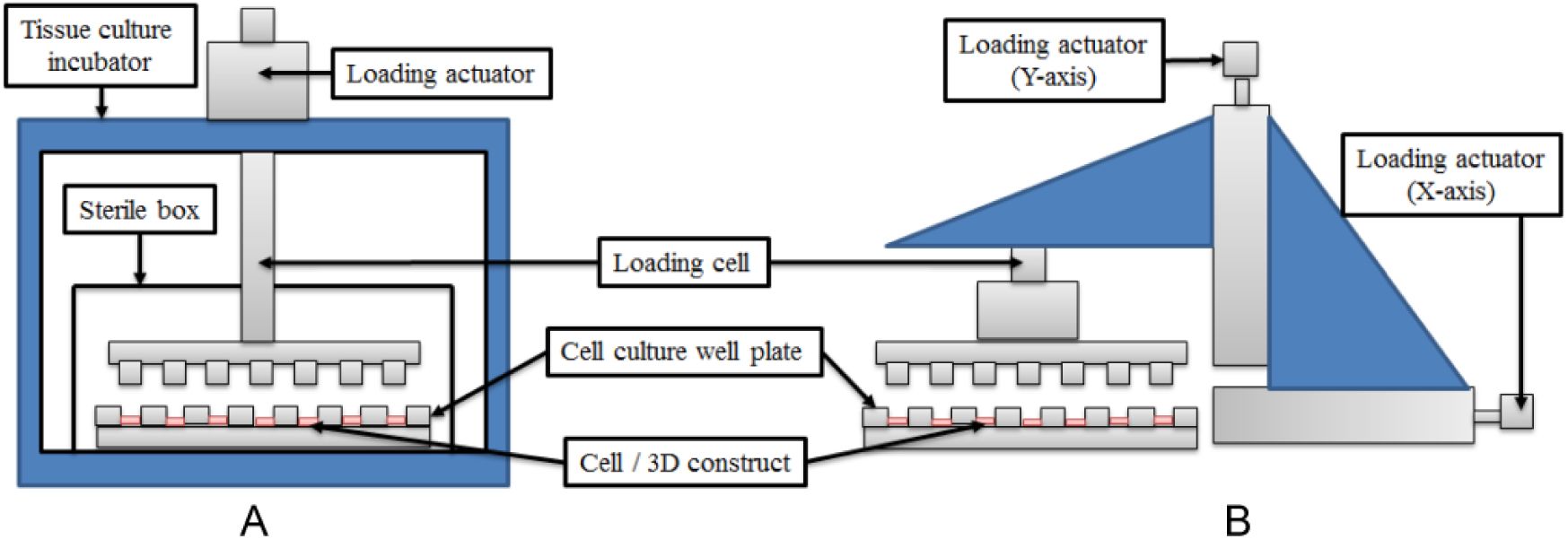
Schematic diagram of the devices used to apply mechanical loads to chondrocytes cultured in 3-dimensional (3D) structures. Both schemes have in common a loading cell, a cell culture well plate and the 3D construct. (
In Vivo Studies
To understand the effect of mechanical loading on chondrocytes behavior within the tissue, some in vivo studies have been performed in both articular cartilage and growth plate. The studies performed in articular cartilage have mainly focused in analyzing the synthesis of ECM molecules as PG, where a direct correlation between loading in the joint and PG levels within the tissue was observed.7,16,84,95 Several studies performed in the growth plate have assessed the effect of mechanical loading as a regulator of bone growth and ossification. Such behavior has been recognized by the Hueter-Volkman law, which establishes that static compression is crucial for bone development; however, excessive loads may inhibit the normal bone growth. Moreover, alterations of mechanical modulation have been associated to several clinical conditions, including the angular progression deformities in tibia (Blount disease), and the development of scoliosis.72,73 Studies using chicks and rodents have demonstrated that mechanical stimulation of bone rudiments during embryological development plays an important role in normal growth and bone morphogenesis. 97
At cellular level, studies using rodents have shown that mechanical loading induces histological changes within the growth plate, specifically in the width of the proliferative and hypertrophic zones.74-77,98 Additionally, it has been observed a decrease in collagen type II and aggrecan concentrations in growth plates subjected to long term static compressions. 82
In Vitro Studies: Tissue Cultures
As an alternative model to study the influence of mechanical loading over the chondrocytes behavior, in vitro protocols applying compressive loads to articular cartilage and growth plate explants have been performed. For articular cartilage, the reported results of ECM molecules synthesis under dynamic loading are contradictory with some studies showing an increase while others indicate either a decrease or no changes after loading.70,71,79,84,99-102 For growth plate, studies using tissue explants have shown differential morphological changes of chondrocytes in the different zones under compression. 80 Additionally, under short-term compressive loads, a decrease in aggrecan, collagen type II and X has been reported. In contrast, under dynamic loading no changes in ECM composition although changes in cell organization were observed. 81
In Vitro Studies: Cell Cultures
Given the difficulties for analyzing the individual effects of mechanical stimuli to which cartilage is subjected in vivo, the response of chondrocytes has been evaluated in monolayer and 3D cultures.16,22,70,71,93,101,102 These analyses not only provide information about the cellular responses and the regulatory mechanisms associated, but also they allow to evaluate the use of mechanical stimuli in tissue engineering approaches. 16 The effect of compressive loads in in vitro cultures of chondrocytes depends up on load’s intensity and duration 7 . In this context, it has been observed that the application of static loads increased the production of several types of MMPs, and also inhibited the synthesis of collagen type II and PG.7,95 In turn, the effects of dynamic loading in cell proliferation and synthesis of PG, GAGs, and collagen type II are contradictory.7,70,71,79,84,95,96,99-118 This behavior may be related to the wide variability of the methodology used for each group, which can be related to different sources of chondrocytes, age of the animal model, characteristics of 3D matrix used, chondrocyte seeding density, frequency of the stimulus, load cycles, and initiation and duration of the stimulation.7,84,96,101,103-108,115
Tension
The chondrocytes response to tensile loading have also been addressed using in vitro monolayer cultures. Such cultures have been performed by seeding cells in membranes that are submitted to uniaxial or biaxial dynamic stretching. Most of the studies have evaluated chondrocyte dynamics in terms of cell proliferation and molecular synthesis of collagens and PG. Similar to that observed for compressive loading, the evidence available suggests that the response of chondrocytes to tensile loading also depends on the duration and intensity of the stimulation.18,119-121 In fact, anabolic effects in chondrocytes are induced after short-term simulation (less than 12 hours); while a decrease in the ECM molecules is only observed after a prolonged stimulation. 18 In addition, some studies have reported an increase of other molecules such as proteases and their inhibitors (e.g., TIMPs, MMPs), soluble factors (e.g., TGFβ, VEGF, PTHrP), and pro-inflammatory factors (e.g., nitric oxide, prostaglandin E, cyclooxygenase 2) after tensile loading stimulation.18,93,119,121-123
However, it is difficult to extrapolate definitive conclusions regarding the stimulus characteristics and chondrocytes response considering that, similarly to compressive loading, there is a high variability in the stimulation protocol used among different studies in terms of strain magnitude, loading duration and frequency (reviewed in Bleuel et al. 18 ).
Hydrostatic Pressure
In Vitro Studies: Tissue Cultures
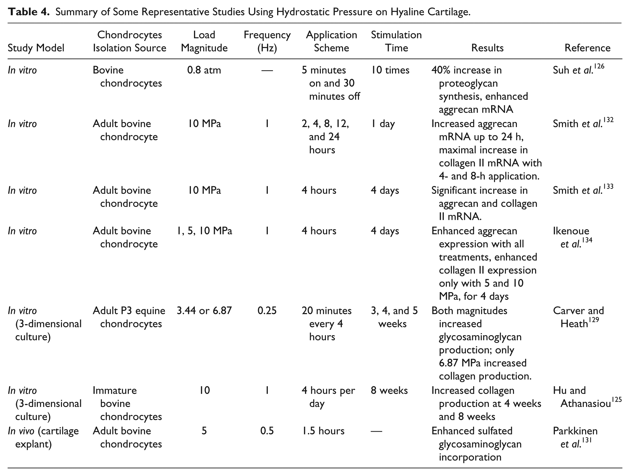
Hydrostatic pressure (HP) has been extensively used as a stimulus to induce changes over hyaline cartilage such as protein expression and matrix production. 124 However, the mechanism by which HP produce those changes is not well understood. HP provides a robust method of chondrocyte stimulation since it can be applied over cells cultured in monolayer, 3D engineered constructs or cartilage explants ( Table 4 ).125,126 Several studies have shown the relation between HP and biological response of hyaline cartilage, such as investigations performed by Parkkinnen et al. have shown a microtubule-dependent compaction of the Golgi apparatus and decrease in GAGs synthesis 127 . In a similar study, Lammi et al. 128 found that static HP resulted in a 37% decrease in GAGs synthesis, reduction of aggrecan mRNA levels and synthesis of atypically large aggrecan molecules. The alteration in aggrecan size demonstrates that HP may affect the production of ECM molecules at posttranslation levels.
Summary of Some Representative Studies Using Hydrostatic Pressure on Hyaline Cartilage.
Studies Performed in Cartilage Explants and 3-Dimensional Constructs
HP also has been applied over 3D scaffolds and explants. For instance, Carver and Heath129,130 showed that chondrocytes cultured in poly glycolic acid (PGA) meshes and stimulated with 6.87 MPa experienced an increase in GAGs and collagen production in adult cells, whereas the same magnitude of HP had an increase only collagen synthesis in juvenile cells. There are no differences of applying HP either in cartilage explants or in chondrocytes cultured in vitro; however, the magnitude of the pressure and the frequency are relevant factors to stimulate PG synthesis. 131
Shear Stress
Within the joint and growth plate chondrocytes are exposed either to compressive HP or deviatory stresses such as shear stress.135 Previous studies have indicated that chondrocytes are exposed to different levels of fluid flow within the tissue,136,137 suggesting that mechanical shear stress has a pathophysiologic relevance in cartilage biology. 138 Within this context, several studies have analyzed the influence of such stimuli on chondrocyte behavior. For instance, Mohtai et al. 139 applied shear stress over human articular chondrocytes seeded in a high monolayer culture. Cell cultures were stimulated with loads of 0.16, 0.41, 0.82, and 1.64 Pa at rotating velocities of 20, 50, 100, and 200 rpm. Results indicated that this stimulation scheme increased the release of pro-inflammatory mediators and nitric oxide, decreased the expression of aggrecan and collagen type II, and induced molecular changes associated with apoptosis. 139 Additionally, it is known that hyaline cartilage tissue engineering constructs, are also affected by shear stress, revealing that this stimulus may alter the intercellular signaling pathways in chondrocytes.129,134 Some reports suggest that fluid shear stress reduces expression of aggrecan and collagen type II. 140
Other studies, like those developed by Waldman et al.141 used a stimulation scheme based on 2% of shear stress amplitude at 1 Hz for 400 cycles every second day, demonstrate that an intermittent application of dynamic shearing forces during 4-week periods improved the quality of the cartilaginous tissue formed in vitro. These data indicate that the nature and magnitude of shear stress may play a significant role in the homeostasis of the structure and function of hyaline cartilage. For this reason, the cellular mechanisms underlying the responses of articular chondrocytes to mechanical stresses are important for understanding the pathogenesis of several hyaline cartilage diseases. 138 Similar to compressive loads, the technique of deliver the shear stress consists of a loading actuator that generate forces along the X and Y axis at the same time ( Fig. 2B ).139,141 In this context, this configuration allows one to apply precise strains on multiple axis similar to the mechanical environment supported by the joint. 142
Cellular Mechanisms Involved in Transduction of Biophysical Stimuli
In chondrocytes, biophysical stimuli are sensed mainly by membrane proteins involved in ECM-cell interactions and ionic channels.7,143,144In addition, some stimuli may induce changes in the cytoskeleton.23,145
ECM-cell interactions are associated mainly with the transduction of mechanical stimuli, since they allow cells to sense conformational changes of the ECM that trigger, intracellularly, the activation of different cell signaling cascades leading to specific cell responses.7,143 The main proteins associated with ECM-cell interaction are integrins, integral membrane proteins that bind to several ECM molecules with different affinities.146-148 The main integrins expressed by chondrocytes are α1β1, α3β1, α5β1, α10β1, αVβ3, and αVβ5, which act as receptors for collagen types VI and II, matrilin-1, fibronectin, osteopontin, COMP, and vitronectin, respectively.87,88,147-149 Cytoplasmic domains of integrins are coupled to kinases that have been implicated in signal transduction through Ras, Rho, and Rac pathways.93,95 In addition to integrins, other membrane proteins like CD44 and annexin V (also known as anchorin CII) have also been associated in ECM-cell interactions in chondrocytes. CD44 is a transmembrane hyaluronan-binding glycoprotein that is involved not only in anchoring but also in sensing and signaling functions.149-151 Annexin V is a member of a family of calcium and phospholipid binding proteins that binds mainly with collagens present in the PCM and ECM, mainly type II and with lesser affinity to types V, IX, X, and XI.150-152
On the other hand, ion channels have been associated to both mechanical and electrical signal transduction. Although a plethora of ion channels have identified in chondrocytes membranes, calcium channels are the main type of ion channels associated to transduction of biophysical stimulation.86,89,95 The calcium channels most associated to these processes include voltage-gated sodium channels, voltage-gated calcium channels, and stretch activated ion channels. In addition, various members of the TRPV (transient receptor potential vanilloid) nonselective cationic channels family, especially TRPV4, have been associated to osmotic stress responses.86,89,95 In fact, it has been described that in chondrocytes, cell deformations caused either by changes in cell volume or due to mechanical loading, lead to activation of different kinds of ionic channels, and some studies have evidenced changes in intracellular calcium levels in chondrocytes after mechanical loading.86,91,92,94,95 Electric signals applied either in direct or indirect contact with the cells, exert their effect on the cell membrane by activating the voltage-gated calcium channels leading to increase in the intracellular Ca2+ levels.51,144,153
Finally, as previously mentioned, some stimuli are associated to conformational changes of the cytoskeleton. That is the case of electrical stimulation, which has proven to induce changes in actin filaments causing cytoplasm elongation followed by a perpendicular alignment with regard to the applied EFs. 145 Furthermore, electric stimulation has shown to promote cell movement, process known as galvanotaxis. 23
Perspectives
This review focuses on the effect of biophysical stimuli in chondrocyte behavior, showing that electric and mechanical stimulation have important roles in physiology of hyaline cartilage. In addition, biophysical stimuli display interesting potential as additional tools for both tissue engineering and regenerative medicine, toward the development of new therapeutic alternatives for cartilage pathologies. The effects of biophysical stimulation on chondrocytes dynamics have been explored in vivo and using several in vitro approximations. In monolayer cell cultures, stimulation with EFs, EMFs, HP, and tension can promote chondrocyte proliferation and molecular synthesis.18,51,61,132,134 Taking this into account, with the current use of scaffolds combined with the application of biophysical stimuli it is possible to obtain tissue constructs that mimic in vivo characteristics of cartilage.
For both electric and mechanical stimuli, different frequencies, intensities, and duration of stimulation schemes have been tested; however, the magnitudes of the stimuli that directly stimulate chondrocytes in those cultures have been poorly explored. Regarding electric stimulation, at present, calculations of cell culture media dielectric constants have been recently reported. These electric constants, such as permittivity and conductivity, are essential parameters to calculate electric and EMFs.54,154 Thus, this leads to a new research area that will allow to implement a better methodology to calculate the EFs and EMFs in a more detailed way. In electrical stimulations, there are many discrepancies in the obtained results because of the fact that one of the limitations is the required high voltage to generate the EFs, the stimulation time to achieve the best rate of proliferation and molecular synthesis, as well as the amount of days that cell cultures need to be under stimulation. For this reason, electrical stimulation for tissue engineering hyaline cartilage still is an open research area. On the other hand, little is known of the mechanical environment and the effects on fluid flow and nutrient availability within cartilage or in vitro 3D cultures of the different types of mechanical stimulation. Recently, there have been some approaches that use computational modeling to approach such issues.155-159 This is a promising field that may improve the understanding of the effects of mechanical stimuli on chondrocyte behavior as well as provide powerful tools for scaffold designs for 3D in vitro culture systems and mechanical stimulation scheme selection. Furthermore, biophysical stimuli could help to enhance cell proliferation, differentiation, and ECM deposition, opening up new possibilities in 3D articular cartilage and growth plate tissues, and allowing to design novel tissue-engineering constructs similar to real life.
Given the good results obtained using some protocols of electrical and mechanical stimulation independently for in vitro hyaline cartilage tissue engineering, the integration of these both methods is currently been studied. 50 This leads to a new field of research that will focus on stimulation of cartilage explants or 3D constructs with 2 biophysical stimuli simultaneously. As a novel and potential tool, it would be possible to create special devices that generate distinct electrical and mechanical intensities according to the necessity of a particular treatment. This contributes to tissue engineering because it is noteworthy that biophysical stimuli are the basis for the next generation of cartilage regeneration technology, since those electrical and mechanical stimuli help the development of biomimetic samples that recreate an environment where the functional, structural, and biological features of hyaline cartilage remain stable at the time of cartilage replacement.
Overall, the use of biophysical techniques provides not only new tools to create in vitro biomimetic tissues, but they can also be novel therapeutic approaches that may be implemented in patients with several chronic hyaline cartilage pathologies. The methodologies used to stimulate cartilage regeneration are getting closer and closer to develop technologies that satisfy the requirements of a successful cartilage healing and make that the concept of cartilage tissue engineering can be extrapolated to a purely clinical environment.
Footnotes
Acknowledgments and Funding
The authors gratefully thank the research support from the Biotechnology Institute of the Universidad Nacional de Colombia and the Administrative Department of Science, Techonology and Innovation in Colombia (COLCIENCIAS).
Declaration of Conflicting Interests
The author(s) declared no potential conflicts of interest with respect to the research, authorship, and/or publication of this article.