
Abstract
Introduction:
Extracellular vesicles (EVs) are lipid bilayer particles released by all cell types, carrying cargos that reflect the cellular states of their origin. Recently, EVs are increasingly recognized as valuable biomarkers and therapeutic vectors in oncology, but their clinical translation is limited by variability in isolation methods and uncertainty regarding long-term storage physical stability.
Methods:
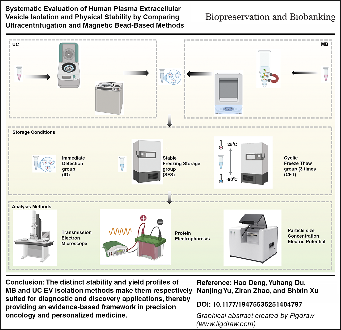
We systematically compared human plasma EVs isolated by ultracentrifugation (UC) or magnetic bead (MB)-based methods under immediate analysis, stable freezing storage, and repeated freeze–thaw conditions. The morphology and protein profiling of EVs were characterized by transmission electron microscopy (TEM) and Western blotting (WB), respectively. EV concentration, particle size, and zeta potential were quantified by particle size analyzer.
Results:
TEM and WB analyses of human plasma EVs confirmed the efficacy of both the UC and MB isolation methods. UC-isolated EVs are of high yield but low physical stability, featuring size reduction and a shift toward more negative zeta potential values after freeze–thaw cycles. Fresh UC-EVs displayed heterogeneous size profiles, whereas freeze–thawed samples shifted to a dominant peak, consisting of small particles with increased counts. Although lower in yield, MB-isolated EVs retained their physical stability across all conditions.
Conclusion:
MB-based EV isolation offers physical stability for standardized diagnostic workflows, whereas UC-based EV isolation provides high yield for discovery studies but vulnerable to freeze–thaw stress. These findings provide an evidence-based framework for selecting EV isolation and storage methods to match downstream applications, guiding the standardization of EVs workflows for future precision oncology and personalized medicine.
Get full access to this article
View all access options for this article.