
Abstract
The musculoskeletal system, essential for mobility, structural support, and organ protection, is frequently compromised by trauma, degenerative diseases, or tumors, profoundly impacting patients’ quality of life. Adhesive hydrogels have emerged as pivotal biomaterials for orthopedic therapies, offering localized treatment with enhanced biocompatibility, tunable mechanics, and sustained bioactive delivery. While systemic drug administration often suffers from off-target effects, adhesive hydrogels enable precise tissue integration and microenvironmental modulation, addressing challenges such as infection control, tissue regeneration, and mechanical reinforcement. However, achieving optimal adhesion strength, dynamic mechanical matching, and selective tissue targeting remains a critical hurdle. Innovative strategies, including dynamic covalent bonds, stimuli-responsive networks, and multifunctional hybridization, have expanded hydrogel applications in diabetic wound healing, load-bearing bone repair, and spinal cord regeneration. For instance, injectable hydrogels with wet adhesion capabilities facilitate minimally invasive delivery, while drug-eluting systems localize chemotherapeutics to tumor sites, reducing systemic toxicity. Despite these advances, scalability, long-term stability, and clinical translation require further exploration. This review systematically examines the design principles, functional mechanisms, and therapeutic applications of adhesive hydrogels in orthopedics, emphasizing their role in bridging biomechanical demands with biological regeneration. We envision that interdisciplinary innovation in smart hydrogels will unlock personalized solutions, transforming the landscape of precision orthopedic medicine.
Impact Statement
Revolutionizing Orthopedic Therapies: Adhesive hydrogels enable localized, precision treatments for musculoskeletal injuries, degenerative diseases, and tumors through precise tissue integration and controlled drug release, overcoming limitations like off-target effects of systemic therapies and significantly improving patient outcomes.
Smart Material Innovation: Dynamic covalent bonds, stimuli-responsive networks, and injectable properties empower hydrogels for complex applications such as diabetic wound healing and load-bearing bone regeneration, reducing the need for invasive surgeries.
Advancing Clinical Translation: While breakthroughs in biocompatibility and functional design highlight potential, challenges in long-term stability and scalable manufacturing demand interdisciplinary collaboration to accelerate personalized precision orthopedic solutions for global clinical impact.
Introduction
Orthopedics, the medical specialty focused on the musculoskeletal system, addresses the prevention, diagnosis, and treatment of disorders affecting bones, joints, tendons, and ligaments—structures vital for mobility, mechanical support, and organ protection. With aging populations and rising trauma incidence, musculoskeletal pathologies such as osteoporotic fractures, degenerative arthritis, tendon ruptures, spinal cord injuries, and chronic wounds have become leading causes of disability worldwide.1–3 These conditions not only impair physical function but also impose substantial socioeconomic burdens, with annual costs exceeding $213 billion due to lost productivity and prolonged health care needs in USA. 4
Current clinical interventions—including autografts, allografts, and synthetic implants—face significant limitations. Autografts, while biocompatible, suffer from donor site morbidity and limited availability. 5 Allografts risk immune rejection and pathogen transmission. 6 Metallic or ceramic implants, despite their mechanical durability, often fail to integrate with host tissues due to interfacial incompatibility in aqueous physiological environments. 7 A critical issue lies in the biomechanical mismatch. Traditional implants (elastic modulus: 50–200 gigapascal (GPa)) starkly contrast with compliant biological tissues (e.g., cortical bone: 15–30 GPa), leading to stress shielding, implant loosening, and secondary tissue damage. 8
Adhesive hydrogels represent a paradigm shift in orthopedic biomaterials. These water-swollen polymer networks, engineered with dynamic covalent bonds and bioinspired adhesives, achieve tissue adhesion strengths of 15–90 kilopascal (kPa), while mimicking the hydrated, porous architecture of native extracellular matrices.9,10 Their tunable viscoelasticity enables seamless integration with mechanically dynamic tissues like tendons and intervertebral discs. 11 Furthermore, their modular design supports localized delivery of therapeutics with spatiotemporal precision, addressing challenges such as bacterial biofilm resistance and chronic inflammation. 12
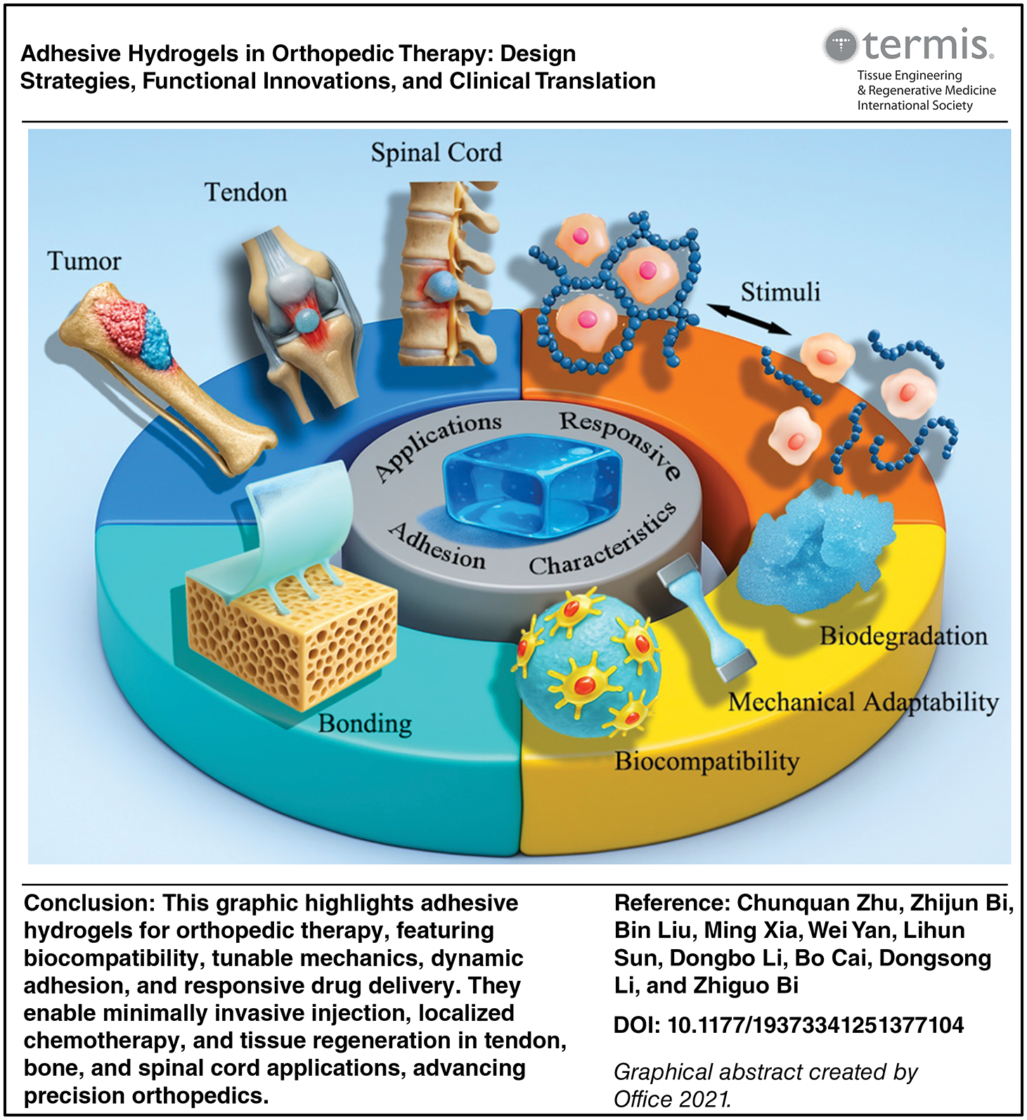
However, clinical adoption remains hindered by unresolved challenges. Key limitations include inadequate fatigue resistance under cyclic loading, unpredictable degradation rates in heterogeneous biological milieus, and insufficient understanding of long-term immune responses. Additionally, achieving selective adhesion—firmly bonding to target tissues while preventing off-target adhesion—requires innovative molecular engineering to avoid postoperative complications like tendon fibrosis. This review critically examines the evolution of adhesive hydrogels across five orthopedic frontiers: bone regeneration, cartilage repair, tendon healing, spinal cord reconstruction, and osteosarcoma therapy. We analyze design strategies that balance adhesion, mechanics, and bioactivity, while spotlighting translational barriers and emerging solutions. By bridging material innovation with unmet clinical needs, adhesive hydrogels hold transformative potential to redefine regenerative orthopedics (Graphical Abstract).
Functional Characteristics of Adhesive Hydrogels
Biocompatibility: the foundation of biological integration
Biocompatibility forms the cornerstone of hydrogel safety and functionality, encompassing the material’s capacity to interact with host tissues without provoking adverse immune responses or chronic inflammation. 13 Natural polymers such as collagen and gelatin inherently exhibit superior biocompatibility due to evolutionary conservation of their molecular motifs. For instance, gelatin methacryloyl (GelMA) hydrogels containing higher Arginine-Glycine-Aspartic Acid (RGD) density demonstrate better adhesiveness compared with lower counterparts. 14 Synthetic systems achieve comparable biocompatibility through biofunctionalization strategies—poly(ethylene glycol) (PEG) hydrogels grafted with fibronectin-mimetic peptides enhance supports nerve growth. 15 Emerging immunomodulatory designs incorporate interleukin-4 (IL-4)-loaded hydrogel, which reduce proinflammatory classically activated macrophage (M1 macrophage) polarization while enhancing tissue remodeling through transforming growth factor beta 1 (TGF-β1) secretion. 16 These advancements highlight the shift from passive biocompatibility to active immune regulation, enabling hydrogels to dynamically participate in tissue repair processes.
Mechanical adaptability: Reconciling strength and flexibility
The mechanical performance of adhesive hydrogels must balance two conflicting demands: sufficient toughness to withstand physiological loads and tissue-like compliance to prevent stress shielding. Architectural innovations address this dichotomy through molecular-level engineering. Double-network hydrogels exemplify this approach, combining rigid poly(2-acrylamido-2-methylpropanesulfonic acid) primary networks with ductile poly(acrylamide) secondary networks to achieve fracture energies up to 700/m2. 17 Moreover, the multireversible connections formed between polyvinyl alcohol, borax, oligomeric procyanidin, and ferric ion exhibited an ultra-stretchability of 100 times, a tissue-adhesive strength of 24 kPa, quick shape adaptation within 2 minutes (min), and a self-healing capability within 40 seconds (s). 18
Recent breakthroughs in photopolymerization have redefined mechanical performance benchmarks. The thioester-based hydrogel developed by Lin et al. achieves unprecedented toughness (138 MJ/m³) through controlled dithiol-diacrylate click chemistry under 405 nm light. 19 Such advancements enable applications in high-load environments such as intervertebral disc replacements, where hydrogels must simultaneously mimic the nucleus pulposus’s compressibility and annulus fibrosus’s tensile strength.
Programmable degradation: Synchronizing with healing phases
Temporal control over hydrogel degradation is critical to align with the phased requirements of musculoskeletal repair. Enzymatically cleavable hydrogels utilize matrix metalloproteinase (MMP)-sensitive sequences (e.g., proline-valine-glycine-leucine-isoleucine-glycine) that degrade at rates proportional to local MMP-2 concentrations (50 nM), ensuring gradual mechanical support reduction as native tissue regenerates. 20 H2O2 concentrations at the wound site range from 100 to 250 μM during the early inflammatory phase, which occurs 2 days after damage, and the later phase, which occurs five days after injury. In wound management, oxidation-responsive systems with thioketal linkages maintain structural integrity until reactive oxygen species levels exceed 250 mM H2O2, allowing controlled epidermal growth factor (EGF) release activity during the inflammatory phase. 21
Hybrid degradation systems integrate multiple mechanisms for staged therapeutic delivery. Dual-crosslinked hydrogels combine gelatin and collagen matrix. Nanometric vesicles based on poly(styrene-b-ethylene oxide) block copolymer (BCPVs) containing adapalene (AD) were prepared using the cosolvent approach. Together with free AD and silver sulfadiazine (SSD), the BCPVs were added to collagen and gelatin matrices. It was demonstrated that the gelatin and collagen matrix-based hybrid delivery systems functioned as a skin dressing that combined a slower, longer-term release of AD to promote skin healing with a gradual release of significant quantities of SSD within the first few hours of usage (to limit infection). 22
Performance validation: Bridging laboratory and clinic
The evaluation of adhesive hydrogel performance involves multiple dimensions, including mechanical properties, physicochemical properties, and biological properties. 23 Mechanical testing within physicochemical properties primarily measures the bonding strength and burst pressure of hydrogels, while physicochemical properties focus on degradation behavior and swelling properties. Biological properties aim to monitor the cell compatibility, tissue compatibility, immune response, and the impact on tissue healing of these hydrogels (Table 1).
Functional Characteristics of Adhesive Hydrogels
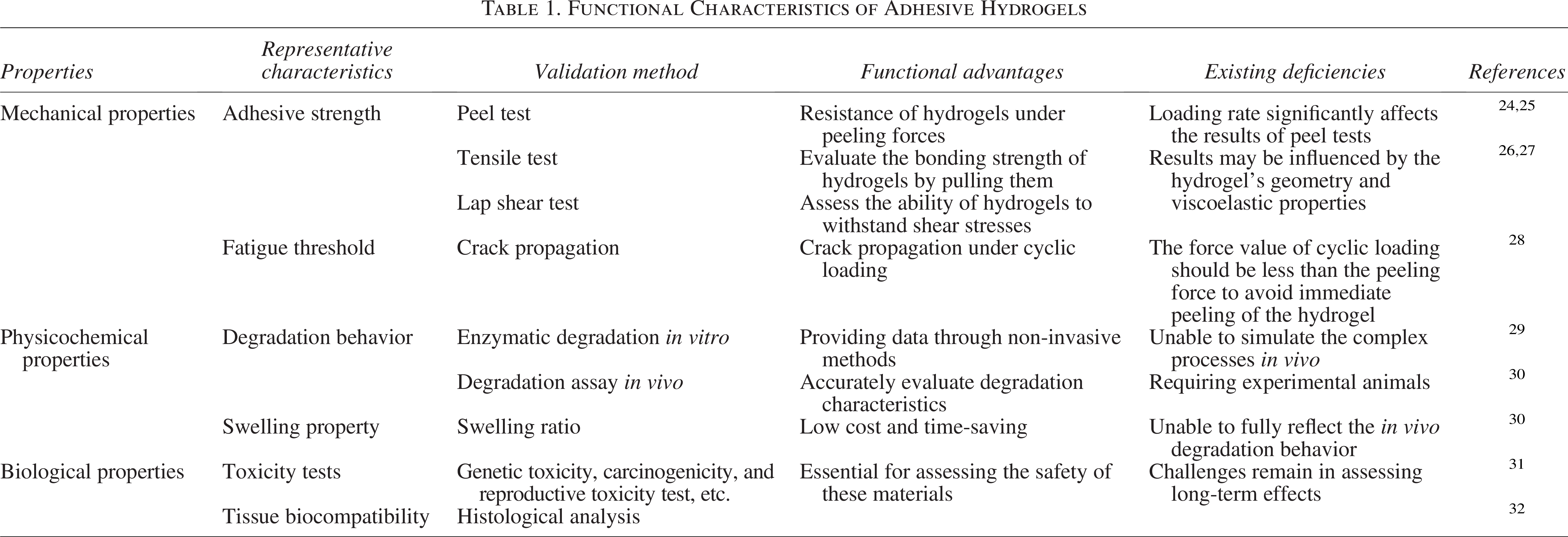
In terms of mechanical properties, bond strength or bonding energy is a key parameter for evaluating adhesive hydrogels, typically obtained through peel, tensile, and lap shear tests. 33 Peel tests measure the resistance of hydrogels under peeling forces, with the T-peel test being popular due to its tensile characteristics at the adhesive interface. 24 It’s worth noting that the loading rate significantly affects the results of peel tests. 25 Tensile tests evaluate the bonding strength of hydrogels by pulling them, while lap shear tests assess the ability of hydrogels to withstand shear stresses, which is crucial for simulating clinical applications.26,27 However, these test results may be influenced by the hydrogel’s geometry and viscoelastic properties and may not fully reflect tissue characteristics and specific patient conditions, thus requiring further evaluation using in vitro models.
The fatigue threshold of hydrogels is evaluated through crack propagation under cyclic loading, commonly employing 90° or T-peel tests. The force value of cyclic loading should be less than the peeling force to avoid immediate peeling of the hydrogel. 28 By calculating the derivative of crack size and cycle count, the interface crack propagation rate and fatigue threshold can be derived. A higher fatigue threshold indicates stronger fatigue resistance of the hydrogel. 34
In terms of physicochemical properties, medical hydrogels must undergo rigorous safety performance evaluations, including degradation behavior and swelling property tests. 29 Degradation performance is typically assessed by placing the hydrogel in a physiological medium containing enzyme. The degradation rate and extent are analyzed by observing and recording changes in volume, surface cracks, etc. Although in vitro experiments can provide initial estimates, they do not fully simulate the complex processes in vivo. 30 Regular sampling and weighing after in vivo implantation can more accurately evaluate degradation characteristics, albeit requiring a large number of experimental animals. To address this issue, researchers have developed non-invasive monitoring techniques such as real-time fluorescence imaging and magnetoelastic resonance sensors. 35 Additionally, by monitoring changes in mechanical properties over time, the degradation degree of hydrogels can be indirectly evaluated.
Swelling properties are another important parameter for evaluating adhesive hydrogels, closely related to the degree of crosslinking. The water absorption rate of hydrogels decreases with increasing crosslinking density, and the swelling ratio is typically calculated by measuring weight or volume changes. 36 Although in vivo implantation can provide more physiologically relevant data, in vitro determination methods are preferred due to their low cost and time-saving characteristics.
Biological performance evaluation involves culturing cells using adhesive hydrogels and monitoring cell viability to assess biocompatibility. At the same time, it is important to have a full understanding of how immune cells work in bone formation and how they interact with hydrogels. 37 The International Organization for Standardization (ISO) has proposed standardized biological assay methods (ISO-10933) to regulate the commercialization and clinical translation of h0ydrogels. These include genetic toxicity, carcinogenicity, and reproductive toxicity tests, in vitro cytotoxicity tests, local reaction tests after implantation, irritation and sensitization tests, and systemic toxicity tests. 31 These in vitro experiments can rapidly detect the biological effects of adhesive hydrogels and are commonly used to evaluate cytotoxicity. Histological analysis is the standard method for determining in vivo tissue biocompatibility, with hematoxylin and eosin staining and Masson’s trichrome staining being the most commonly used staining techniques. These can detect the degree of tissue inflammation and changes in tissue structure caused by hydrogels. 32
Adhesion Mechanisms of Adhesive Hydrogels
Physical interactions at the interface
Hydrogen bonding networks
The interfacial adhesion between hydrogels and biological tissues involves sophisticated molecular interactions that overcome the inherent challenges posed by hydrated environments and tissue heterogeneity. This section delineates the physicochemical principles governing hydrogel-tissue integration, with emphasis on emerging strategies to enhance adhesion durability and biological specificity.
Adhesive hydrogels can form adhesion with biological tissues through hydrogen bonding and hydrophobic interactions. 38 Functional groups like hydroxyl and carboxyl groups in viscous hydrogels commonly used in orthopedics can interact with corresponding groups on the tissue surface through the formation of hydrogen bonds, achieving adhesion. Hydrogen bonding (bond energy 5–40 kJ/mol) serves as a primary adhesion mechanism, particularly between hydrogel functional groups (–OH, –COOH) and tissue macromolecules.39–42 Besides, synthesized naphthalene-diimide derivative achieves 15 to 20 kJ/mol interaction strength through π-π stacking. 43 These mechanisms enable autonomic self-healing and shear-thinning injectability, critical for minimally invasive delivery. 44 However, time-dependent mechanical relaxation limits their utility in load-bearing scenarios. 45
Chemical crosslinking, in contrast, employs covalent bonds formed via photopolymerization, enzyme-mediated conjugation, or click chemistry. 46 These methods provide robust mechanical integrity (fracture energy up to 4686 J/m2 in double networks) 47 and precise control over degradation kinetics, as exemplified by triblock copolyether. 48 Dynamic covalent chemistry further bridges the gap between stability and adaptability, for example, disulfide exchange networks respond to physiological glutathione levels, 49 while imine bonds enable pH-triggered reconfiguration. 50
Hydrophobic synergy
The presence of interfacial water can hinder the adhesion of hydrogels. When two hydrophobic substances approach each other, the conformational rearrangement of water molecules leads to hydrophobic interactions. 51 Compared to other reversible interactions such as ionic bonds and hydrogen bonds, hydrophobic interactions may exhibit attractive forces over longer ranges (100–200 Å), potentially conferring superior stretchability to the hydrogel material. 52 Cellulose nanocrystal-reinforced systems demonstrate this principle, achieving elongation through aligned hydrophobic domains while maintaining strong adhesion to tissue surfaces. 53
Covalent bonding strategies
Adhesive hydrogels can achieve robust tissue integration through covalent bonding mechanisms. Specifically, functional groups within hydrogels undergo covalent reactions with surface moieties on biological tissues to form stable chemical linkages, including disulfide bonds, Schiff bases, N-hydroxysuccinimide esters (NHS-esters), and thiol-ene bonds. 54 Among these, Schiff bases formed between aldehyde/keto groups and amino groups represent dynamic covalent bonds. 55 Given the abundance of amino groups in tissue proteins, aldehyde-functionalized compounds have emerged as promising candidates for designing body-adhesive hydrogels. For instance, Du’s team developed dual-aldehyde cellulose (DAC) through cellulose functionalization and constructed a double-network hydrogel (PAM/DAC-2Gel) by crosslinking DAC with gelatin (Gel) via Schiff base chemistry, followed by integration with polyacrylamide (PAM). By modulating free and bound water content, they engineered hydrogel materials with enhanced interfacial adhesion in humid environments and conductivity for sensing applications. 56
Disulfide bonds, another covalent strategy, leverage thiol groups present in natural proteins or thiol-modified polymers to achieve tissue adhesion. 57 Building on this principle, Ren et al. engineered a self-healing hydrogel adhesive using hydrazide-modified hyaluronic acid (HA) and o-phthalaldehyde (OPA)-terminated four-armed polyethylene glycol (4aPEG-OPA). The OPA/nucleophile condensation generated dynamic hydrazone bonds for intrinsic self-healing, while stable phthalimide bonds formed between OPA and tissue amines ensured strong adhesion. Incorporating disulfide bonds enabled controlled hydrogel degradation over 6–22 weeks. In animal models, this adhesive outperformed commercial fibrin and cyanoacrylate glues in sealing hepatic/vascular injuries and accelerating full-thickness skin wound healing. 54
NHS-esters provide a distinct covalent approach by reacting with tissue amines to form stable amide linkages. YZheng et al. devised a multifunctional hydrogel combining NHS-conjugated alginate (Alg-NHS), poly(ethylene glycol) diacrylate (PEGDA), tannic acid, and Fe³+ ions. Through synergistic covalent (NHS-amide) and noncovalent (metal-phenolic/ionic) interactions, they achieved an ultra-stretchable (924% strain) and tough (4697 kJ/m³) hydrogel capable of rapid (<5 s) wet tissue adhesion while supporting physiological functions. 58
Thiol-ene click chemistry offers rapid reaction kinetics and minimal byproducts for tissue adhesion. Qi et al. engineered a glucose-responsive hydrogel using boronic acid-modified gelatin and thiolated HA to encapsulate AuPt@melanin nanoparticles. Initial thiol-ene crosslinking formed a weak gel covering wounds, which was subsequently strengthened by UV-induced radical polymerization. Combined with NIR-triggered thermotherapy, this system promoted macrophage polarization from M1 to M2 phenotypes, effectively treating diabetic foot ulcers in rats. 59 Among these covalent strategies, Schiff bases and NHS-esters dominate current clinical applications due to their reliability and adaptability.
Beyond intrinsic covalent mechanisms, hydrogel adhesion can be augmented through physicochemical modifications without altering chemical compositions. Temporal control of chain interactions (e.g., hydrogen bond “time-domain regulation”) and “Janus network” designs improve mechanical properties while enabling functionalization. 60 Introducing reactive groups (e.g., carboxyls for electrostatic adhesion or thiolated chitosan for dynamic disulfide bonds) enhances interfacial bonding.61,62 Notably, incorporating hydrophobic poly(vinyl butyral) into poly(acrylic acid) hydrogels reduced water permeation without dense crosslinking, achieving 211.4 kPa adhesion strength and a low swelling ratio (1.2). 63
Emerging hybrid systems synergize both approaches to resolve historical trade-offs. For instance, poly(vinyl alcohol)-polydopamine-tannin acid hydrogels combine high fracture toughness with strong underwater adhesion, addressing the competing demands of mechanical resilience and interfacial bonding in dynamic musculoskeletal environments. 64 Advanced fabrication techniques now integrate topographical cuesand spatiotemporal control over bioactive factor release. 65 These innovations underscore the critical balance required between adhesion-cohesion optimization (via polymer chain length and crosslink density tuning) 66 and degradation-bioactivity coupling (controlled through hydrolyzable segments like ester groups with tunable hydrolysis rates). 67 Such design principles are pivotal for engineering hydrogels capable of withstanding cyclic physiological loads while promoting tissue regeneration (Fig. 1).
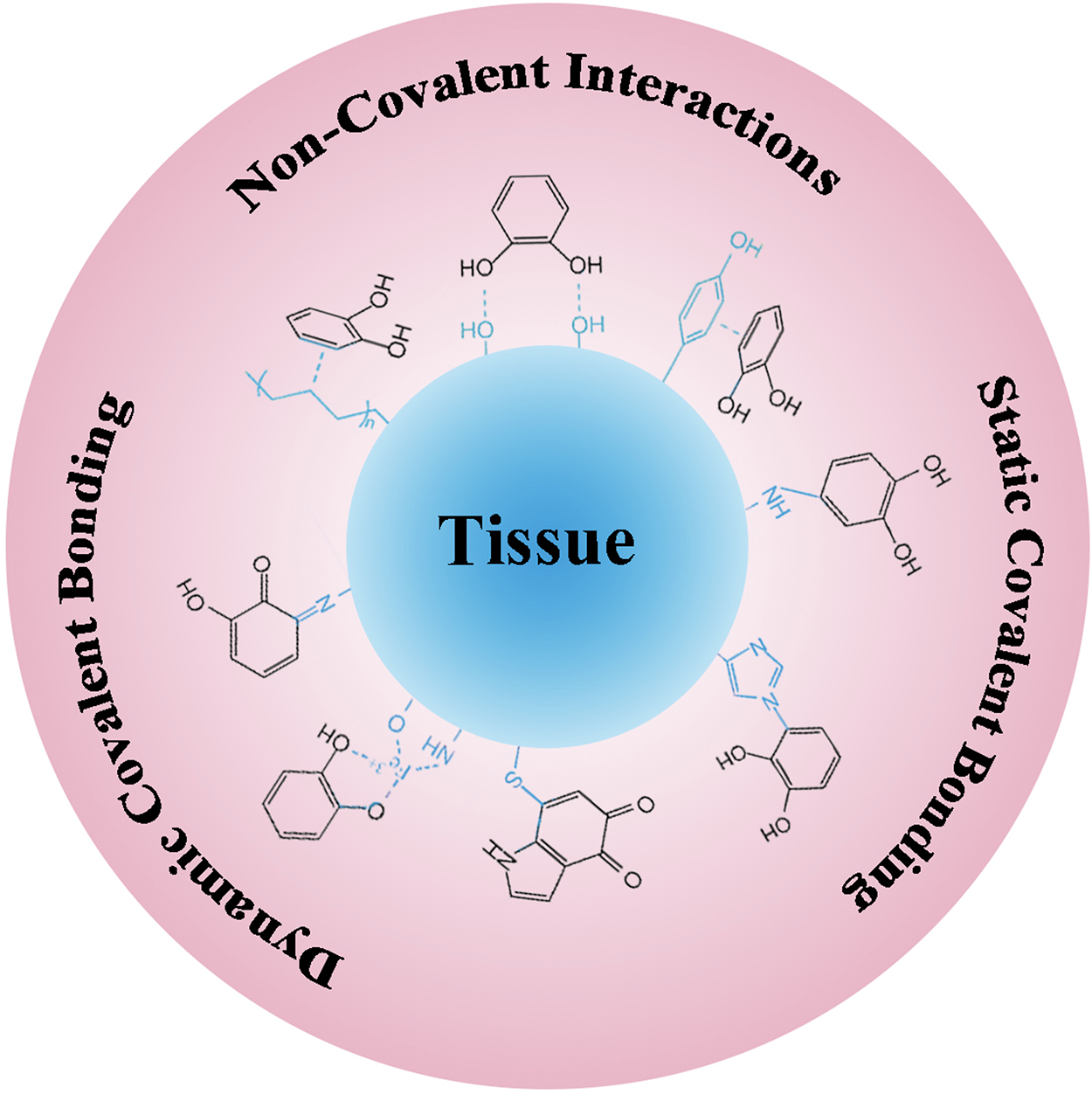
Design strategies of tissue adhesion based on various adhesion mechanisms.
Stimuli-Responsive Adhesive Hydrogels: Mechanisms and Applications
Temperature- and pH-Responsive systems
The responsiveness of adhesive hydrogels refers to their capacity to sense physicochemical stimuli (e.g., temperature, pH, light, redox signals) and undergo triggered transformations in 3D morphology, phase state, or functional properties, enabling injectability, self-healing, shape memory, and dynamic adhesion. 68
Thermoresponsive hydrogels leverage ambient-to-physiological temperature gradients to modulate adhesion. For instance, poly(N-isopropylacrylamide) derivatives exhibit a lower critical solution temperature (LCST) near 37°C, undergoing sol-gel transitions suitable for thermally targeted bone regeneration. 69 Similarly, polyphenol-protein coordination hydrogels maintain structural stability at body temperature while enhancing molecular chain mobility, achieving rapid adhesion with sustained interfacial integrity. 70
pH-sensitive hydrogels rely on protonation/deprotonation of functional groups (e.g., carboxyl, amino) to alter charge states and intermolecular interactions. 71 Guo et al. developed a dual pH/glucose-responsive hydrogel where Schiff base dissociation under acidic conditions and competitive glucose binding enable controlled drug release. L-arginine-enhanced catechol groups further ensured robust tissue adhesion. 72
Redox- and Light-Responsive strategies
Redox-responsive systems utilize dynamic covalent bonds (e.g., disulfide linkages) or catalytic nanoparticles for tunable adhesion. 73 A notable example involves Ag-lignin nanoparticle (Ag-Lignin NP)-embedded hydrogels, where continuous catechol regeneration via redox cycling provides durable adhesion (>14 days) alongside antibacterial activity. 74 Reversible disulfide-based oxidation-reduction reactions further enable on-demand interfacial adhesion/deadhesion across diverse substrates. 75
Light-responsive hydrogels achieve spatiotemporal control through photoactive moieties (e.g., coumarin, o-nitrobenzyl groups). 76 Photodimerization allows reversible crosslink density modulation, while photolytic cleavage enables irreversible network degradation. 77 Strategic incorporation of these groups into polymer backbones or side chains permits precise adhesion tuning under specific wavelengths. 78
Synergistic adhesion mechanisms
Strong hydrogel adhesion arises from synergistic polymer chemistry, topological design, and energy dissipation. Adhesive strength depends on both bulk cohesion and interfacial bonding. Ren et al. demonstrated this via catalyst-free o-phthalaldehyde/amine (hydrazide) crosslinking, achieving rapid tissue adhesion with injectable HA-PEG hydrogels. Lap shear tests revealed concentration-dependent adhesion strengths (18.0 ± 1.8 kPa at 4% to 30.3 ± 4.6 kPa at 10% w/v), surpassing commercial fibrin glue and benzaldehyde/hydrazide systems. 54
Multistimuli-Responsive integration
Combining multiple responsiveness overcomes limitations of single-stimulus systems. Chen et al. engineered tough heterogeneous nanocomposite hydrogels using clay nanosheets as crosslinkers for mechanical robustness, with graphene oxide (GO) and Fe3O4 nanoparticles imparting multistimuli sensitivity. Hydrogen bond-mediated interfacial assembly enabled complex architectures responsive to heat, light, and magnetic fields. 79
These combinatorial strategies allow spatiotemporal regulation of hydrogel properties to address dynamic microenvironments during tissue healing. For instance, thermoresponsive hydrogels enable minimally invasive delivery for bone defect repair, while redox/pH dual-responsive systems adapt to inflammatory phase changes in chronic wounds. Future designs may integrate biosensing elements for real-time microenvironment adaptation.
Biomedical Applications of Adhesive Hydrogels in Orthopedics
Tendon repair
Tendons, composed of hierarchically organized collagen fibers within an anisotropic extracellular matrix (ECM), exhibit remarkable mechanical strength through specialized tendon-to-bone insertion sites, enabling robust force transmission between muscles and skeletal structures.80,81 However, their hypovascular nature and limited cellularity severely restrict intrinsic repair capacity, particularly under pathological conditions such as overuse, trauma, or age-related degeneration. 82 Current surgical interventions, including suturing and grafting, often lead to incomplete functional restoration, postoperative adhesions, or infection risks, with a high recurrence rate of tendon rupture.83,84 To address these limitations, advanced adhesive hydrogels have been engineered to mimic tendon-specific biomechanical properties while enabling targeted therapeutic delivery.
A breakthrough was achieved by Choi et al., who developed a triple-network (TN) hydrogel combining imidazole-functionalized polyasparamide (providing multiple hydrogen bonds for bone adhesion) with energy-dissipating alginate-PAM networks. Through uniaxial stretching and secondary crosslinking, this anisotropic hydrogel achieved tendon-like tensile modulus (12 MPa) and interfacial adhesion strength (>800 J/m2) without chemical modification of bone surfaces. Remarkably, it successfully replicated native ligament’s bone-to-ligament-to-bone architecture, demonstrating clinical potential for load-bearing applications 85 (Fig. 2). Complementing structural mimicry, Benjamin R. Freedman’s Janus hydrogel integrated a tough PAM matrix with chitosan-based adhesive, achieving record tendon adhesion energy (>1,000 J/m2) in live rat models. Sustained release of triamcinolone acetonide from this system reduced peritendinous fibrosis by 60% compared to controls, addressing the critical challenge of postoperative scar formation. 86 These innovations exemplify the dual strategy of biomechanical optimization and precision pharmacotherapy in next-generation tendon repair systems.
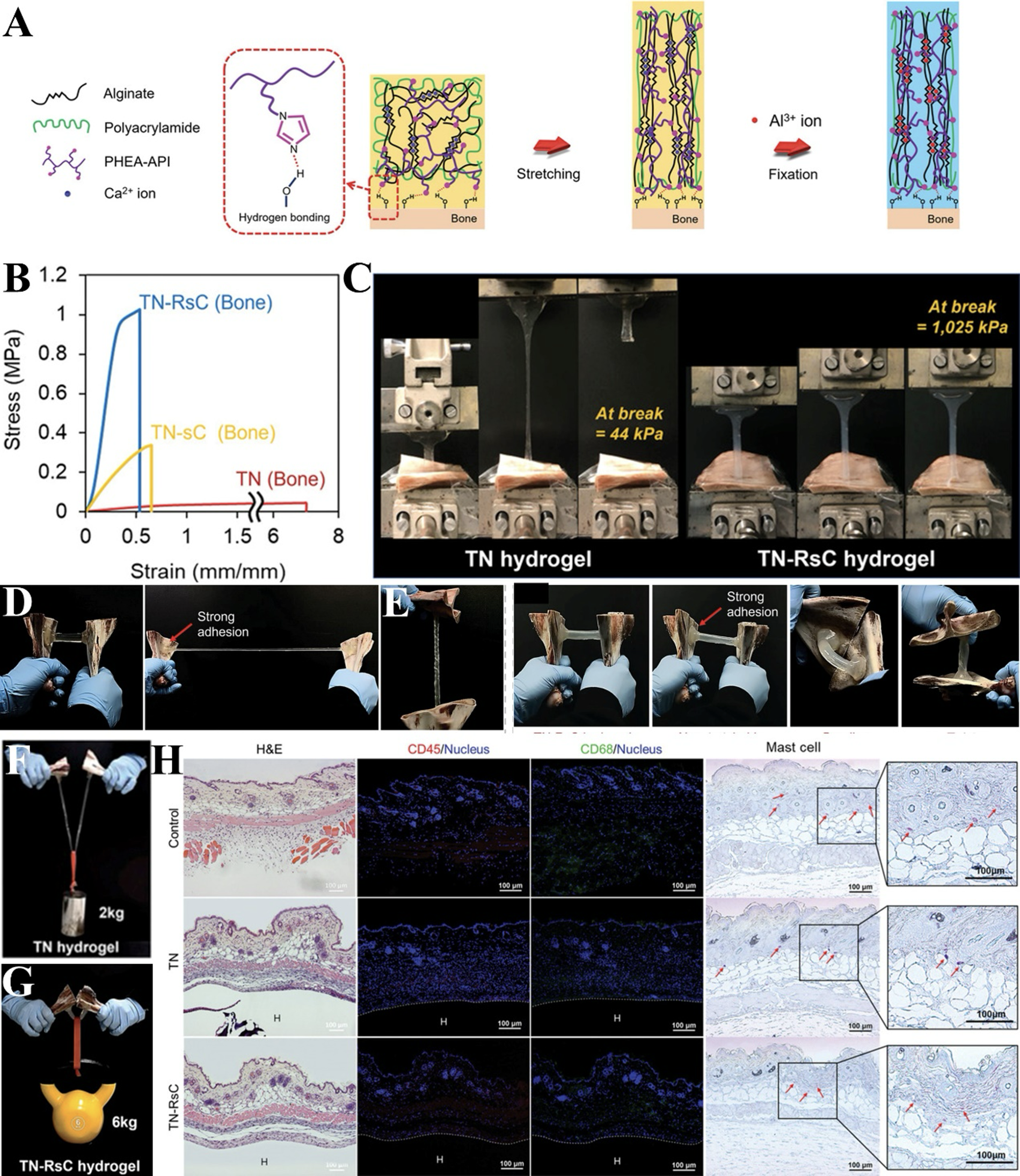
Bioinspired Anisotropic Hydrogel with Bone Adhesion Mimics Tendon Enthesis.
Bone regeneration
Bone tissue engineering faces significant challenges in addressing critical-sized defects caused by trauma, tumors, or pathological fractures, where conventional therapies such as metal fixation and autografts often lead to complications like immune rejection, donor site morbidity, or limited adaptability to irregular geometries.87–90 Adhesive hydrogels have emerged as transformative solutions by combining injectability, ECM-mimetic microenvironments, and programmable drug release. Their high-water content (>90%) and tunable mechanical properties (elastic modulus: 1–50 kPa) facilitate cell infiltration and osteogenic differentiation, while robust tissue adhesion (>50 kPa) ensures stable integration with moist bone surfaces, reducing postoperative displacement risks.91,92
Inspired by mussel adhesion mechanisms, Hu et al. engineered an osteogenic hydrogel incorporating polyvinyl alcohol, L-dihydroxyphenylalanine (DOPA), and catechol-Fe³+ coordinated zeolitic imidazolate framework-8 (ZIF-8). This system achieved unprecedented bone adhesion strength (10 MPa)—10-fold higher than commercial cyanoacrylate adhesives—and successfully stabilized comminuted femoral fractures in rabbits. The sustained release of Ca2+/PO4³− from anchored Bio-Oss particles further enhanced fracture repair by promoting osteoblast mineralization 93 (Fig. 3).

A Mechanically Reinforced Hydrogel Enables Strong Hard Tissue Adhesion and Enhanced Bone Regeneration.
With their excellent biocompatibility and porous structure, hydrogels can be loaded with a variety of drugs and bioactive substances, while their tunable properties allow for customized release kinetics and targeting to specific sites. Among them, exosomes are membrane-bound vesicles secreted by cells that act as intercellular messengers carrying proteins, lipids, and nucleic acids to other cells, where they play a role in a variety of physiological and pathological processes. Exosome donor cells can be cocultured with material, lentivirally engineered, overexpressing or knocking down functionally relevant miRNAs, and stimulated by hypoxia to modulate exosome production, targeting capacity, and function related to bone regeneration. A biphasic hydrogel, featuring interpenetrating GelMA and DNA networks, synergized static mechanical stability with dynamic stress relaxation. Functionalized with bone marrow-derived mesenchymal stem cell (BMSC)-recruiting exosomes, this system sustained vascular endothelial growth factor (VEGF) release over 21 days, increasing neovascularization by 2.5-fold in rat calvarial defects compared to acellular controls. 94 Chen et al. advanced this paradigm with an injectable HA-based hydrogel coloaded with stromal cell-derived factor 1α (SDF-1α) and M2 macrophage-derived exosomes (HA@SDF-1α/M2D-Exos), which reduced bacterial load by 3 logs through inherent antimicrobial activity while enhancing human umbilical vein endothelial cell (HUVEC) migration and collagen deposition via exosome-mediated PI3K-Akt signaling. 95
Cartilage repair
Cartilage, an avascular tissue comprising chondrocytes embedded within a collagen-proteoglycan matrix, is essential for load-bearing and joint mobility but exhibits limited regenerative capacity due to its sparse cell density, slow matrix turnover, and absence of vascularization.96,97 While cell-based therapies such as autologous chondrocyte implantation offer partial solutions, challenges persist in donor cell availability, fibrocartilage formation, and functional restoration.98,99 Adhesive hydrogels have emerged as multifunctional platforms to address these limitations by integrating localized drug delivery, stem cell recruitment, and genetic engineering within a biomimetic microenvironment.
Conventional anti-inflammatory therapies using NSAIDs or corticosteroids, though effective in pain management, carry risks of systemic complications including osteonecrosis (4–6% incidence) and muscle atrophy.100–103 Adhesive hydrogels mitigate these risks through spatially controlled release mechanisms. For instance, platelet-rich plasma (PRP)-loaded hydrogels reduce cartilage-degrading MMP expression while enhancing matrix viscosity via fibrin network formation, demonstrating dual therapeutic and structural benefits 104 (Fig. 4). Beyond small molecules, mesenchymal stem cells (MSCs) encapsulated within methacrylated hyaluronic acid hydrogels have shown remarkable efficacy, suppressing proinflammatory IL-1β levels and increasing collagen II deposition. 105 To circumvent the scarcity of primary articular chondrocytes, nasal chondrocytes—which exhibit 85% similarity in glycosaminoglycan (GAG) content—have been successfully incorporated into adhesive hydrogels, maintaining phenotypic stability for over four weeks in vitro. 106
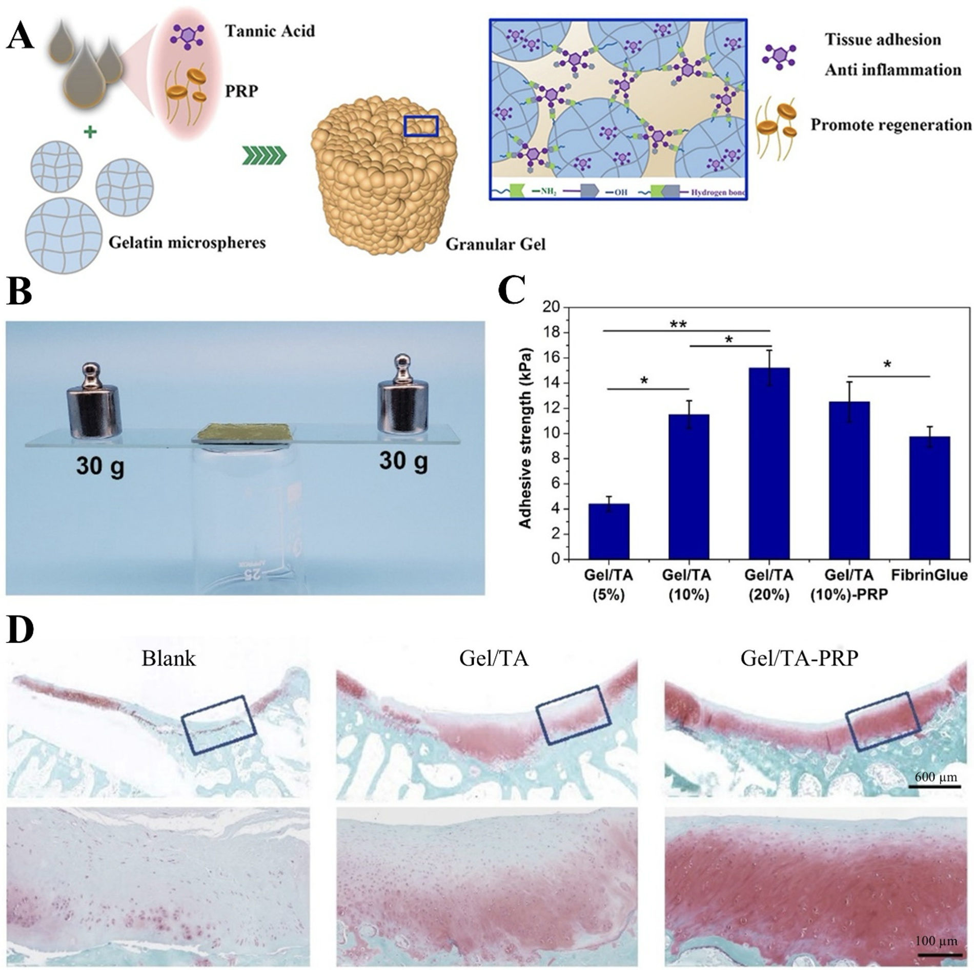
Injectable platelet-rich plasma (PRP)-Based Granular Hydrogel Combines Tissue Adhesion and ROS Scavenging to Promote Cartilage Repair.
Advancements in gene-activated hydrogels further expand therapeutic possibilities. A pioneering study demonstrated that Sox9 plasmid DNA-loaded hydrogels achieved sustained transfection efficiency, upregulating aggrecan expression and enhancing cartilage repair. 107 These systems synergize with mechanically adaptive networks mimicking native cartilage’s compressive modulus, essential for replicating dynamic joint loading. However, clinical translation faces hurdles such as scalable cell sourcing and mismatched degradation rates. 108 Future designs must integrate patient-specific mechanics, dynamic adhesion, and feedback-controlled drug release to bridge the gap between preclinical promise and clinical reality.
Spinal cord injury repair
The spinal cord, a critical component of the central nervous system, extends from the brainstem to the lumbar region (terminating at L1-L2 in adults) and serves three primary functions: transmitting motor commands, relaying sensory input, and coordinating reflexes.109,110 Traumatic injuries, infections, or congenital anomalies can cause spinal cord injury (SCI), leading to axonal degeneration, neuronal loss, demyelination, and chronic inflammation. These pathological changes result in permanent motor, sensory, and autonomic dysfunction, often accompanied by systemic complications such as neuropathic pain and respiratory impairment.111,112 The limited regenerative capacity of adult neurons in the spinal cord makes functional recovery after SCI a formidable challenge. 113
Adhesive hydrogels have emerged as promising platforms to address these challenges by reconstructing the neural microenvironment. Their ability to mimic the extracellular matrix facilitates nutrient exchange with surrounding tissues, while their adhesive properties enable stable integration with irregular lesion sites, creating a growth-permissive niche for axonal regeneration.114–116 For instance, Liu et al. developed an in situ-forming hydrogel composed of glycidyl methacrylate-modified silk fibroin and laminin acrylate. Upon UV exposure, this system rapidly polymerized, forming topological entanglements with spinal tissue at the molecular level. In rat models, the hydrogel promoted significant neural regeneration and restored hindlimb motor function within 8 weeks. 117 Xiao et al. further advanced this approach using a chitosan-DOPA/peptide adhesive hydrogel, which bridged spinal cord stumps through robust wet adhesion while secreting neurotrophic factors, enhancing axonal sprouting compared to nonadhesive controls 118 (Fig. 5).
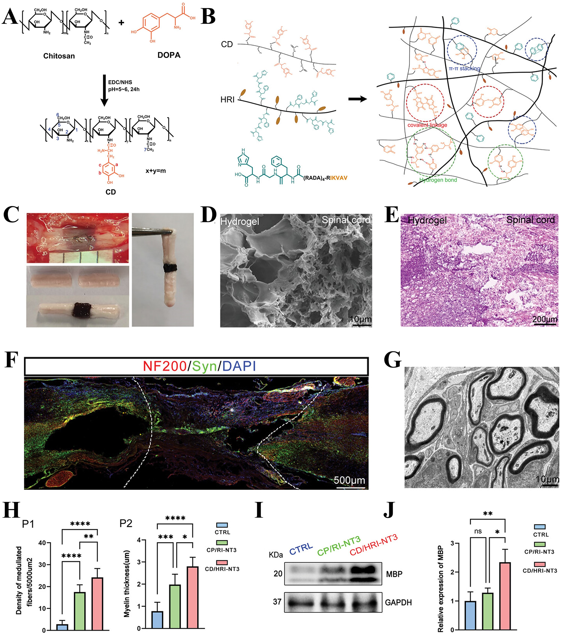
Injectable, Adhesive, and Self-Healing Dual-Network Hydrogel Promotes Neural Regeneration after Spinal Cord Injury.
Beyond structural support, adhesive hydrogels enable precision delivery of therapeutic agents. A dopamine-functionalized HA hydrogel demonstrated dual functionality: its mussel-inspired adhesion mechanism achieved strong interfacial strength in wet conditions, while sustained ibuprofen release reduced proinflammatory TNF-α levels. 119 MSC-derived exosomes, encapsulated within adhesive hydrogels, have shown exceptional immunomodulatory potential. These exosome-laden systems polarized macrophages toward the anti-inflammatory M2 phenotype and enhanced synaptic plasticity markers, significantly improving locomotor recovery. 120
Bone tumor therapy
Osteosarcoma, the most prevalent primary malignant bone tumor, predominantly affects adolescents and young adults (10–20 years old) with an annual global incidence of 2–3 cases per million. 121 Achieving complete tumor eradication remains challenging due to poorly defined margins in 30%–40% of cases, leading to recurrence rates of 80%–90% postsurgery and a dismal 5-year survival rate (<28%) for recurrent disease.122,123 While current standard therapies combining wide resection and neoadjuvant chemotherapy have improved 5-year survival to 68%, systemic toxicity and suboptimal intratumoral drug distribution limit their efficacy.124–126 Adhesive hydrogels address these limitations by enabling localized, sustained drug delivery directly to tumor sites, enhancing therapeutic precision while minimizing off-target effects.127,128
Paclitaxel (PTX), a cornerstone chemotherapeutic agent, exemplifies the challenges of conventional administration. Taxol®—a PTX formulation solubilized in polyoxyl castor oil and ethanol—induces severe hypersensitivity reactions (incidence: 20%–40%), necessitating premedication with high-dose corticosteroids.129,130 Although albumin-bound PTX (nab-PTX) reduces allergic risks, it incurs higher costs and increases peripheral neuropathy incidence. 131 Wang et al. circumvented these issues using an o-phthalaldehyde-terminated PEG hydrogel that covalently binds tissue amines, achieving stable tumor adhesion and sustained PTX release over 30 days. This approach reduced systemic exposure compared to intravenous PTX while increasing tumor apoptosis, demonstrating superior efficacy in murine osteosarcoma models 132 (Fig. 6).
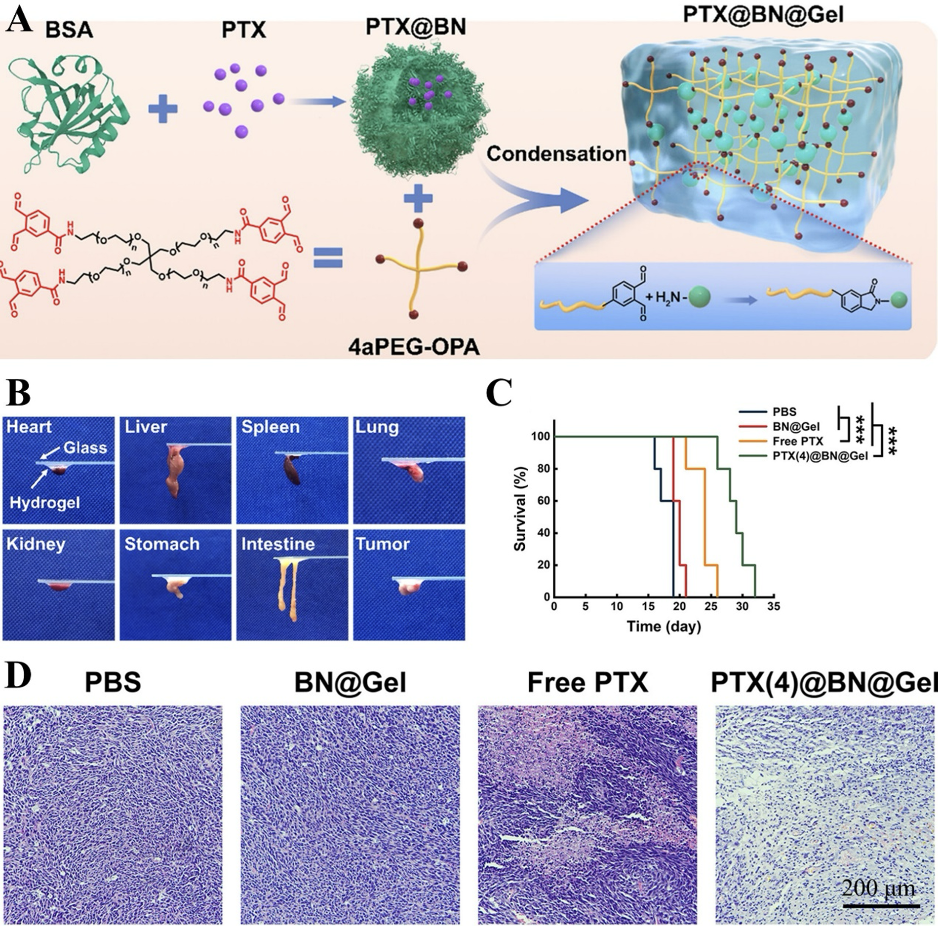
Injectable Adhesive Hydrogel with Drug-Loaded Nanoparticles Enables Sustained Tumor Therapy.
Emerging strategies synergize chemotherapy with immunomodulation. Chu et al. developed a nanocomposite hydrogel codelivering Mg2+, anti-PD-L1 antibodies, and the Hedgehog inhibitor vismodegib. The sustained Mg2+ release enhanced osteogenic differentiation, while localized PD-L1 blockade increased tumor-infiltrating CD8+ T cells, achieving dual osteogenesis and immunotherapeutic effects in rat models. 133 Such multifunctional systems exemplify the potential of adhesive hydrogels to transform osteosarcoma management by integrating precise drug delivery, immune activation, and bone regeneration within a single platform.
Conclusions and Future Perspective
Adhesive hydrogels have emerged as transformative tools in orthopedics, driven by advances in bioinspired design, dynamic chemical bond engineering, and multifunctional integration. These materials combine mechanical compatibility with robust tissue adhesion and bioactive functionality, enabling them to mimic native extracellular microenvironments while promoting cell recruitment, differentiation, and targeted therapeutic delivery. Innovations such as injectability, wet adhesion, and localized sustained-release mechanisms have unlocked minimally invasive applications and personalized treatment strategies for complex conditions, including chronic wounds, spinal cord injuries, and osteosarcoma, demonstrating superior efficacy over conventional therapies.
Despite these breakthroughs, critical challenges impede clinical translation. Persistent issues include optimizing dynamic mechanical matching between hydrogels and host tissues, ensuring long-term interfacial stability under physiological loads, and mitigating immune responses during chronic implantation. A paramount unmet need lies in achieving spatiotemporal control over adhesion strength—ensuring durable integration with target tissues while preventing postoperative adhesions—a goal that demands innovative material designs, such as stimuli-responsive systems adaptable to biochemical or mechanical cues. Furthermore, the growing demand for personalized therapies necessitates intelligent hydrogels capable of autonomous drug release modulation or patient-specific structural customization via 3D bioprinting.
Future progress hinges on interdisciplinary collaboration across materials science, biology, and clinical medicine. Priorities include overcoming technical barriers in scalable manufacturing, establishing standardized protocols for long-term biosafety evaluation, and fostering industry-academia partnerships. By addressing these challenges, adhesive hydrogels are poised to transition from laboratory prototypes to clinical mainstays, revolutionizing orthopedic care through precision-driven solutions for tissue regeneration and functional restoration.
Ethics Approval and Consent to Participate
All participants provided their informed consent; the study was reviewed and approved by the First Hospital of Jilin University’s Ethics Committee, and all methods involving human subjects were performed out in accordance with the Declaration of Helsinki.
Consent for Publication
All the participants in our study consent for publication.
Authors’ Contributions
Z.B. conceived and presented the idea. C.Z. and Z.B. processed data and article writing. B.L. participated in the acquisition and interpretation of data. All the authors contributed to the article and approved the submitted version. All listed authors have made a significant scientific contribution to the research in the article approved its claims and agreed to be an author.
Footnotes
Funding Information
National Key Research and Development Program of China (2022YFC2405805). Jilin Provincial Scientific and Technological Development Program (20230204077YY).
Availability of Data and Materials
The data used in the study could be accessible from the correspond author.
Disclosure Statement
The authors declared no conflict of interest.