
Abstract
Central nervous system (CNS) injuries are a major cause of neurological impairment and long-term disability, driven by intertwined processes including neuroinflammation, oxidative stress, apoptosis, and disruption of the blood–brain barrier (BBB) or blood–spinal cord barrier (BSCB). This review summarizes the physicochemical features and neuroprotective mechanisms of tanshinone IIA (TanIIA), a lipophilic diterpene quinone from Salvia miltiorrhiza, and appraises preclinical evidence across CNS injury models. By synthesizing recent experimental findings from in vivo and in vitro models of cerebral ischemia and spinal cord injury, we focus on mechanistic pathways and functional outcomes. TanIIA shows BBB permeability and multi-target actions, including suppression of neuroinflammatory signaling, attenuation of oxidative damage, preservation of mitochondrial function, restoration of BBB/BSCB integrity, mitigation of aberrant glial activation, and promotion of neurological recovery. Emerging studies also suggest potential synergy with glucocorticoids, which may allow steroid dose reduction while maintaining efficacy in selected settings. While preclinical results are encouraging, key translational gaps remain, particularly regarding target specificity, pharmacokinetics, formulation, and clinically relevant dosing windows. Future work integrating multi-omics approaches with advanced disease-relevant models may accelerate the development of TanIIA-based therapeutics for CNS injuries.
Keywords
Introduction
The central nervous system (CNS) is highly vulnerable to endogenous and exogenous insults, and subsequent inflammatory responses can trigger interconnected pathological cascades that drive neurological dysfunction and long-term disability. Ischemic stroke and spinal cord injury (SCI) are among the most prevalent and clinically challenging forms of CNS injury. 1
Despite extensive research, effective disease-modifying treatments remain limited. 2 CNS injury involves multifactorial mechanisms, including oxidative stress, neuroinflammation, neuronal apoptosis, and dysregulated calcium homeostasis, which collectively restrict neuronal repair and regeneration. 3 In addition, disruption of blood–brain barrier (BBB) integrity and persistent expression of axonal growth-inhibitory molecules further aggravate neurodegeneration and impede functional recovery.
Traditional Chinese medicine (TCM) has long been used for CNS-related disorders and is increasingly investigated as a source of neuroactive natural products. Tanshinone IIA (TanIIA), a lipophilic diterpene quinone isolated from Salvia miltiorrhiza, has attracted attention for its multi-target pharmacology and its ability to penetrate the BBB. 4 Preclinical studies indicate that TanIIA confers neuroprotection in diverse CNS injury models, potentially through modulation of inflammation and oxidative injury as well as regulation of emerging cell-death pathways such as ferroptosis. 4 Given that many natural products (eg, curcumin and resveratrol) exhibit neuroprotective potential yet differ in pharmacokinetics and molecular targets, Table 1 summarizes key distinctions, emphasizing BBB penetration and mechanistic features relevant to TanIIA.
Comparative Overview of Antioxidant Mechanisms of Representative Traditional Chinese Medicine (TCM) Active Ingredients in CNS Injury Models.

In this review, we summarize the physicochemical characteristics and pharmacological mechanisms of TanIIA, appraise evidence from cerebral ischemia and SCI models, and outline translational priorities to facilitate the development of TanIIA-based therapies for CNS injuries.
Physicochemical Characteristics and Pharmacokinetics of TanIIA
Physicochemical Properties
TanIIA is a lipophilic diterpene quinone isolated from the roots and rhizomes of Salvia miltiorrhiza, a widely used medicinal herb in TCM. It has the molecular formula C19H20O3 and a molecular weight of 296 Da (Figure 1). 5 Historically, TanIIA has been used in TCM for promoting blood circulation and resolving blood stasis. Also known as tanshinone II, TanIIA forms reddish, needle-like crystals. 3 Due to its high lipophilicity (logP ≈ 5.8), TanIIA is practically insoluble in water but readily soluble in several organic solvents, including dimethyl sulfoxide (DMSO), ethanol, acetone, diethyl ether, and benzene. 5

Chemical structure of TanIIA.
The major pharmacologically active constituents of S. miltiorrhiza are broadly categorized into lipophilic diterpene quinones and hydrophilic phenolic acids. 6 Within the diterpene quinone fraction, TanIIA has attracted particular interest because of its diverse bioactivities, including improvement of microcirculation, anti-inflammatory effects, and antimicrobial properties. 7
TanIIA is able to cross the BBB, a feature commonly attributed to its lipophilicity; however, brain exposure can be limited by efflux transporters, including P-glycoprotein and multidrug resistance-associated proteins. Inhibition of these transporters has been reported to increase brain distribution of TanIIA from approximately 31% to 77%. 8 Furthermore, TanIIA has been designated as a quality-control marker for S. miltiorrhiza in the Chinese Pharmacopoeia, underscoring its importance for standardization of herbal preparations. 9
Pharmacokinetic Profile and Metabolism
Despite its diverse pharmacological activities, TanIIA shows pharmacokinetic limitations that may constrain clinical translation. TanIIA is classified as a Biopharmaceutics Classification System (BCS) class II compound, characterized by low aqueous solubility and high permeability. In rats, the absolute oral bioavailability of TanIIA is low (≈3.5%), which has been attributed to extensive hepatic first-pass metabolism. 10 After intravenous administration, TanIIA exhibits a relatively short elimination half-life (t1/2) of approximately 1–2 h. 11 TanIIA is primarily metabolized in the liver, with hydroxylation reported as a major biotransformation route mediated by cytochrome P450 enzymes, including CYP2A6. In addition, TanIIA has been reported to inhibit CYP1A2, suggesting a potential for pharmacokinetic drug–drug interactions when co-administered with CYP1A2 substrates. 12
Multi-Target Neuroprotective Mechanisms of TanIIA
Salvia miltiorrhiza has been reported to exert tissue-protective effects across ischemia–reperfusion (I/R) injury models in multiple organ systems, with proposed mechanisms including attenuation of oxidative stress, preservation of mitochondrial function, regulation of cellular energy metabolism, and maintenance of intracellular calcium homeostasis. 13 In addition, extracts of S. miltiorrhiza have been suggested to modulate the thromboxane A2/prostacyclin I2 balance, thereby supporting vascular tone and hemodynamic stability. 14 Given that I/R injury is driven by converging processes—excess reactive oxygen species (ROS) generation, mitochondrial dysfunction, metabolic derangements, and calcium overload—multi-target interventions may be advantageous for limiting irreversible cellular injury. 4
Among the bioactive constituents of Salvia miltiorrhiz, TanIIA is frequently implicated as a key contributor to these protective effects. TanIIA has been reported to scavenge ROS and inhibit lipid peroxidation, actions that are particularly relevant to secondary injury cascades after spinal cord injury (SCI). 15 In experimental SCI models, preparations derived from Salvia miltiorrhiz (eg, extracts or injections) have been associated with improved microcirculation and tissue perfusion and with mitigation of vasospasm and hypoxia, which together may attenuate secondary damage progression. Mechanistically, TanIIA-mediated neuroprotection has been linked to suppression of oxidative injury and calcium overload, including reduced ROS generation and inhibition of intracellular Ca2 + influx. Although TanIIA is not expected to regenerate necrotic neurons, it may help preserve reversibly injured neural cells, thereby limiting functional loss and supporting endogenous repair processes. 16
Advances in Models of Central Nervous System Injury
Ischemic Brain Injury
Experimental studies in ischemic stroke models suggest that TanIIA mitigates secondary brain injury through coordinated regulation of neuroinflammation, redox imbalance, ferroptosis-related pathways, microglial phenotypes, and astrocyte reactivity.
Neuroinflammatory Modulation
Ischemic stroke is a leading cause of adult mortality and long-term disability and accounts for approximately 80% of all stroke cases. It is primarily caused by transient or permanent occlusion of cerebral vessels, resulting in ischemia and subsequent infarction.17–19 Although stroke pathophysiology is multifactorial, neuroinflammation is increasingly recognized as a key driver of secondary brain injury and adverse outcomes.20,21 TanIIA has shown anti-inflammatory activity in experimental ischemia models. Su et al 22 reported that TanIIA improved neurological outcomes in a dose-dependent manner (4, 8, and 12 mg/kg) in rat I/R models, potentially via the miR-124-5p/FOXO1 axis with concomitant suppression of pro-inflammatory mediators. In addition, TanIIA has been reported to modulate the miR-449a/ACSL4 axis, thereby attenuating ferroptosis-associated injury and reducing the accumulation of ferrous iron (Fe2+), ROS, and malondialdehyde (MDA). 23 Collectively, these findings support a role for TanIIA in dampening inflammatory and oxidative components of ischemia-induced tissue injury.
Regulation of Oxidative Stress and Energy Metabolism
Oxidative stress and impaired energy metabolism are major contributors to ischemic neuronal injury. In a focal cerebral ischemia model, TanIIA improved motor performance, accompanied by enhanced antioxidant defenses and reduced lipid peroxidation. Specifically, TanIIA increased superoxide dismutase (SOD) and glutathione peroxidase (GPx) activity, decreased MDA levels, and modulated neurotransmitter disturbances in the cortex and hippocampus, including reductions in glutamate and γ-aminobutyric acid (GABA), which may help limit excitotoxic injury and preserve neural integrity. 24 Supporting evidence from other CNS I/R settings further suggests metabolic benefits of TanIIA. In a spinal cord I/R model, Zhang Li et al 25 observed increased serum glutathione (GSH) and elevated Na+/K + -ATPase activity following TanIIA administration, consistent with improved redox balance and membrane/energy homeostasis.
Inhibition of Ferroptosis
Ferroptosis, an iron-dependent form of regulated cell death, has emerged as a contributor to tissue damage after CNS ischemia. Xu et al 26 demonstrated that TanIIA promoted functional recovery after spinal cord injury by inhibiting ferroptosis in neurons and oligodendrocytes. Mechanistically, TanIIA upregulated GPx 4 and downregulated acyl-CoA synthetase long-chain family member 4 (ACSL4), thereby reducing lipid peroxidation and mitochondrial damage. In cerebral ischemia models, TanIIA has also been reported to suppress the TLR4/NF-κB signaling cascade 27 and inhibit the TGM2/PANX1 channel axis, with downstream attenuation of neuroinflammation and microglial activation (Figure 2). 28

TanIIA crosses the blood-brain barrier and exerts neuroprotection via multiple targets.
Regulation of Microglial Polarization
Microglial activation and phenotype switching critically shape post-ischemic inflammation. In a middle cerebral artery occlusion model, TanIIA (3 and 9 mg/kg) significantly reduced infarct volume at 24 h, associated with inhibition of NF-κB signaling and a shift in microglial polarization from the pro-inflammatory M1 phenotype toward the anti-inflammatory M2 phenotype. 29 Furthermore, Tang et al 30 reported that TanIIA monotherapy yielded greater neuroprotection than tetramethylpyrazine (TMP) alone or a TanIIA–TMP combination, potentially involving activation of PI3 K/Akt signaling, suggesting TanIIA as a principal neuroactive component in this setting.
Suppression of Glial Cell Proliferation and Glial Scar Formation
Reactive astrocytes contribute to glial scar formation and release inhibitory cues that restrict axonal regeneration. In a cerebral I/R model, TanIIA suppressed proliferation of GFAP-positive astrocytes, improved neuronal metabolic homeostasis, and attenuated glial scar formation. 31 Consistently, Wang et al 32 showed that TanIIA reduced infarct volume, decreased NF-κB nuclear translocation, downregulated GFAP expression, and reduced neuronal apoptosis, supporting a role for TanIIA in limiting astrocyte-associated inflammation and neurotoxicity.
Spinal Cord Injury
Disruption and Repair of the Blood–Spinal Cord Barrier
SCI causes acute mechanical disruption of the blood–spinal cord barrier (BSCB), which can trigger pronounced inflammatory responses, edema, and secondary tissue damage. 33 Sodium tanshinone IIA sulfonate (STS), a water-soluble derivative of TanIIA with improved systemic exposure, has been reported to reduce BSCB permeability, attenuate edema and hemorrhage, and inhibit matrix metalloproteinase (MMP) activity.34,35 These actions may preserve tight-junction and adherens-junction proteins, thereby facilitating BSCB repair and functional recovery. Notably, STS has also shown efficacy in models of neuromyelitis optica spectrum disorders, suggesting broader applicability across inflammatory CNS disorders.
Regulation of Inflammation and Glial Activation in SCI
Secondary SCI is characterized by sustained neuroinflammation, glial activation, oxidative injury, and neuronal apoptosis. 36 In experimental SCI, TanIIA has been reported to improve motor recovery, reduce oxidative stress indices, and suppress astrocytic proliferation.36,37 In neuropathic pain models, TanIIA attenuated pain-related behaviors, decreased pro-inflammatory cytokines (TNF-α and IL-1β), increased SOD activity, and reduced MDA levels. 38 In SCI without radiographic abnormality models, TanIIA promoted microglial polarization toward an M2-like phenotype and inhibited NF-κB activation. 39 Beyond its direct neuroprotective actions, TanIIA has also been linked to enhanced mesenchymal stem cell (MSC)-based repair, including facilitation of MSC neuronal differentiation and mitigation of glial pathology, thereby supporting tissue regeneration. 40
Oxidative Stress Regulation and Anti-Apoptotic Actions
TanIIA exhibits antioxidant and anti-apoptotic activities in SCI models. It has been reported to upregulate Bcl-2, downregulate caspase-3, and decrease the Bax/Bcl-2 ratio, consistent with reduced neuronal apoptosis.36,40 In cellular injury settings, TanIIA alleviated H2O2-induced neuronal damage by limiting intracellular calcium ([Ca2+]i) accumulation and restoring hippocampal long-term potentiation, suggesting a potential role in maintaining synaptic plasticity. 41 Together with increased SOD activity and reduced ROS and MDA levels, these effects may contribute to preservation of spinal cord structure and mitigation of oxidative injury.
Combined Application with Glucocorticoids
Mechanistically, TanIIA shares several neuroprotective features with methylprednisolone (MP), including enhancement of antioxidant defenses (eg, SOD activation and reduced ROS/MDA) and attenuation of apoptosis (eg, reduced caspase-3 activity). Co-administration of TanIIA and MP has been reported to confer greater neuroprotection than either agent alone. 40 Yao et al 42 further showed that the combination improved motor recovery and reduced inflammatory and apoptotic markers in both in vivo and in vitro models. These observations support the concept that TanIIA may serve as an adjunct to glucocorticoids and potentially enable a dose-sparing strategy to mitigate steroid-related adverse effects.
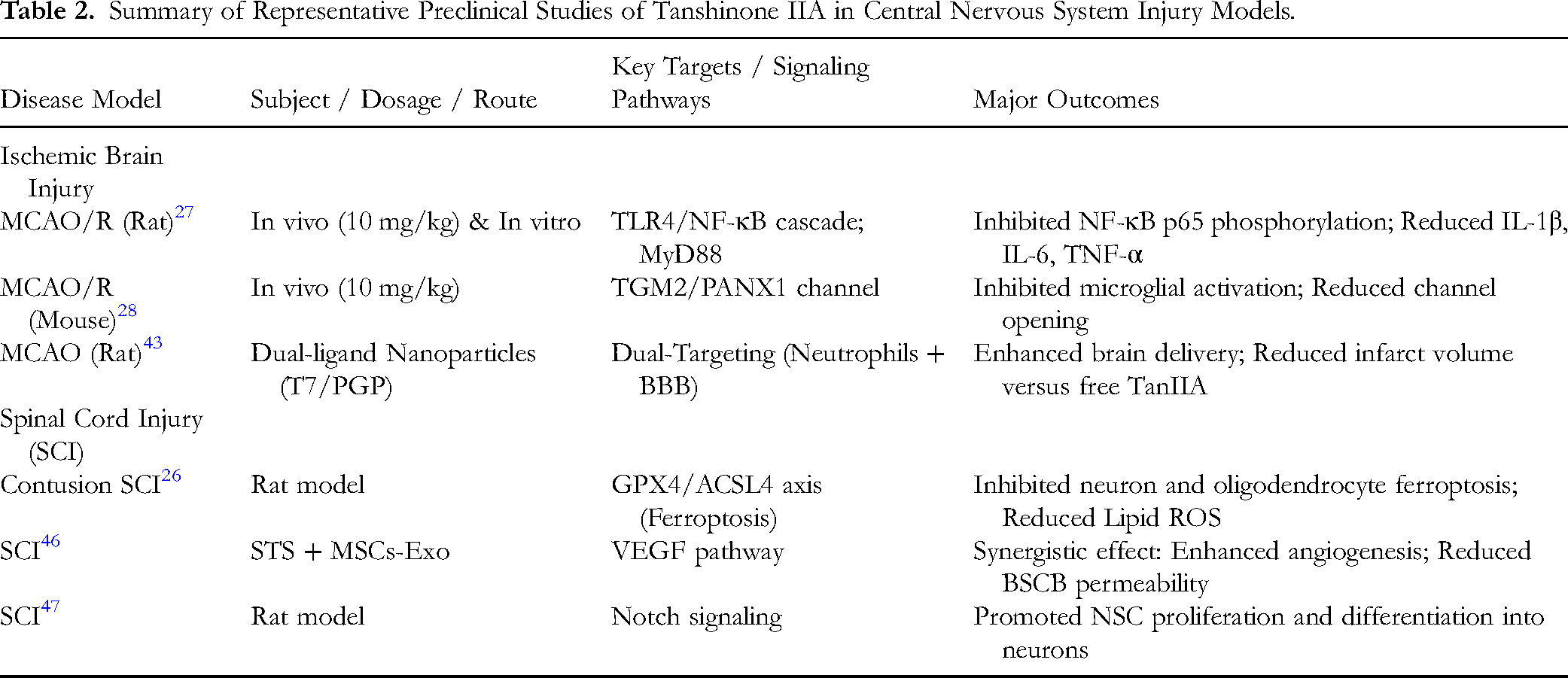
Collectively, these preclinical studies suggest that TanIIA and its derivatives exert pleiotropic neuroprotective actions after SCI, spanning barrier stabilization, anti-inflammatory and glia-modulating effects, redox control, and anti-apoptotic signaling. Table 2 summarizes representative studies, including disease models, dosing regimens, and key molecular pathways such as the GPX4/ACSL4 ferroptosis axis and TLR4/NF-κB signaling.
Summary of Representative Preclinical Studies of Tanshinone IIA in Central Nervous System Injury Models.
Future Perspectives
Although preclinical studies support the neuroprotective potential of TanIIA, clinical translation is likely to be constrained by its unfavorable pharmacokinetic profile, particularly low aqueous solubility, limited oral bioavailability, and rapid metabolism. Escalating the dose alone is unlikely to be an optimal solution and may increase variability in exposure as metabolic pathways become saturated. Accordingly, formulation- and delivery-based strategies represent a priority. Recent work suggests that brain-targeted delivery platforms—such as borneol-modified nanoparticles or dual-ligand functionalized nanoformulations (eg, T7 peptide- and PGP-targeting systems)—may enhance cerebral accumulation and improve CNS exposure (Figure/Table as applicable). 43
Beyond delivery, several translational gaps remain. First, target engagement should be strengthened by integrating computational prediction (eg, docking) with experimental validation to define direct molecular targets and actionable pathways. Second, pharmacokinetic and bioavailability studies in humans are needed to establish exposure–response relationships and to guide clinically feasible dosing. Third, standardized regimens—including dose, timing window, and route of administration—should be defined across representative CNS injury settings. Fourth, rational combination strategies (eg, with glucocorticoids, stem cell-based approaches, or neurotrophic agents) warrant evaluation using prespecified hypotheses and clinically relevant endpoints. Finally, multi-omics profiling and advanced in vitro/in vivo models may help resolve system-level effects, identify biomarkers of response, and clarify safety signals. Future clinical trials should incorporate functional outcomes, biomarker-based mechanistic readouts, and long-term safety assessments to determine whether TanIIA-based interventions can provide meaningful benefit for patients with CNS injuries.
Conclusion
TanIIA, a major bioactive constituent of Salvia miltiorrhiza, has shown multi-target neuroprotective effects across experimental models of CNS injury, including ischemic stroke and spinal cord injury. Reported actions span suppression of inflammatory signaling, attenuation of oxidative stress, preservation of mitochondrial function, modulation of glial activation, and inhibition of apoptosis-related pathways. TanIIA and clinically used derivatives may achieve CNS exposure via the blood–brain and blood–spinal cord barriers and have been explored as adjuncts to established therapies (eg, glucocorticoids). Nevertheless, translating these findings will require improved formulation and exposure control, clarification of target engagement, and well-designed clinical studies using clinically meaningful outcomes and safety endpoints.
Footnotes
Acknowledgements
The authors thank Dr Jie Huang for preparing ![]() and for editorial assistance during the revision. Written permission to use Figure 2 has been obtained from Dr Jie Huang.
and for editorial assistance during the revision. Written permission to use Figure 2 has been obtained from Dr Jie Huang.
Ethical Approval
Not applicable.
Patient Consent for Publication
Not applicable.
Author Contribution
Weiting Chen conceived and supervised the study. Min Tang drafted the manuscript. Xixi Guo and Xiaoshuang Jiang contributed to literature retrieval and synthesis and participated in manuscript revision. Jiuzhou Lin contributed to literature screening, data extraction, and manuscript revision. All authors read and approved the final manuscript.
Funding
The authors disclosed receipt of the following financial support for the research, authorship, and/or publication of this article: This work was supported by the Zhejiang Provincial Natural Science Foundation of China, (grant number LTGY24H150007) and Zhejiang Province Medical and Health Science and Technology Plan Project under Grant No (2024KY555).
Declaration of Conflicting Interests
The authors declared no potential conflicts of interest with respect to the research, authorship, and/or publication of this article.
Data Availability
The data supporting the findings of this study are available from the corresponding author upon reasonable request.