
Abstract
Objectives
This study was conducted in the Kemisie district, Oromia special zone of the Amhara region, Ethiopia. The present research aimed to study in vitro antibacterial activities of methanol crude extracts of medicinal plants, namely, Croton macrostachyus Hochst. ex Delile. Leaves and Clematis hirsuta Perr. et Guill. root and phytochemical analysis of methanol and hexane crude extracts.
Methods
The plant samples were collected and extracted with methanol and hexane after being dried and powdered. The in-vitro antibacterial activities were examined with the isolates of Clostridium chauvoei and Pasteurella multocida bacterial pathogens of cattle. The antibacterial activity of the methanol extract of the leaves of C. macrostachyus and the root of C. hirsuta against C. chauvoei and P. multocida pathogens was determined by using agar well diffusion, Minimum inhibitory concentration, and minimum bactericidal concentration methods.
Results
The methanol crude extracts of the plants were promisingly effective against the bacterial isolates, as shown by concentrations ranging between 1 to 2 mg/mL for C. macrostachyus leaves extracts and 1.5 to 2 mg/mL for C. hirsuta root extracts compared to the standard control. The MIC value was recorded for both bacteria at 1 mg/mL and MBC at 2 mg/mL with C. macrostachyus leaves extracts, and the MIC and MBC values for C. hirsuta roots extracts were recorded as 1.5 and 2 mg/mL, respectively.
Conclusion
The potency of these extracts, based on their zones of inhibition and MIC values, was higher in C. macrostachyus, which shows that its extract can be used after respective in vivo and clinical trials against P. multocida bacteria capable of causing huge mortality and morbidity with a very serious economic impact in the animal industry, including Ethiopia. This study provides the first evidence of the antibacterial efficacy of these extracts against C. chauvoei and P. multocida.
Introduction
Through scientific proof of their therapeutic potential, biological activities are essential in confirming the traditional usage of herbal medicines. Researchers can find the bioactive substances that give healing capabilities by screening for activities like antibacterial, antioxidant, anti-inflammatory, and anticancer activities. In addition to addressing issues like antibiotic resistance and promoting the development of novel therapeutic approaches, this also improves the efficacy, safety, and uniformity of herbal products. Also, knowing about these practices encourages the sustainable use and conservation of medicinal plants and aids in the preservation of indigenous knowledge.1–3
Despite the large number of livestock in Ethiopia, the sector is characterized by low productivity, and hence, income derived from this sector of agriculture cannot play a significant role in the development of the country's economy. The low productivity is raised by poor nutrition and reproductive performance, inadequate management, high disease incidence, and parasite burden. Livestock diseases are the major cause of economic losses to the peasant farmers and pastoralists in Ethiopia, amounting to hundreds of millions of birr annually. Although many of the diseases could be controlled by using available vaccine technology, due to the use of non-selective medicines, many diseases developed a drug-resistant nature. 4 Medicinal plants are a basic source of traditional medicine and synthetic medicine. Many researchers devoted their time to searching for and synthesizing new drugs for emerging diseases by using plant extracts. However, still more Ethiopian indigenous people engage in plant-based traditional medicine to get cured of diseases arising from bacteria, worms, fungi, viruses, protozoa, and so on. 5
In animal husbandry, the control and prevention of transmissible diseases are basic for the economic development of the country. The introduction of antibiotics to treat bacterial infections almost 50 years ago led to a dramatic improvement in animal production. However, the emergence of antibiotic-resistant strains demonstrates that the treatment of bacterial infections can’t rely on the use of antibiotics without some critical consideration. Special attention has been paid to the use of antibiotics in animals, including antimicrobial growth promoters, because these can contribute to the problems with antibiotic resistance in humans. The demand for medicinal plants is increasing in both developing and developed countries, and the bulk of their material trade is still from wild-harvested plants due to their safe nature, effectiveness, and low cost. Indigenous remedies are gaining popularity among people, especially in developing countries, where modern health services are limited. 6 Besides their importance in health care, medicinal plants have high socio-cultural and socio-economic values, providing off-farm income and employment opportunities to local people. 7
The roots of C. hirsuta have a history of traditional use in ethnoveterinary medicine, particularly in Ethiopia. Literature survey revealed that C. macrostachyus is traditionally used to treat or manage a lot of human and animal diseases and ailments, including black leg 8 and general healing purposes. 9 The use of C. hirsuta roots in traditional veterinary medicine highlights the reliance on local plant resources for animal healthcare in certain African communities.10–12 C. macrostachyus is a plant with numerous ethnoveterinary uses in traditional African medicine, particularly in Ethiopia and other East African countries. It's used to treat various ailments in animals, including wound healing, 10 infections, 9 and parasitic infestations. 13 The plant's bark, leaves, and sap are commonly used in preparations like poultices, decoctions, and extracts. 11
Phytochemical studies on the leaves of C. macrostachyus have demonstrated the presence of a rich diversity of secondary metabolites, including terpenoids, flavonoids, saponins, 14 phenolic compounds, 15 and alkaloids. 16 These compounds likely contribute to the plant's significant ethnomedicinal applications in treating various ailments. Similarly, phytochemical studies on the root of C. hirsuta have identified a range of secondary metabolites, including fatty acids, 17 sterols, 18 and lignans. 19 These compounds, along with the crude extracts, exhibit antibacterial and antioxidant activities, supporting the traditional use of this plant for various ailments.
The studies of traditional medicine should have continued due to their significant values, such as being a source for the pharmaceutical industry and creating motives for conserving biodiversity, as well as providing sufficient access to synthesize modern medicines. C. hirsuta of the root and C. macrostachyus of the leaves are the common medicinal plants used for the treatment of livestock diseases in Ethiopia.20,21 The authors of this work anticipated that these two plants contained the active components of secondary metabolites. Therefore, extracting, phytochemical screening, and antibacterial investigation are vital. To the best of our knowledge, the extraction of ingredients and their antibacterial activities of C. macrostachyus leaves and C. hirsuta plants in this study area against cattle pathogens like C. chauvoei and P. multocida have not yet been investigated. This will be the first report on C. macrostachyus leaves and C. hirsuta root methanol crude extracts against C. chauvoei and P. multocida bacterial isolates. Therefore, the present study aimed to extract ingredients from C. macrostachyus leaves and C. hirsuta root, phytochemical activities, and then determine the in vitro antibacterial activities against selected bacterial isolates and compare their efficacy with commonly used antibiotics against standard isolates of pathogenic bacterial isolates.
Materials and Methods
Description of the Study Area
The present study was conducted in the Kemisie district, Oromia special zone of Amhara regional state, northeast Ethiopia (Figure 1). Kemisie is located 325 km far to north of Addis Ababa and 75 km away from Dessie city, Ethiopia, in the southern direction. The geographical location of the area is between 10° 01’ to 11° 25’ N and 39° 41’ to 40° 24’ E. The altitude is from 1000 to 2500 m.a.s.l., with a temperature range of 12–33 °C. The mean annual rainfall of the area ranges from 600 to 900 mm. 22 According to the Kemisie town pastoral, agriculture, and rural development office, the livestock population in 2022 was estimated to be 728,054, of which goats had the highest population (321,232), cattle (278,965), followed by sheep (127,857). Blackleg and bovine pasteurellosis were reported as the most common disease types affecting the district. 23 The local communities of this study area have long been dependent on traditional medicinal plants accessed from natural forest patches, cultivated land, and field margins to manage various health problems for their animals, as we confirmed during sample collection by oral interviews with resident arbiters.
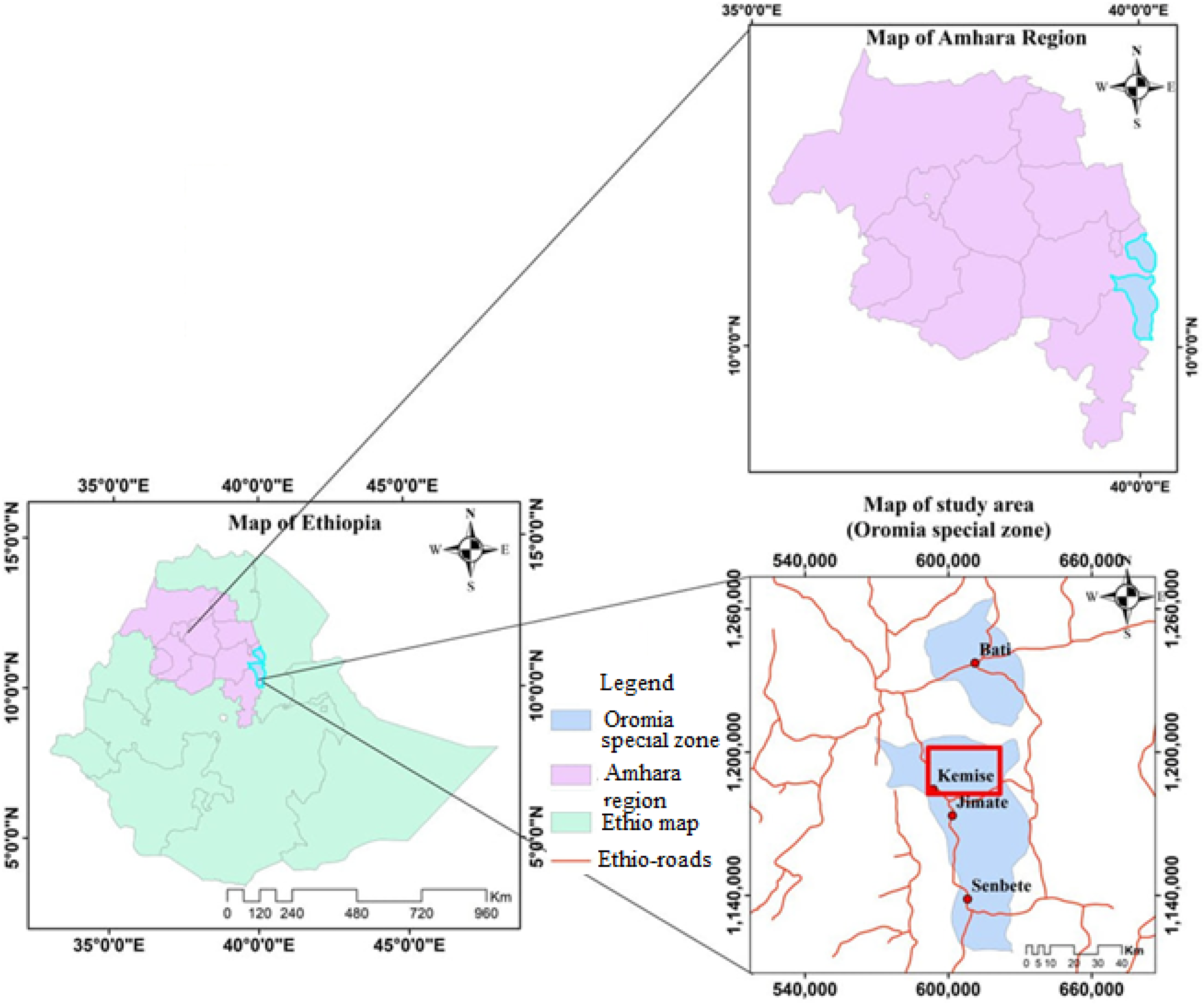
Map of Ethiopia, Amhara Region, and the Study Area (Oromia Special Zone, Kemise) (Drawn by Addisu Tamir Wassie Using ArcGIS 10.8).
Plant Collection, Authentication, and Preparation
The medicinal plants, namely Croton macrostachyus Hochst. ex Delile (Bisana) leaves and Clematis hirsuta Perr. et Guill. (Azohareg) root (Figure 2a &b respectively) were collected in and around forest areas of Kemisie district, Oromia special zone of Amhara regional state, Ethiopia, in February 2024. The plants were identified and authenticated by Assistant Professor Belay Melese, Department of Biology, Wollo University, Ethiopia. Herbarium specimens (voucher Mul 0004/2024) were prepared after the plants were identified and confirmed, and then deposited in the herbarium at Wollo University Biology Department, Dessie, Ethiopia. The selectively collected plants were cut and packed into sacks. The tops of sacks were tied, labeled, and transported to the Chemistry department laboratory, College of Natural Sciences, Wollo University, Dessie, Ethiopia. Before further processing, the samples were washed by running tap water to eliminate organisms, fungi, dust, and other foreign organic and inorganic matter, and then chopped into pieces. The chopped samples (Figure 3a) were dried under moderate conditions for two weeks. The dried leaves and roots were ground by using an electric grinder machine into an ordinary mesh size (Figure 3b), and the amount of each was measured by using an analytical balance. The measured powders were preserved in air-tight polyethylene plastic bags for the next analysis.

(a) C. macrostachyus leaves; (b) C. hirsuta (Ranunculaceae) root (Photo taken by Fekade Beshah Tessema (a) and Addisu Tamir Wassie (b), both in Feb, 2024).
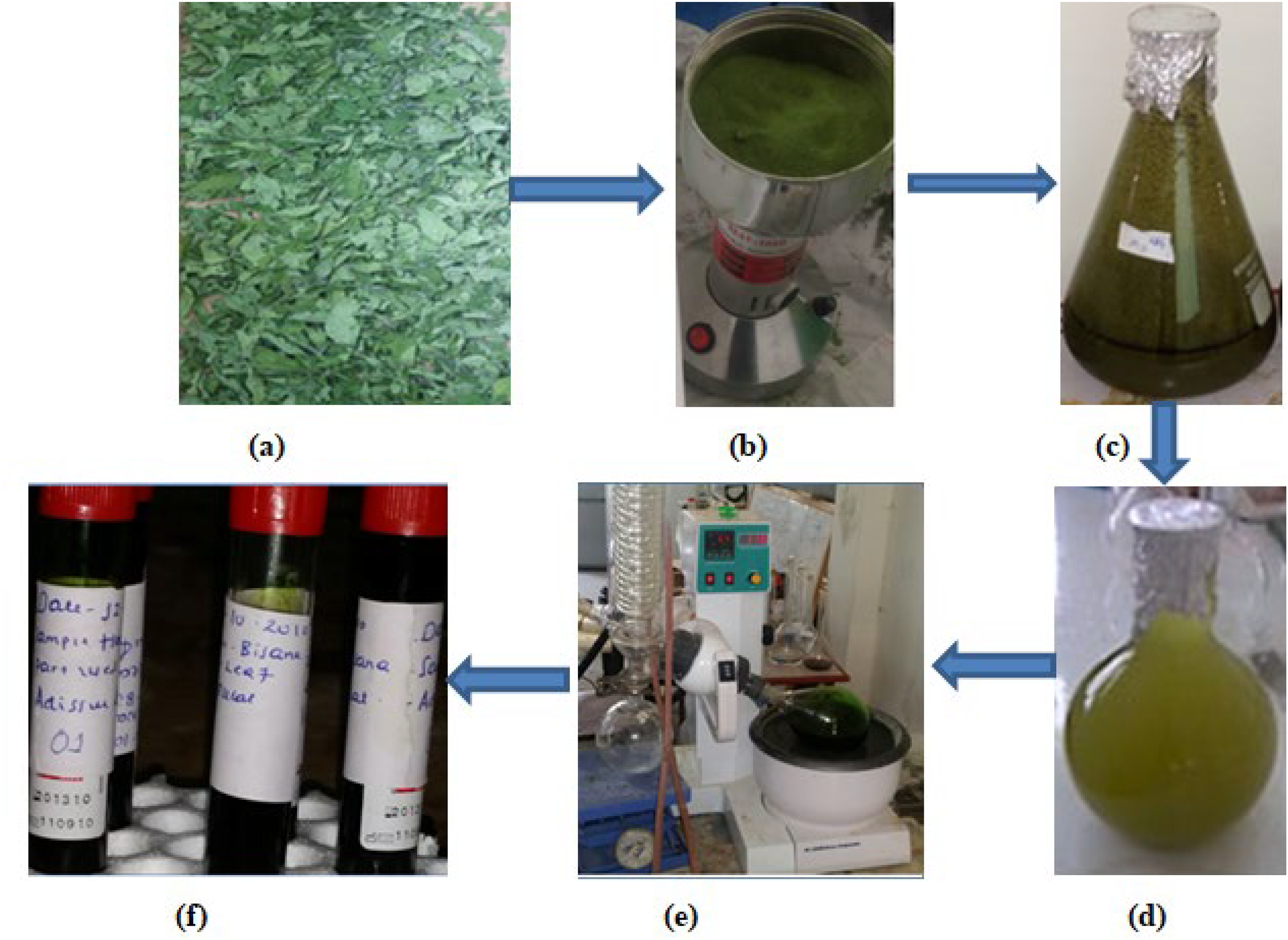
Chopped Leaves of C. macrostachyus (a), Powder of C. macrostachyus (b), Soaked C. macrostachyus (c), Extracted C. macrostachyus Leaves (d), Concentrating C. macrostachyus Extracts Using a Rotary Evaporator (e), Concentrated C. macrostachyus Leaves Extracts (f).
Extraction of Plant Materials
Methanol (0.762 polarity) and n-Hexane (0.009 polarity) were used as solvents to represent the polar and nonpolar solvents, respectively, and from an availability point of view, and also the ability of their easy evaporation from crude extracts. The four 2.5 L Erlenmeyer flasks were washed with clean tap water and then rinsed with distilled water, followed by 95% methanol. Four 250 g powdered samples of both plants (2 * 250 g each) were measured separately by using an analytical balance and added into four cleaned Erlenmeyer flasks. 1L methanol (95%) was added to one Erlenmeyer flask (Figure 3c), which contained powder of C. macrostachyus leaves, and another 1L methanol (95%) was added to the second flask, which contained C. hirsuta root powder. In addition, the other two flasks with 250 g of powder from each plant sample were mixed with 1 L of hexane (96%). The flasks were closed with aluminum foil and macerated for three days (72 h). The macerated materials were occasionally shaken manually three times per day for five minutes while standing at room temperature. After the extraction was completed (observed persistent color of soaks, Figure 3d), the extracts were separated from their marc by gravity filtration with the support of ashless Whatman filter paper No.1. 24 Subsequently, the solvents were removed from crude extracts by using a rotary evaporator, Figure 3e, (Stone Staffordshire, England ST15 OSA) at 45 °C under reduced pressure, with the help of a vacuum pump, and the constant weight method (Gravimetric) was used to ensure the complete removal of the solvents. Solvent-free extracts were stored in airtight glass vials (Figure 3f) at 4 °C for phytochemical screening and antibacterial activities.
Phytochemical Constituent Analysis of Glycosides, Quinones, Steroids, Phlobatannins, Proteins, and Carbohydrates.25–27
The results of the crude extract of phytochemical constituents were tabulated in Table 1 after conducting the following chemical tests.
Phytochemical Analysis Results of the Crude Methanol and Hexane Extracts of C. macrostachyus Leaves and C. hirsuta Roots.

Test for Phenolic Compounds and Tannins
Test for Saponins (Froth Test)
0.5 g of plant extract was shaken vigorously with 5 mL of distilled water in a test tube for 10 min and warmed in the water bath. The formation of stable foam indicated the presence of saponins.
Test for Phlobatannins
About 2 mL of the plant extract solution was added to 2 mL of hydrochloric acid (1% HCl), and the mixture was boiled. The deposition of red precipitate provided evidence for the presence of phlobatannins.
Tests for Flavonoids
5 mL of diluted ammonia solution was added to the portion of 0.5 g plant extract containing the test tube, followed by the addition of a few drops of concentrated sulfuric acid. The formation of yellow color precipitate confirms the presence of flavonoids.
Test for Terpenoids (Salkowski Test)
0.5 g of the plant extract was dissolved in 2 mL of chloroform and evaporated till dry. Then, after 2 mL of concentrated sulfuric acid was added and heated for 2 min. The appearance of a greyish color indicated the presence of terpenoids.
Test for Glycosides (Keller Killiani Test)
0.5 g of extract was diluted in 5 mL of distilled water, 2 mL of glacial acetic acid was added, and then two drops of 5% ferric chloride solution were added. Later, 1 mL of concentrated sulfuric acid was added along the wall of the test tube. The formation of a brown ring at the junction of two liquids indicated the presence of glycosides.
Test for Steroids
After dissolving 2 mg of extract in 2 mL of chloroform in a clean test tube, 2 mL of concentrated sulfuric acid was added to the solution along the wall of the test tube; a red color produced in the lower chloroform layer indicated the presence of steroids.
Tests for Alkaloids
Tests for Quinones
The 5 mL plant extract solution was mixed with 1 mL concentrated sulfuric acid. The development of a red color showed the existence of quinines.
Tests for Proteins
Tests for Carbohydrates
Antibacterial Activities
Standard Bacterial Organisms and Standardization of the Inoculums
Clostridium chauvoei (Blackleg) and Pasteurella multocida (bovine pasteurellosis) bacterial isolates were obtained from the National Veterinary Institute (NVI), Bishoftu, Ethiopia. 0.2 mL of a 24-h-old culture of each organism was dispensed into 20 mL of sterile nutrient broth and incubated for 5 h to standardize the culture to 106 cfu/mL with 0.5 McFarland (0.5 McFarland = ∼1.5 × 10^8 CFU/mL) standards. 28
Determination of Antibacterial Activity
The antibacterial activity of C. macrostachyus leaves and C. hirsuta root extracts was determined against the C. chauvoei and P. multocida bacterial pathogens of cattle. The Agar Well Diffusion, minimum inhibitory concentration (MIC), and minimum bactericidal concentration (MBC) methods were used to determine antibacterial activities of extracts. 0.5 mL of overnight broth culture of each clinical isolate containing 106 cfu/mL was aseptically transferred to the solidified nutrient agar and spread evenly on the agar surface using a sterile glass spreader. Four 6 mm wells were bored into the agar and filled with the extracts, while the solvent (0.5 mL ethanol, 98%) served as the control. The Petri dishes were incubated at 37 °C for 18 to 24 h, and the inhibition zones were measured. 29
Antibiotic sensitivity of the C. chauvoei and P. multocida bacterial isolates was determined by the Standard Disc Diffusion Method. Different antibiotics were used in the present work, viz. Erythromycin (E)(15 µg); Gentamicin (G) (10 µg); Tetracycline (T) (30 µg) and Penicillin G sodium (standard powder from Eli Lilly and Co., Indianapolis, Ind.; 2- and 10-unit discs from BBL) were melted and cooled nutrient agar media were poured in sterile petri-dishes and swabbed with overnight culture of bacterial strains. Under aseptic conditions, antibiotic discs were placed on the surface of the inoculated plates. Following overnight incubation at 37 ± 0.2 oC, the zone of inhibition (mm) for each drug was measured, and values were compared by using standards to determine the sensitivity pattern of C. chauvoei and P. multocida bacterial isolates. 30 C. chauvoei of the bacterial strains were collected in sterile nutrient agar slants and sub-cultured on Oxoid nutrient agar media.
Agar Well Diffusion Method
In the agar well diffusion method, the nutrient agar was inoculated with the selected microorganisms, and wells of 6 mm diameter were punched in the agar and filled with plant extracts. A control well without plant extract made of 1% ethanol solution at 100 microliters (µL) was also run along with wells having plant extracts in the same plate. The plates were incubated at 37 °C for 24 h, and the antimicrobial activity was evaluated by measuring the diameter of the zone of inhibition. The diameter of the zone of inhibition was measured using a ruler and recorded in mm. The antibacterial activities of the crude extracts were evaluated by comparing their zones of inhibition with standard antibiotic agents. 31
Determination of Minimum Inhibitory Concentration
The MIC of the extracts was also determined using the agar well diffusion technique. Two-fold serial dilutions were prepared to achieve a decreasing concentration range of 200 to 12.5% (V/V). A 0.5 mL volume of each solution was added aseptically into the wells of Mueller-Hinton agar plates that were already seeded with standardized inoculums (106 cfu/mL) of the bacterial isolates. The plates were incubated at 37 °C for 24 h. The lowest concentration of the extracts showing a clear zone of inhibition was considered as the MIC,32,33
Determination of Minimum Bactericidal Concentration
The test tubes that didn’t show any visible turbidity after incubation of the batch of test tubes were sub-cultured on nutrient agar plates and incubated at 37 °C for 18 to 24 h. The concentration that shows no visible growth after incubation was considered the minimum bactericidal concentration. 34
A Statistical Analysis
The data obtained from laboratory results of the collected samples were entered into Excel databases. (Version 16; Microsoft). All statistical analyses were performed using SPSS (version 28.0; IBM Corp., Armonk, NY). Descriptive statistics were calculated for all quantitative measurements, including zone of inhibition diameters (mm) and minimum inhibitory concentrations (MIC), where determined. Experiments were done in triplicate, and data are presented as means ± standard deviations (SD). One-way ANOVA with post-hoc tests was used for comparing inhibition zones. For qualitative phytochemical screening results, observations are presented descriptively “+’’ for presence and “-’’ for absence.
Results
Qualitative Phytochemical Analysis of the Crude Extracts of C. macrostachyus Leaves and C. hirsuta Root
The test result presented in Table 1 shows that the qualitative phytochemical investigations of the crude extracts of C. macrostachyus leaves and C. hirsuta root by using methanol and hexane solvents. The results presented in Table 1 disclose that the methanolic crude leaves extract of C. macrostachyus was rich in plentiful types of phytochemicals like tannins, phenolic compounds, phlobatannins, flavonoids, carbohydrates, alkaloids, quinones, proteins, and glycosides, but in hexane crude extracts, flavonoids, terpenoids, steroids, alkaloids, and protein phytochemicals were found. Whereas, the methanol crude extract of C. hirsuta root contained a small number of types of phytochemicals when it is compared with C. macrostachyus leaves crude extract. Tannins, saponins, phlobatannins, glycosides, alkaloids, and carbohydrates were present in the methanol crude extract of C. hirsuta root. Only terpenoid and carbohydrate phytochemicals were present in the hexane crude extract of C. hirsuta root.
Antibacterial Activity of Crude Extracts
Antibacterial Activity of C. macrostachyus Leaves Crude Extracts
The crude extract of C. macrostachyus was active against the gram-negative bacterial strains, which showed the growth inhibition zones of 12 ± 0.22 mm at concentrations of 2 mg/mL, but there was no activity (NA) or no zone of inhibition was observed at the concentration of 0.5 mg/mL for P. multocida. The inhibition zone (mm) of C. hirsuta roots extract, as shown in Table 2, was recorded as 8.6 ± 0.28 at 2 mg/mL, and there was no activity or no zone of inhibition was observed at the concentration of 0.5 and 1 mg/mL for P. multocida.
The Susceptibility Pattern of Crude Methanol Extract of C. macrostachyus Leaves and C. hirsuta Root Against P. multocida Isolate.

Antimicrobial Activities of C. hirsuta Root Crude Extract
The root extract of C. hirsuta caused a growth inhibition zone of 8 ± 0.24 mm against the gram-positive bacterial strain C. chauvoei at the concentration of 2 mg/mL and 6 ± 0.24 mm at 1.5 mg/mL, whereas no activity was observed at the concentration of 0.5 to 1 mg/mL, as presented in Table 3. A treatment with more potent or efficient antibacterial activity is shown in plate (II), but plate (III) has a diminished antibacterial effect, indicating plant extracts with lesser potency or activity (Figure 4). When it came to the test organism on the plate, the test chemicals showed antibacterial action. One can use this plate to compare the relative efficacy of various extracts or concentrations; the inhibitory zone diameters (in millimeters) must be measured and documented for accurate analysis.

Prepared growth control plate (I), Zone of inhibition for P. multocida (II), and Zone of inhibition for C. chauvoei (III) with different concentrations of extract & control.
the Susceptibility Pattern of Crude Methanol Extract of C. hirsuta Root and C. macrostachyus Leaves Against C. chauvoei Bacterial Isolate.

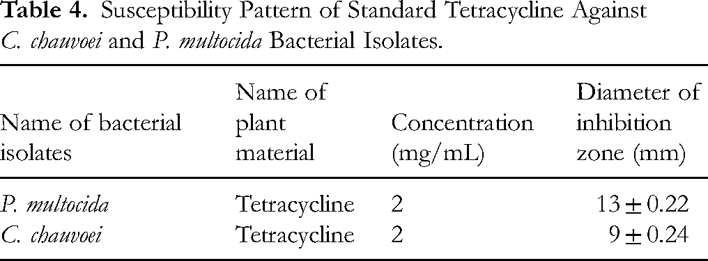
In terms of antibacterial activity against P. multocida, C. macrostachyus exhibits moderate promise. The action of C. hirsuta against C. chauvoei is weak, as shown in Table 4, and it might not be effective by itself at the tested concentration. As Minimum Inhibitory Concentration (MIC) and Minimum Bactericidal Concentration (MBC) indicated in Table 5, the stronger antibacterial activity is shown by C. macrostachyus, which inhibits P. multocida at a lower dose (1 mg/mL), but the antibacterial effectiveness of C. hirsuta is lower, and a greater dose (2 mg/mL) is required to inhibit P. multocida. A similar pattern was observed for the C. chauvoei with the same concentration of crude extracts as shown in Table 5.
Susceptibility Pattern of Standard Tetracycline Against C. chauvoei and P. multocida Bacterial Isolates.
The Minimum Inhibitory Concentration (MIC) and Minimum Bactericidal Concentration (MBC) of Crude Methanolic Extract of Plants Against C. chauvoei and P. multocida Bacterial Isolates.

NA: not applicable, G: growth
As per the authors’ knowledge, no prior report has been found for the respective extracts against the specific isolates for comparison.
Discussion
Plants and plant products have been used extensively throughout history to treat medical problems. Medicinal plants are widely used in African communities to treat different types of bacterial diseases. 35 The traditional therapeutic methods, especially the use of medicinal plants, still play a vital role in covering the basic health needs in developing countries, and moreover, the use of herbal remedies has risen in developed countries in the last decades. In this connection, plants continue to be a rich source of therapeutic agents. 36
Leaves were the most frequently sought plant part, accounting for 47% of the reported medicinal plant species, followed by root (22%), whole plant (16%), and aboveground part (16%). Similarly, in studies conducted elsewhere in Ethiopia, leaves were indicated to be the most frequently used plant part to treat livestock ailments. 37 In the present study, C. macrostachyus leaves methanolic extract effectively inhibited P. multocida growth; this finding is in line with Sharma et al (2010), who reported that gram-negative bacteria lack a peptidoglycan layer, which is permeable to these substances. The observations from this study are consistent with previous reports describing the antibacterial activity of P. multocida. 38 It is well accepted that C. macrostachyus leaves extracts play an important role in its antimicrobial activity in vitro. 39 The crude extract of C. macrostachyus was active against P. multocida strains with growth inhibition zones of 9 ± 0.41, 10 ± 0.36, and 12 ± 0.22 mm at concentrations of 1, 1.5, and 2 mg/mL, respectively, but no activity was observed at the lowest concentration of 0.5 mg/mL.
In the present study, the large differences between the minimum inhibitory concentration and the minimum bactericidal concentration indicated that the bioactive components may be present in much lower concentrations. 40 Studies in the Shirka district 41 in northern Ethiopia and the central plateau and rift valley of Ethiopia 42 documented the root to be the most extensively used plant part in the preparation of herbal remedies. Ranunculaceae could be related to their wider distribution and use in Ethiopia/the flora area. These families were also reported to have the largest share of ethnomedicinal species in other ethnobotanical inventories. 43 Ranunculaceae were reported to be of frequent use in the local ethnoveterinary medical system. Roots (65%, 33 species) were most often utilized for remedy preparation. 20 Infections caused by anaerobes can also be divided according to the origin of the microorganisms. Exogenous anaerobic infections are predominantly caused by Gram-positive spore-forming bacilli (Clostridium spp.). These microorganisms are the causative agents of serious infections like visceral gas gangrene, lockjaw, myonecrosis, and botulism. These infections are monobacterial in almost all cases, and the effects of bacterial toxins are the prime causes of these pathologies. 44 The organic extracts of C. hirsuta showed activities against the C. chauvoei bacterial strains, with the growth inhibition zone 8.6 ± 0.28 mm at 2 mg/mL. Disc diffusion assay revealed maximum inhibition zones against P. multocida with C. macrostachyus leaves extracts, suggesting the antibacterial efficacy of the tested organism. Furthermore, it compared favorably with the standard antibacterial drug tetracycline. However, the C. hirsuta root extract will show maximum inhibition zones at high concentrations, as shown by our trial progress.
Our qualitative analysis of the phytochemicals in both extracts revealed the presence of phenolic compounds, such as tannins and flavonoids. These groups of phytochemicals disrupt the structure and function of bacterial cells and have a major impact on antimicrobial action. In addition, flavonoids can interfere with energy metabolism, break down bacterial membranes, and prevent the formation of nucleic acids. Tannins can also disrupt enzyme activity and denature bacterial proteins. More specifically, flavonoids are known for their ability to inhibit microbial growth and virulence through several key actions. Their specific effects are often dependent on their chemical structure, such as the number and position of hydroxyl groups. 45 Disrupting the microbial cell membrane, 46 and inhibiting microbial enzymes 47 are among the common mechanisms. Flavonoids can also inhibit crucial enzymes necessary for microbial metabolism and survival, 45 suppressing biofilm Formation, 48 and inhibiting nucleic acid synthesis. 49
Similarly, tannins are large, complex polyphenolic compounds known for their ability to bind to and precipitate proteins. This “astringent” property is central to their antimicrobial effects. Some of the mechanisms of its action include: binding to cell walls and membranes, 49 Inhibiting enzymes and transport proteins, and chelating metal ions. 50 Tannins can also prevent microorganisms from adhering to host tissues, a key step in the infection process. 51 This makes them particularly useful for treating infections of the mucous membranes, such as those in the oral cavity or digestive tract. The potential of both phytochemicals to prevent bacterial infections and biofilm development is being investigated. 48 This could serve as a benchmark for future research on the components of each extract's mechanism of action.
Conclusion
In conclusion, the methanolic extracts of the C. macrostachyus leaves and C.hirsuta roots showed more important primary and secondary metabolites than hexane extracts and considerable antibacterial activity against P. multocida and C.chauvoei bacterial strains used at a concentration of 2 mg/mL with inhibition zones of 12 ± 0.22 and 8.6 ± 0.28, respectively. The result obtained at 2 mg/mL was comparable with the standard control value (13 ± 0.22, 9 ± 0.24, respectively) and thus promising for potential use in the treatment of infectious diseases caused by bacteria after respective in vivo and then clinical studies. The extracts of C. hirsuta with slightly less value of inhibition zone (11.6 ± 0.22 and 8 ± 0.24, respectively) than C. macrostachyus has also shown considerable antibacterial activity against the P. multocida and C. chauvoei bacterial strains at a concentration of 2 mg/mL and thus can be used after clinical trials in the treatment of infectious diseases caused by the bacteria. The MIC value was recorded for both bacteria at 1 mg/mL and MBC at 2 mg/mL with C. macrostachyus leaves extracts, and the MIC and MBC values for C. hirsuta root extracts were recorded as 1.5 and 2 mg/mL, respectively, which also shows that the selected plant materials can be good candidates for antibacterial activity. In Ethiopia, research on herbal remedies for human ailments is well-established, but similar studies for livestock diseases remain limited.
Lack of cytotoxicity testing, narrow bacterial strain selection, proposing a mechanism for the biological activities attempted, and isolation of major phytochemicals from the extracts examined are among the limitations of this study. Therefore, this plant can be considered for further investigation, including isolation of active compounds, in vivo trials, and clinical studies.
Footnotes
Acknowledgments
The authors would like to acknowledge Wollo University and the National Veterinary Institute (NVI) for allowing access to the reagents and bacterial organism isolates.
Ethical Approval
Ethical Approval is not applicable to this article.
Funding
The authors received no financial support for the research, authorship, and/or publication of this article.
Declaration of Conflicting Interests
The authors declared no potential conflicts of interest with respect to the research, authorship, and/or publication of this article.
Data Availability
All data generated or analyzed during this study are included in this published article.
Statement of Human and Animal Rights
This article does not contain any studies with human or animal subjects.
Statement of Informed Consent
There are no human subjects in this article and informed consent is not applicable.