
Abstract
Aims
This study investigates the antimicrobial potential of aqueous extracts from the leaves and stems of Cymbopogon citratus on the root canal pathogens of E. faecalis and S. sanguinis.
Material and Methods
The sample extraction was constructed by reflux condensation, frozen at −20 °C and lyophilised under a freeze dryer at −50 °C to obtain a powder form. The chemical structure was analysed under Fourier-Transform Infrared Spectroscopy (FTIR) in transmission mode, wavenumber range from 400 to 4000 cm−1 with a spectral resolution of 4 cm−1. The C. citratus extract powder was observed and viewed under a Scanning Electron Microscope (SEM). The extract powder was subjected to serial dilutions of 480 mg/mL, 240 mg/mL, 120 mg/mL, 60 mg/mL and 30 mg/mL. These concentrations were evaluated against Enterococcus faecalis and Streptococcus sanguinis under minimum inhibitory concentration (MIC) and minimum bactericidal concentration (MBC) assays.
Result
Both leaf and stem extracts demonstrated concentration-dependent antibacterial activity, with efficacy observed against S. sanguinis than E. faecalis in concentrations ranging from 120 mg/mL to 480 mg/mL.
Conclusions
These findings suggest that C. citratus extracts have promising potential as a natural antibacterial agent for root canal irrigation. However, further studies are necessary to explore its bactericidal mechanisms, optimise formulation, and assess its clinical applicability in root canal disinfection.
This is a visual representation of the abstract.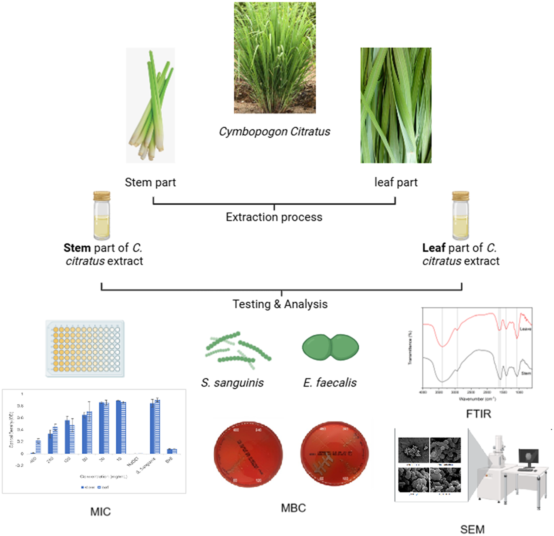
Keywords
Introduction
The primary objective of root canal treatment is to achieve chemo-mechanical debridement of the root canal area of the tooth while facilitating the effectiveness of an antimicrobial disinfection solution.1,2 During the procedure, the infected and damaged pulp tissue in the root canal will be removed, followed by cleaning and disinfecting the canal space with an irrigation solution before restoring the area with a biocompatible material. 3 Sodium hypochlorite (NaOCl), in concentrations of 0.5% to 5.25%, is the most effective root canal irrigation with strong antimicrobial and tissue-dissolving capabilities. 4
However, NaOCl was shown to be associated with cytotoxicity, where its’ chlorine content may cause irritation to the stem cell and apical papillae at the surrounding root area. 5 Furthermore, the persistence of bacterial isolates such as Enterococcus faecalis (E. Faecalis) and Streptococcus sanguinis (S. sanguinis) following NaOCl irrigation presents a major challenge in root canal treatment. These bacteria can develop tolerance to alkaline environments and resistance to irrigation solutions such as NaOH and chlorhexidine. 6 This underscores the need for alternative therapeutic approaches, particularly plant-based materials that remain underexplored in clinical applications.
Cymbopogon citratus (C. citratus) or lemongrass is known for its broad spectrum of pharmacological properties, including antibacterial, antifungal, anti-inflammatory, and dental caries inhibition activities. 7 Given its wide array of biological activities and its proven efficacy against multidrug-resistant bacteria like methicillin-resistant Staphylococcus aureus (MRSA), Staphylococcus epidermidis (MRSE), and various gram-negative pathogens. 8 C. citratus warrants investigation as a natural alternative to NaOCl antimicrobial irrigation for root canal treatment. However, none of the studies have yet explored the effect of aqueous extract of C. citratus on multispecies biofilms composed specifically of Streptococcus sanguinis (S. sanguinis) and Enterococcus faecalis (E. faecalis), which are predominant as root canal pathogens.
Therefore, this study aimed to investigate different concentrations of aqueous extract of C. citratus targeting the root canal pathogens E. faecalis and S. sanguinis. The present study's benefit is providing an opportunity for further research to produce herbal-based material for root canal treatment. Furthermore, using an aqueous extract of C. citratus solution will provide safer treatment procedures by eliminating the risk of toxicity and oral soft tissue injury.
Materials and Methods
Materials and Test Organisms
Fresh C. citratus plants were harvested from local residents and verified by the Herbarium Unit, School of Biological Sciences, Universiti Sains Malaysia. E. faecalis (ATCC 10231) and S. sanguinis (ATCC 10556) were used in this study, and both were cultured onto two separate blood agar plates (Oxoid, UK) at 37 °C and incubated for 24 h.
The negative control was an untreated bacterial suspension and a blank brain-heart infusion, whereas the positive control was a treated bacterial suspension with 2.5% Sodium Hypochlorite (NaOCl).
Preparation of Extracts and Stock Concentration
Aqueous extracts were prepared based on a modified protocol from Halabi and Sheikh 2014. 9 Fresh C. citratus leaves and stems were cut and separated, thoroughly washed, rinsed with distilled water, and oven-dried at 50 °C for three days until the weight was constant. They were kept in sterile plastic containers at room temperature (26 ± 1 °C). The dried C. citratus was ground into powder form and added to distilled water at a 1:10 powder-to-water ratio. The extraction was constructed by reflux condensation for 4 h. The mixture was left overnight for cooling before filtering through Whatman No. 1. The filtered sample was frozen at −20 °C and lyophilised using a freeze dryer at −50 °C until a powder was obtained. The powder was weighed at 9600 mg. They were mixed with 10 mL of sterile distilled water, placed in a flask, and stirred continuously for 24 h using a magnetic stirrer, ensuring complete dissolution to 960 mg/mL of C. citratus. Serial dilution with sterile distilled water was constructed to achieve the final concentration of 480 mg/mL, 240 mg/mL, 120 mg/mL, 60 mg/mL and 30 mg/mL.
Determination of Minimum Inhibitory Concentration (MIC)
The minimal inhibitory concentration (MIC) of aqueous C. citratus extracts was determined using the micro broth dilution method, according to Clinical and Laboratory Standards Institute (CLSI) guidelines. 10 100 μL of each extract concentration and 2.52% NaOCl were pipetted into 96-well plates. 100 μL of a bacterial suspension (1 × 10⁶ CFU/ mL) of S. sanguinis and E. faecalis (prepared using the 0.5 McFarland Standard) was added to each well to make it a 1:1 ratio. As controls, 200 μL of bacterial suspension and blank brain-heart infusion (BHI) were pipetted separately into additional wells. The plates were incubated anaerobically at 37 °C with 5% CO2 for 48 h. Bacterial growth was assessed by measuring optical density at 660 nm using a microplate reader (Varioskan Flash at 0 h at the Centre Research Laboratory). The plates were incubated at 37 °C overnight, and 24-h measurement was recorded the next day. All assays were performed in triplicate.
Determination of Minimum Bactericidal Concentration (MBC)
The minimal bactericidal concentration (MBC) was determined immediately after MIC quantification at 24 h. Using a flamed metal loop, samples from 96-well plates containing C. citratus extract concentrations were each combined with a 1 × 10⁶ CFU/mL bacterial suspension and then streaked onto six separate blood agar plates. As for control, a sample of 2.52% NaOCl combined with a 1 × 10⁶ CFU/mL bacterial suspension was streaked onto a separate blood agar plate. All plates were incubated anaerobically at 37 °C with 5% CO2 for 24 h. Bacterial growth was then evaluated by naked eye observation to determine the MBC.
Fourier-Transform Infrared Spectroscopy (FTIR)
The chemical structure of C. citratus extraction from leaf and stem was evaluated by FTIR using a Cary 600 Series FTIR Spectrometer. 2 mg of the freeze-dried extract was accurately weighed and mixed thoroughly with 100–200 mg of spectroscopic grade potassium bromide (KBr). The mixture was finely ground and compressed into a thin, transparent pellet using a hydraulic press under vacuum conditions. The spectra were recorded in the mid-infrared range of 4000–400 cm−1 with a resolution of 4 cm−1 and averaging 16 scans per sample. A background spectrum was recorded using a pure KBr pellet prior to each analysis to eliminate any atmospheric interference.
Scanning Electron Microscopy (SEM) Analysis
To evaluate the morphological alterations in S. sanguinis induced by the aqueous leaf extract of C. citratus, scanning electron microscopy (SEM) was employed following standard sample preparation protocols with minor modifications.
A pure culture of S. sanguinis was grown overnight in Brain Heart Infusion (BHI) broth at 37 °C under anaerobic conditions. The bacterial suspension was adjusted to an optical density equivalent to 0.5 McFarland standard (approximately 1.5 × 10⁸ CFU/mL). Aliquots of the bacterial suspension were treated with the minimum inhibitory concentration (MIC) of C. citratus leaf extract and incubated for 24 h at 37 °C. Untreated bacterial cultures served as the negative control.
After the incubation, 1 mL of each bacterial suspension was centrifuged at 5000 rpm for 10 min to collect the cells. The pellets were washed twice with 0.1 M phosphate-buffered saline (PBS, pH 7.4). Fixation was performed using 2.5% glutaraldehyde in PBS for 2 h at 4 °C. The samples were washed thrice with PBS and then subjected to a graded ethanol dehydration series (30%, 50%, 70%, 90%, and 100%) for 10 min each.
The dehydrated bacterial pellets were mounted onto aluminium stubs and allowed to air-dry completely in a desiccator. Samples were then sputter-coated with a thin layer of gold using a sputter coater (Quorum Q150R ES) to enhance conductivity.
Results
Linear Regression Analysis of MIC Data
Linear regression analysis was employed to evaluate the inhibitory effects of C. citratus stem and leaf extracts on S. sanguinis and E. faecalis.
The OD-concentration response curves revealed that S. sanguinis was more susceptible to both extracts, with a sharper decline in optical density observed for the stem extract. The estimated MIC for the stem extract was approximately 240 mg/mL, whereas the leaf extract demonstrated an MIC closer to 480 mg/mL (Figure 1a).

(a) MIC Linear Regression Curve of C. citratus Against S. sanguinis. (b) MIC Linear Regression Curve of C. citratus Against E. faecalis. (c) Comparison of MIC and MBC Values for Treatment Against S. sanguinis and E. faecalis Compared to NaOCl (Positive Control).
In contrast, E. faecalis exhibited greater resistance, as evidenced by a more gradual decline in OD values across the tested concentrations. The leaf extract achieved near-complete inhibition at 480 mg/mL, while the stem extract failed to reach the defined inhibition threshold (OD ≤ 0.1), suggesting an MIC exceeding 480 mg/mL (Figure 1b).
The optical density (OD) results from the MIC assay demonstrated a clear concentration-dependent inhibitory effect of C. citratus extracts on S. sanguinis. Both the stem and leaf extracts significantly reduced bacterial growth, particularly at concentrations ≥240 mg/mL. At 480 mg/mL, both extracts exhibited nearly complete inhibition (OD < 0.05), consistent with the defined MIC threshold. Notably, the leaf extract showed slightly lower OD values at intermediate concentrations (240 and 120 mg/mL), suggesting a marginally higher potency compared to the stem extract. These findings indicate that C. citratus extracts possess bacteriostatic potential, particularly against S. sanguinis. The differential response between the two bacterial species underscores the importance of microbial susceptibility profiling when evaluating plant-based antimicrobials.
MIC and MBC Values
MIC and MBC values were determined via broth microdilution and subsequent subculturing. NaOCl exhibited complete bactericidal activity at 0 mg/mL for both species (Figure 1c).
The leaf extract of C. citratus showed bactericidal activity at 240 mg/mL for S. sanguinis and 480 mg/mL for E. faecalis, while the stem extract demonstrated limited antimicrobial activity with no confirmed MBC under the tested conditions. The positive control (2.5% NaOCl) resulted in complete inhibition, while both the S. sanguinis and BHI controls showed normal growth, confirming the assay's validity. These observations indicate that both extracts possess measurable antibacterial activity, with the leaf extract showing slightly superior inhibitory potential Table 1.
MIC and MBC of C. citratus Leaf and Stem Extracts to the 2.5% Sodium Hypochlorite (NaOCl) Against E. faecalis and S. sanguinis.

Note: MIC = Minimum Inhibitory Concentration; MBC = Minimum Bactericidal Concentration.
FTIR Spectrum
The FTIR spectra of both C. citratus leaf and stem extracts reveal the presence of important phytochemical functional groups such as hydroxyls, carbonyls, aliphatic chains, and aromatic structures (Figure 2). The leaf extract displays more prominent O–H and C–O peaks, indicating a higher concentration of polyphenolic and carbohydrate-based compounds, which could explain its better inhibitory effect against E. faecalis. Meanwhile, the stem extract appears richer in aliphatic chains and shows a moderate presence of bioactive functional groups (Table 2).
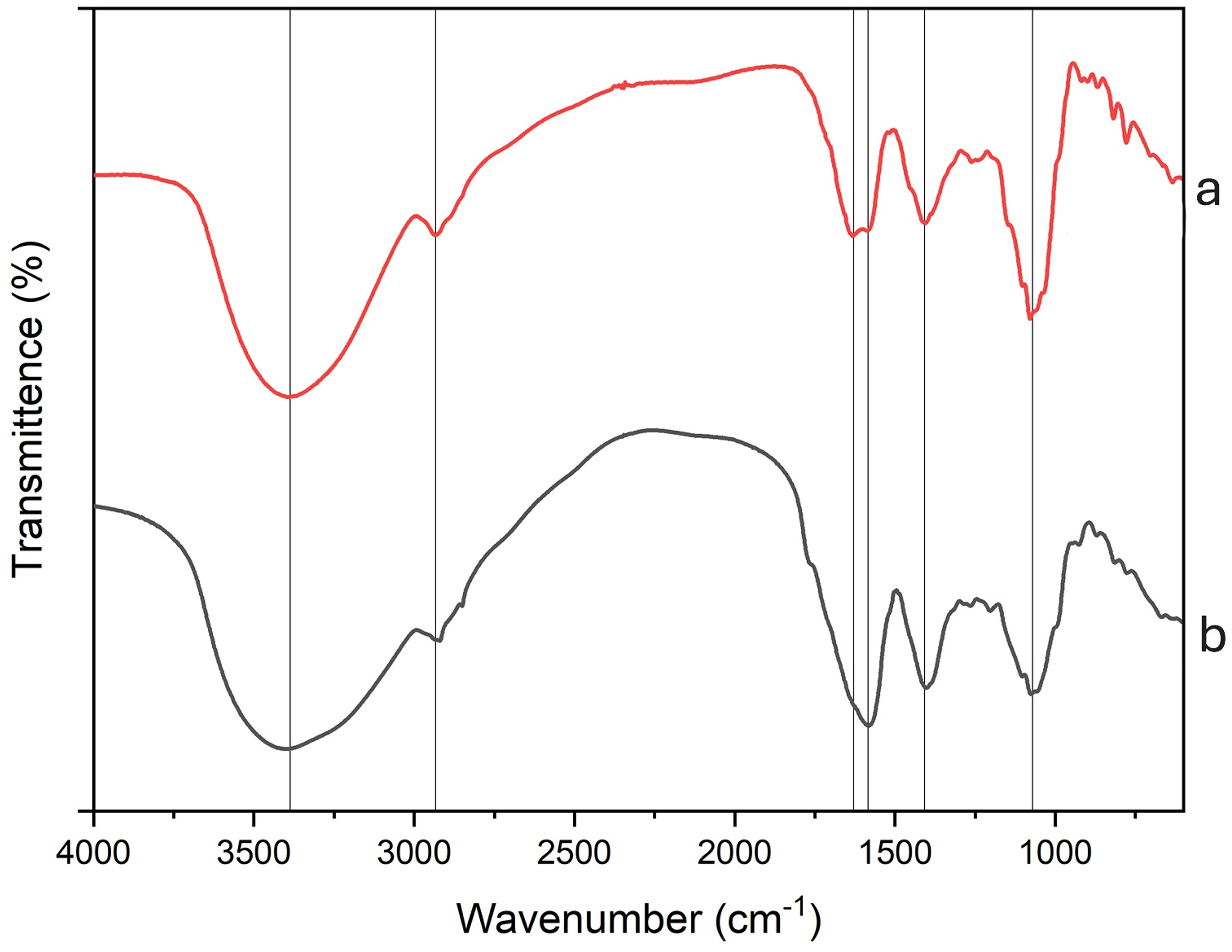
FTIR Spectroscopy of C. citratus Extract from Leaf and Stem Parts.
FTIR Interpretation of C. citratus Leaf and Stem Extracts.

Scanning Electron Microscopy (SEM)
Scanning Electron Microscopy (SEM) (Figure 3) revealed distinct morphological changes in S. sanguinis cells treated with 120 mg/mL of aqueous C. citratus leaf extract. Treated cells appeared swollen and displayed notable abnormalities in shape compared to the untreated control. Several cells exhibited ruptured membranes with visible discontinuities in their cell walls, suggesting structural compromise. Additionally, evidence of impaired replication was observed, with multiple cells appearing deformed and non-replicating, indicating the extract's disruptive effect on bacterial cell division and integrity.
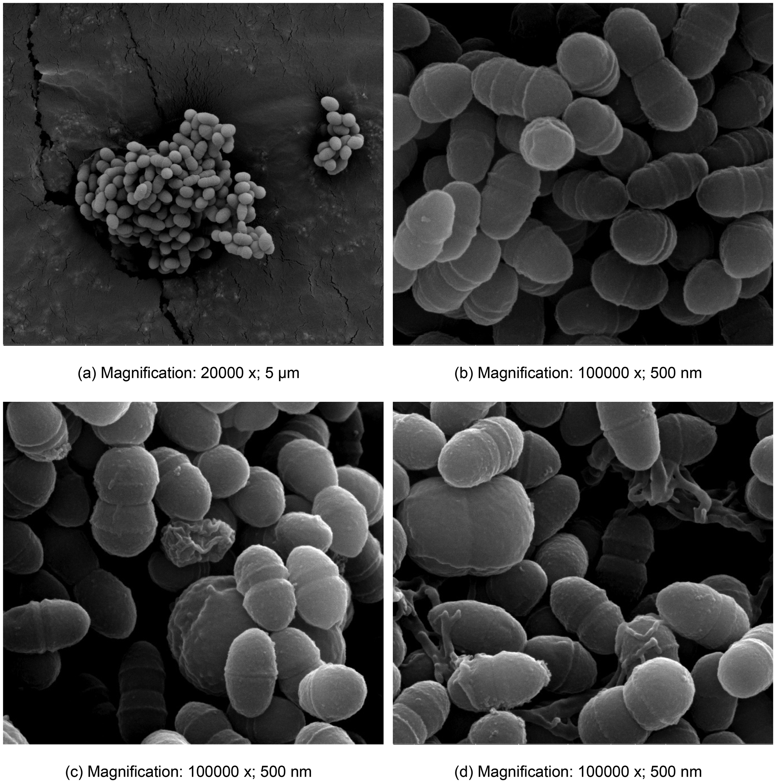
SEM Micrograph of S. sanguinis Control Condition (a) and Reacted with Aqueous Extract of C. citrus Leaf.
Discussion
This study highlights the potential of aqueous extract of C. citratus as a natural antibacterial irrigation solution for root canal treatment. The extracts demonstrated concentration-dependent antimicrobial activity, with notable efficacy against S. sanguinis and E. faecalis, key pathogens in root canal infections. These findings support the development of C. citratus as a safer herbal alternative to sodium hypochlorite, addressing the need for effective and biocompatible solutions in root canal (endodontic) treatment.
The antimicrobial activity of C. citratus leaf and stem extracts was evaluated against S. sanguinis and E. faecalis through MIC and MBC assays and further supported by phytochemical characterizations using FTIR spectroscopy. The results demonstrate differential efficacy of the extracts, with distinct patterns of susceptibility observed between the two bacterial species.
The MIC and MBC data indicate that both extracts were more effective against S. sanguinis than E. faecalis. The stem extract exhibited the lowest MIC and MBC against S. sanguinis, at approximately 240 mg/mL, yielding a MBC/MIC ratio of 0.25, indicative of strong bactericidal activity. Conversely, the leaf extract showed both MIC and MBC values around 480 mg/mL against S. sanguinis, also classifying it as bactericidal but comparatively less potent.
Against E. faecalis, both extracts were markedly less effective; the leaf extract demonstrated a MIC of approximately 480 mg/mL with no complete bactericidal activity at this concentration, while the stem extract failed to reach inhibitory thresholds even at 480 mg/mL, suggesting possible resistance or insufficient potency.
In this study, FTIR is only used for preliminary screening, and not for definitive compound identification, since a single FTIR band might suggest the presence of citral's functional group (eg, aldehyde), but it cannot specifically identify citral and other compounds. The presence of citral and other compounds was referred to and supported by other studies, which have identified it in C. citratus extracts.11,12 In addition, the presence of several other key functional groups that are associated with antimicrobial properties, including the broad O–H stretching peaks (∼3350-3400 cm1), characteristic of phenolic and flavonoid compounds, which are known for their antibacterial activity.
The potential antibacterial activity of the leaf extract may be attributed to its higher concentrations of geranial (18.8%) and neral (16.3%) compared to the stem, which contains 14.9% and 12.9%, respectively, as reported by Olayemi 2017. 13 These compounds, also known as α-citral and β-citral, are phenolic components recognized for their antimicrobial properties. Their combined effects likely contribute to the activity of C. citratus against both Gram-positive and Gram-negative bacteria, as supported by previous studies.14,15
The presence of phenolic compounds or alcohols of bioactive antimicrobials such as citral and geraniol. 16 The peak at 2932 cm−1 is indicated by C–H stretching vibrations from aliphatic chains, typically found in methyl (-CH3) and methylene (-CH2-) groups. This refers to the extract's fatty acids or long-chain hydrocarbons. At the peak of 1628 cm1, the C = O stretching vibration is observed in carbonyl groups in aldehydes, ketones, and carboxylic acids. This peak could be associated with antimicrobial bioactive citral. 17 The peak at 1583 cm−1 corresponds to the C = C stretching vibration of aromatic rings, referring to phenolic acids or flavonoid compounds in the extract. 18 The band at 1407 cm−1 is associated with C–H bending vibrations, particularly from alkanes or alkyl side chains, contributing to the extract's overall stability and hydrophobic nature. The absorption at 1071 cm−1 is C–O stretching vibrations associated with alcohols, ethers, or carboxyl groups. 12
The superior activity of the stem extract against S. sanguinis, despite its comparatively lower O–H content, may suggest a synergy between its lipid-based constituents and minor phenolic components. Meanwhile, the broader spectrum of functional groups in the leaf extract could account for its comparatively better, though still limited, efficacy against the more resistant E. faecalis. The results also highlight the importance of bacterial susceptibility profiling, as Gram-positive cocci such as E. faecalis often exhibit intrinsic resistance mechanisms, including efflux pumps and biofilm formation, which may hinder phytochemical penetration. 11
However, neither extract showed bactericidal effects against E. faecalis, even at the highest concentration tested (480 mg/mL), suggesting that this bacterial species exhibits substantial resilience to C. citratus extracts. Notably, the MBC values observed in this study were higher than those typically reported for established agents such as sodium hypochlorite, indicating that the current concentrations may not yet be clinically feasible for effective root canal disinfection.
As root canal treatment requires completely eradicating viable bacteria to ensure long-term success and prevent reinfection. While the aqueous extract of C. citratus demonstrated antibacterial activity, the MBC results highlight the need for further optimisation. The antimicrobial activity of the extract could be optimised by refining extraction parameters (temperature, time, pH) to enhance the yield of active compounds. Bioassay-guided fractionation and structural characterisation (using techniques like LC-MS or NMR) may help isolate and standardise the most potent constituents. Additionally, incorporating the extract into advanced delivery systems or exploring synergistic combinations with other antimicrobials could further enhance its efficacy against E. faecalis and S. sanguinis.
Enhancing the concentration and retention of the extract within the root canal system, potentially through improved delivery methods, could increase its efficacy. Additionally, evaluating its performance in biofilm models and clinical settings will be critical to determining its suitability as an antibacterial root canal irrigation solution. These findings underscore the potential of C. citratus extracts as antimicrobial agents but emphasise the necessity for formulation adjustments or combination therapies to achieve effective bactericidal activity.
Overall, the combination of MIC, MBC, and FTIR data provides a comprehensive understanding of the antibacterial potential and phytochemical basis of C. citratus extracts. While both leaf and stem possess bioactive compounds, their effectiveness is species-specific, and further isolation of active constituents may enhance their therapeutic potential.
The SEM analysis was conducted on S. sanguinis cells treated with the leaf extract of C. citratus at a concentration of 120 mg/mL. The observations revealed significant morphological changes in the bacterial cells, indicating the extract's potential antibacterial activity, as reported in similar studies.8,19,20 The treated S. sanguinis cells appeared swollen compared to untreated cells, suggesting that the extract likely caused disruption of the bacterial cell membrane. This morphological change can be attributed to the bioactive compounds, such as citral, identified through FTIR analysis by the C = O stretch at 1628 cm−1. Citral is known to have the ability to disrupt lipid bilayers, leading to changes in membrane permeability, which may result in cellular swelling through the influx of water and ions. 21
The antimicrobial effects of the extract led to the rupture of the bacterial cell envelope, likely due to the action of the aliphatic compounds within the extract. These compounds disrupt lipid components of bacterial membranes, causing cell lysis. Furthermore, abnormalities were observed in the treated cells, which appeared unable to divide or reproduce. Normally, bacterial cells undergo binary fission to proliferate; however, the treated cells displayed abnormal, deformed shapes and, in some cases, appeared to be non-replicating. 22 This may be attributed to the bioactive compounds interfering with essential bacterial functions, such as cell division.
The phenolic and aromatic compounds in C. citratus extracts may also significantly inhibit the bacterial cell cycle. These compounds have been shown to form complexes with bacterial proteins and enzymes, disrupting their activity and preventing DNA replication and protein synthesis. 10 Consequently, this could halt bacterial replication and interfere with cellular functions necessary for proliferation, leading to the observed deformations and lack of cell division.
Conclusion
This study highlights the potential of C. citratus extracts as natural antimicrobial agents for root canal treatments. The limitations of MIC and MBC assays in distinguishing bacteriostatic from bactericidal effects indicate the need for further research. Future studies should incorporate time-kill assays to evaluate bactericidal kinetics, biofilm disruption assays to assess the extract's ability to penetrate and disrupt biofilm structures, and advanced mechanistic studies using scanning electron microscopy or flow cytometry techniques. Clinical validation and exploration of synergistic interactions with established disinfectants, such as sodium hypochlorite or chlorhexidine, will be crucial for optimising the application of C. citratus extracts in root canal disinfection.
Footnotes
Acknowledgements
The authors thank the multidisciplinary laboratory staff, School of Dental Sciences, Universiti Sains Malaysia, and Forest Research Institute Malaysia (FRIM) for their technical assistance and resources. Their support was invaluable in the successful completion of this study.
ORCID iDs
Funding
The authors disclosed receipt of the following financial support for the research, authorship, and/or publication of this article: The authors sincerely thank Universiti Sains Malaysia for supporting this research under RGS 203.PPSG.6171342 (FRGS/1/2022/SKK0/USM/02/17).
Declaration of Conflicting Interests
The authors declared no potential conflicts of interest with respect to the research, authorship, and/or publication of this article.
Data Availability Statement
Data will be made available on request.