



Abstract
Objective/Background
The medicinal plant Rhamnus ussuriensis J.J. Vassil. (R. ussuriensis) has been widely used in folk medicine, but its composition and antioxidant potential remain poorly studied. This research aimed to evaluate the effects of drying methods and extraction solvents on the active ingredients and antioxidant capacity of R. ussuriensis fruit, and to explore its potential therapeutic applications.
Methods
Freeze-drying and oven-drying were compared for active ingredient preservation. Four solvents were tested for extraction efficiency and antioxidant properties. Phenolic stability was assessed under heating, varying pH, and simulated human digestion. UHPLC-ESI-Q-TOF-MS identified chemical constituents. Oil stability tests, acute oral toxicity studies, and animal experiments evaluated safety and potential applications.
Results
Freeze-dried fruit showed significantly higher active component levels than oven-dried (total carbohydrate: 781.35 ± 7.46 mg/g; total protein: 829.08 ± 9.69 mg/g; total phenolics: 67.52 ± 1.78 mg/g; total flavonoids: 90.45 ± 0.93 mg/g; total tannins: 62.12 ± 1.84 mg/g). Antioxidant assays (IC50 for DPPH: 56.63 ± 1.84 μg/mL; ABTS: 30.18 ± 0.96 μg/mL) confirmed superior bioactive compound preservation. Adding ethanol to the solvent improved extraction efficiency and antioxidant properties, with ethanol and 80% ethanol extracts from freeze-dried fruit performing equally well. Phenolics in the ethanol extract showed stability in heating, pH variation, and simulated digestion tests. HPLC-ESI-Q-TOF-MS identified 51 compounds, primarily flavonoids and phenolics. The ethanol extract stabilized sunflower oil and showed no acute oral toxicity. Animal experiments demonstrated significant hepatoprotective effects against D-galactosamine-induced liver damage in rats.
Conclusion
Freeze-drying is the preferred method for preserving active ingredients in R. ussuriensis fruit. The ethanol extract, rich in flavonoids and phenolics, exhibits strong antioxidant properties, stability, and hepatoprotective effects, highlighting its potential for treating oxidative stress-related conditions.
Keywords
Introduction
There are approximately 200 species of plants belonging to the genus Rhamnus globally, distributed across temperate to tropical regions. They are primarily found in Eastern Asia and southwestern North America, with a limited presence in Europe and Africa. In China, this genus boasts 57 species and 14 varieties.1,2 The plants of the Rhamnus genus are primarily used to treat constipation, facilitate diuresis, exert anti-inflammatory effects, promote digestion, and lower blood pressure. 3 Rhamnus plants are predominantly characterized by their abundance of two primary chemical constituents: anthraquinones and flavonoids, along with phenolics and various other compounds. Pharmacological investigations have primarily focused on their antioxidant, anti-tumor, anti-inflammatory, laxative, and antibacterial properties,4-7 highlighting their significant medicinal value and extensive application potential. Furthermore, certain components within Rhamnus plants, such as apigenin, emodin, quercetin, and orientin, exhibit antioxidant, antibacterial, and anti-inflammatory effects. 8
Rhamnus ussuriensis J.J. Vassil. (R. ussuriensis) (Figure 1) is a shrub that belongs to the Rhamnus genus. It frequently grows along riverbanks, in mountain forests, or among shrubs on mountain slopes below an altitude of 1600 m. 9 Oil can be extracted from its seeds for use in lubricating oil production. Both the bark and fruit contain tannins, which can be utilized for extracting tannin compounds and yellow dyes. Furthermore, its branches and leaves can be used as pesticides to combat soybean aphids and treat rice blast disease. 9 Currently, no studies have been reported on isolating and characterizing the constituents of R. ussuriensis. However, its pharmacological effects, particularly its antioxidant and anti-inflammatory properties, have been extensively documented. 10 In this experiment, extracts derived from the leaves, fruits, and branches of R. ussuriensis were prepared using 70% ethanol. The results indicated that the 70% ethanol extract from the fruits contained higher levels of phenolics and flavonoids and exhibited the strongest antioxidant capacity among the tested extracts. 10 Furthermore, the research team conducted in-depth studies and found that the water extract of the fruits demonstrated the most promising immune-enhancing activity. 11 Therefore, the experimental results suggest that further research on the fruit of R. ussuriensis is warranted.

Morphology of Rhamnus ussuriensis Fruit.
Plants of the Rhamnus genus have been widely used in clinical practice for their anti-inflammatory, anticancer, and antioxidant activities. While R. ussuriensis shares taxonomic classification with these species, its phytochemical composition and pharmacological properties remain largely unexplored, warranting systematic investigation. This study is the first systematic investigation of antioxidant activities in R. ussuriensis fruit. While previous studies have characterized antioxidative properties in other Rhamnus species (eg, Rhamnus prinoides, 12 Rhamnus lycioides 13 ), no prior reports exist regarding either the phytochemical profile or bioactivity of R. ussuriensis. Furthermore, the comparative analysis of freeze-drying versus oven-drying provides novel methodological insights for preserving thermo-sensitive antioxidants in understudied Rhamnus fruits.
Experimental Section
Plant Materials
In October 2023, the fruit of R. ussuriensis, with voucher specimen number 2023-10-11-001, was harvested in Tonghua, located in Jilin Province, China. The specific geographical coordinates were latitude N 41°44′47.56″ and longitude E 125°56′53.85″, with an altitude of 470 m. The specimen, which is currently housed in the Herbarium of Tonghua Normal University, was identified by professor Junlin Yu.
Two Drying Methods of R. ussuriensis Fruit
The R. ussuriensis fruit is processed using two drying methods. One method involves freeze-drying for a duration of 24 h, while the other method consists of oven-drying at a temperature of 50 °C for 48 h. In both methods, drying was continued until samples reached constant weight, ensuring minimal residual moisture. However, precise moisture content were not quantified in this study.
Phytochemical Composition Analysis
An analysis of phytochemical composition was performed on fifteen distinct chemical components, employing a previously validated methodology. 14 For a comprehensive understanding of the experimental methods, see the Supplemental Material for full details.
Production of Various Extracts from Oven-Dried R. ussuriensis Fruit (ORUF) and Freeze-Dried R. ussuriensis Fruit (FRUF)
The objective of this experiment was to explore how different solvents affect the efficiency of extracting active components from FRUF and ORUF. To achieve this, four different polar solvents were utilized: water, ethanol, 80% ethanol, and methanol. The extraction protocol was performed according to established methodology. 14 The detailed experimental procedure was as follows:
The collected samples of Rhamnus ussuriensis fruit were dried using two methods: freeze-drying and oven-drying. The freeze-dried samples were dried at −60 °C for 24 h, while the oven-dried samples were dried at 50 °C for 48 h. After the drying process was completed, both sets of samples were pulverized into powder separately. The powder (20 g) of FRUF or ORUF was added to a single-neck round-bottomed flask (glass, 500 mL), followed by addition of 200 mL of various solvents (double-distilled water, absolute methanol, absolute ethanol, or 80% ethanol; in the following text, the first three are referred to as water, methanol, and ethanol, respectively) and refluxing using a hotplate magnetic stirrer employing methyl silicone oil as the heating medium for 6 h at the respective boiling points of the solvents. Extracts were filtered through a Whatman No.1 filter paper and evaporated under reduced pressure at < 50 °C until dry using a rotary evaporator. All dried extracts were weighed and stored at −20 °C until use. Yield was calculated as % yield = (weight of dry extract/initial weight of dry sample) × 100.
Phytochemical Quantification Analysis
A quantitative analysis of phytochemicals was conducted to determine the contents of several compounds, such as total protein content (TProC), total phenolic content (TPheC), total flavonoid content (TFC), total carbohydrate content (TCC), total tannin content (TTanC), gallotannin content (GC), condensed tannin content (CTC), total phenolic acid content (TPAC) and total alkaloid content (TAC). The methods employed for this analysis were previously described in references.14-15 The detailed experimental procedure was as follows:
TCC
Briefly, 250 μL of FRUF or ORUF extract (0.02-0.5 mg/mL) in distilled water, 125 μL of phenol solution (5%), and 625 μL of H2SO4 were mixed in an Eppendorf tube and incubated for 30 min. Subsequently, 200 μL of the sample was pipetted from each Eppendorf tube onto a microplate. A calibration curve was produced based on glucose (0-200 mg/L) as a standard. The absorbance of the sample was recorded at 490 nm against a blank sample consisting of FRUF or ORUF extract with distilled water. The mean of three readings was used and TCC was expressed in milligrams of glucose equivalents (GE)/g of FRUF or ORUF extract.
TProC
Briefly, 200 μL of bicinchoninic acid working solution and 20 μL of FRUF or ORUF extract (0.1-2.0 mg/mL) in distilled water were mixed in a microplate and incubated at 37 °C for 30 min. A calibration curve was produced based on bovine serum albumin (0-500 mg/L) as a standard. The absorbance of the sample was recorded at 562 nm against a blank sample consisting of FRUF or ORUF extract with distilled water. The mean of three readings was used and TProC was expressed in milligrams of BSA equivalents (BSAE)/g of FRUF or ORUF extract.
TPheC
Briefly, 100 μL of FC reagent (1 M) and 200 μL of FRUF or ORUF extract (0.5-2.0 mg/mL) in distilled water were mixed in an Eppendorf tube and incubated for 5 min. Subsequently, 500 μL of Na2CO3 solution (20%) was added and allowed to stand at room temperature for 40 min in the dark (with mixing every 10 min). Subsequently, 200 μL of the sample was pipetted from each Eppendorf tube onto a microplate. A calibration curve was produced based on gallic acid (0-100 mg/L) as a standard. The absorbance of the sample was recorded at 750 nm against a blank sample consisting of FRUF or ORUF extract with distilled water and Na2CO3. The mean of three readings was used and TPheC was expressed in milligrams of gallic acid equivalents (GAE)/g of FRUF or ORUF extract.
TFC
Briefly, 100 μL of AlCl3 (2%) in methanol and 100 μL of FRUF or ORUF extract (0.5-5.0 mg/mL) in methanol were mixed in a microplate and incubated at room temperature for 10 min. A calibration curve was produced based on quercetin (0-100 mg/L) as a standard. The absorbance of the sample was recorded at 415 nm against a blank sample consisting of FRUF or ORUF extract with methanol. The mean of three readings was used and TFC was expressed in milligrams of quercetin equivalents (QE)/g of FRUF or ORUF extract.
TPAC
Briefly, 20 μL of FRUF or ORUF extract (0.2-2.0 mg/mL) in distilled water, 20 µL of Arnow reagent, 20 µL of HCl solution (0.1 M), 120 µL of distilled water and 20 µL of NaOH solution (1 M) were mixed in a microplate and recorded immediately at 490 nm against a blank sample (Arnow reagent was replaced with distilled water). A calibration curve was produced based on caffeic acid (0-100 mg/L) as a standard. The mean of three readings was used and TPAC was expressed in milligrams of caffeic acid equivalents (CAE)/g of FRUF or ORUF extract.
TTanC
Briefly, 200 μL of FC reagent (1 M) and 200 μL of FRUF or ORUF extract (0.2-2.0 mg/mL) in distilled water were mixed in an Eppendorf tube and incubated for 5 min. Subsequently, 100 μL of Na2CO3 solution (20%) and 1500 μL of distilled water were added and allowed to stand at room temperature for 30 min in the dark (with mixing every 10 min). Subsequently, 200 μL of the sample was pipetted from each Eppendorf tube onto a microplate. A calibration curve was produced based on tannic acid (0-200 mg/L) as a standard. The absorbance of the sample was recorded at 725 nm against a blank sample consisting of FRUF or ORUF extract with distilled water and Na2CO3. The mean of three readings was used and TTanC was expressed in milligrams of tannic acid equivalents (TAE)/g of FRUF or ORUF extract.
GC
Briefly, 875 µL of FRUF or ORUF extract (0.2-2.0 mg/mL) in methanol and 375 µL of saturated KIO3 solution were mixed in an Eppendorf tube and incubated at 15 °C for 120 min. A calibration curve was produced based on gallic acid (0-400 mg/L) as a standard. The absorbance of the sample was recorded at 550 nm against a blank sample (KIO3 was replaced with distilled water). The mean of three readings was used and GC was expressed in milligrams of GAE/g of FRUF or ORUF extract.
CTC
Briefly, 4 mg of phloroglucinol was added to 2 mL of FRUF or ORUF extract (0.5-10.0 mg/mL) in distilled water. Subsequently, 1 mL of HCl solution and 1 mL of formaldehyde solution were added and mixed in an Eppendorf tube and incubated at room temperature overnight. The precipitate was separated by filtration, the unprecipitated phenolics were measured in the filtrate according to the method of TPheC.
TAC
Berberine hydrochloride (1.24-12.36 mg/L) was used as a reference material to construct a standard curve. The absorbance was obtained at 420 nm against a blank sample of chloroform. The mean of three measurements was calculated. The total alkaloid content is expressed in milligrams of berberine hydrochloride equivalents (BHE)/g of FRUF or ORUF extract.
Assays for Antioxidant Capability
Multiple methods were employed to evaluate antioxidant capability, encompassing the 2,2'-azino-bis(3-ethylbenzothiazoline-6-sulfonic acid) diammonium salt (ABTS), 1,1-diphenyl-2-picrylhydrazyl radical (DPPH), hydroxyl radical, superoxide radical, cupric ion reducing antioxidant capacity (CUPRAC), Ferric ion reducing antioxidant power (FRAP), metal chelating, hydrogen peroxide (H2O2), singlet oxygen, β-carotene bleaching, hypochlorous acid (HClO), and nitric oxide (NO) assays. These assessments adhered to previously validated protocols.14-15 The detailed experimental procedure was as follows:
DPPH Assay
Briefly, 100 µL of FRUF or ORUF extract (0.01-2.0 mg/mL) in methanol and 100 µL of DPPH in methanol (50 µM) were mixed in a microplate and allowed to stand at room temperature for 20 min in the dark. The absorbance of the sample was recorded at 515 nm. The Half-maximal inhibitory concentration (IC50) values were calculated and expressed as the mean ± standard deviation (SD) in μg/mL.
ABTS Assay
Briefly, 190 μL of diluted ABTS solution and 10 μL of FRUF or ORUF extract (0.5-5.0 mg/mL) in DMSO were mixed in a microplate and incubated for 20 min in the dark. The absorbance of the sample was recorded at 734 nm. The IC50 values were calculated and expressed as the mean ± SD in μg/mL.
Hydroxyl Radical Assay
Briefly, 50 µL of FRUF or ORUF extract (0.1-10.0 mg/mL) in DMSO, 50 µL of FeSO4 solution (3 mM) and 50 µL of H2O2 solution (3 mM) were mixed in a microplate and incubated for 10 min. After then 50 µL of salicylic acid solution (6 mM) was added and incubated at room temperature for 30 min in the dark. The absorbance of the sample was recorded at 492 nm. The IC50 values were calculated and expressed as the mean ± SD in μg/mL.
Superoxide Radical Assay
Briefly, 45 µL of FRUF or ORUF extract (0.1-10.0 mg/mL) in DMSO (10 mg/mL), 15 µL of NBT in DMSO (1 mg/mL) and 150 µL of NaOH in DMSO (50 μM) were mixed in a microplate and the absorbance of the sample was recorded immediately at 560 nm against a blank sample (NBT was replaced with DMSO). The scavenging activity was expressed as % scavenging rate and was calculated as follows:

FRAP Assay
Briefly, 20 µL of FRUF or ORUF extract (0.16 mg/mL) in DMSO and 180 µL of FRAP reagent were mixed in a microplate and incubated at 37 °C for 30 min in the dark. A calibration curve was produced based on FeSO4 (0-600 mg/L) as a standard. The absorbance of the sample was recorded at 595 nm. Trolox was used as positive reference. The FRAP was expressed as the Trolox Equivalent Antioxidant Capacity (TEACFRAP).
CUPRAC Assay
Briefly, 20 µL of CuCl2 solution (100 mM), 50 µL of neocuproine in 96% ethanol (7.5 mM), 50 µL of NH4Ac solution, 20 µL of FRUF or ORUF extract (0.2-2.0 mg/mL) in DMSO, and 30 µL of distilled water were mixed in a microplate and incubated at 50 °C for 20 min. This mixture was allowed to stand at room temperature for 10 min. The absorbance of the sample was recorded at 450 nm. The CUPRAC was expressed as the Trolox Equivalent Antioxidant Capacity (TEACCUPRAC).
Iron Chelating Assay
Briefly, 50 µL of FRUF or ORUF extract (0.5-10.0 mg/mL) in methanol, 110 µL of ultra-pure water, and 20 µL of FeCl2 solution (0.5 mM) were mixed in a microplate and incubated for 5 min. Subsequently, 20 µL of ferrozine solution (2.5 mM) was added and incubated for 10 min. The absorbance was recorded at 562 nm against a blank sample (ferrozine solution was replaced with water). The IC50 values were calculated and expressed as the mean ± SD in μg/mL.
Copper Chelating Assay
Briefly, 40 µL of FRUF or ORUF extract (0.5-10.0 mg/mL) in ultra-pure water, 140 µL of acetic acid-sodium acetate buffer solution (pH 6.0, 50 mM), and 10 µL of CuSO4 solution (5 mM) were mixed in a microplate and incubated for 30 min. Subsequently, 10 µL of pyrocatechol violet solution (4 mM) was added and incubated for 30 min. The absorbance was recorded at 632 nm against a blank sample (pyrocatechol violet was replaced with water). The IC50 values were calculated and expressed as the mean ± SD in μg/mL.
H2O2 Assay
Briefly, 70 µL of phenol solution (pH 7.0, 12 mM, in 84 mM phosphate buffer (PBS)), 20 µL of 4-aminoantipyrine solution (pH 7.0, 0.5 mM, in 84 mM PBS), 32 μL of H2O2 solution (pH 7.0, 0.7 mM, in 84 mM PBS), 8 µL of horseradish peroxidise (EC 1.11.1.7) solution (pH 7.0, 1 U/mL, in 84 mM PBS) and 70 µL of FRUF or ORUF extract (0.5-10.0 mg/mL) (pH 7.0, in 84 mM PBS) were mixed in a microplate and the absorbance of the sample was recorded immediately at 504 nm against a blank sample (phenol solution was replaced with PBS). The IC50 values were calculated and expressed as the mean ± SD in μg/mL.
Singlet Oxygen Assay
Briefly, 40 µL of FRUF or ORUF extract (0.5-10.0 mg/mL) (pH 7.4, in 45 mM PBS), 50 µL of N,N-dimethyl-4-nitrosoaniline (pH 7.4, 0.2 mM, in 45 mM PBS), 20 μL of histidine solution (pH 7.4, 0.1 mM, in 45 mM PBS), 20 µL of NaClO solution (pH 7.4, 0.1 mM, in 45 mM PBS), 20 μL of H2O2 (pH 7.4, 0.1 mM, in 45 mM PBS) and 50 µL of PBS (pH 7.4, 45 mM) were mixed in a microplate and allowed to stand at room temperature for 40 min. The absorbance of the sample was recorded at 440 nm against a blank sample (FRUF or ORUF extract was replaced with PBS). The IC50 values were calculated and expressed as the mean ± SD in μg/mL.
β-Carotene Bleaching Assay
Briefly, β-carotene solution was prepared by dissolving β-carotene (2 mg) in CHCl3 (10 mL). Then, 2 mL of the solution was pipetted into a flask and vortex-mixed with linoleic acid (40 mg) and Tween 40 (400 mg). After the removal of CHCl3, 100 mL of oxygenated ultrapure water was added, and the emulsion shaken vigorously. Aliquots (2.4 mL) of the emulsion were pipetted into different test tubes containing 0.1 mL of FRUF or ORUF extract (5 mg/mL) in methanol. Butylated hydroxytoluene and butyl hydroxyanisole were used as positive controls. In the control group, FRUF or ORUF extract was replaced with methanol. When the sample was added to the emulsion, it was recorded as t = 0 min. The tubes were capped and placed in a water bath at 60 °C. The absorbance was recorded at 470 nm every 15 min until 120 min. Antioxidant activity coefficient (AAC) was calculated according to the following equation:

HClO Assay
HClO was freshly prepared by adjusting the pH of a 1% (v/v) of NaClO to 6.2 with 1% H2SO4. The concentration of HClO was determined by reading the absorbance at 235 nm and using the molar extinction coefficient of 100 M−1·cm−1. Briefly, 20 µL of FRUF or ORUF extract (10 mg/mL) aqueous solution, 20 µL of 150 mM taurine aqueous solution, 20 µL of 0.5 mM HClO solution and 140 μL of PBS (pH 7.4, 50 mM) were mixed in a microplate and incubated for 10 min. Subsequently, 2 µL of 2 M KI aqueous solution was added and mixed. The absorbance was recorded at 350 nm against a blank sample (taurine and HClO were replaced with water). IC50 values were calculated and expressed as the mean ± SD in μg/mL.
NO Assay
Briefly, 3 mL of FRUF or ORUF extract (1 mg/mL) in methanol and 3 mL of sodium nitroprusside solution (pH 7.4, 5 mM, in 0.1 M PBS) were mixed in an Eppendorf tube and incubated at 25 °C for 150 min. At intervals, 100 μL of the sample was pipetted from each Eppendorf tube onto a microplate containing 100 µL of Griess reagent. In the control group, FRUF or ORUF extract was replaced with methanol. The absorbance was recorded at 546 nm against a blank sample (Griess reagent was replaced with distilled water).
Investigations on the Stability of Ethanol Extract and 80% Ethanol Extract Derived from FRUF
The pH, thermal, and gastrointestinal tract model system stability of both the ethanol extract and 80% ethanol extract of FRUF, using methods previously outlined in. 14 The detailed experimental procedure was as follows:
pH Stability
The stability in acidic and basic environments was investigated using ethanol or 80% ethanol extract (2 mg/mL) of FRUF dissolved in deionized water with the pH adjusted to 1, 3, 5, 7, 9, or 11 using 1 M HCl or 1 M NaOH. The final concentration of ethanol or 80% ethanol extract was 50 mg/mL. After incubation at room temperature for 1 h, the pH of the mixture was adjusted to 7 and the TPheC and the ABTS scavenging abilities were examined.
Thermal Stability
To evaluate the thermal stability, ethanol or 80% ethanol extract (2 mg/mL) of FRUF dissolved in deionized water (50 mg/mL, pH 7) was placed in test tubes with screw caps. The test tubes were placed in a boiling water bath (100 °C). Samples were removed after 0, 15, 30, 60, 120, 180, and 240 min and cooled in an ice-water bath, and the TPheC and the ABTS scavenging abilities were examined.
Modeling of the Stability in the Gastrointestinal Tract
100 mL of ethanol or 80% ethanol extract (1 mg/mL) of FRUF in distilled water were mixed with 10 mL of PBS (pH 6.8, 10 mM) and incubated at 37 °C for 2 min (oral condition). Then 0.5 mL of 1 M HCl-KCl buffer (pH 1.5) and 5 mL of pepsin solution (pH 1.5, 32 U/mL in 1 M HCl-KCl buffer) were added to samples. The mixtures incubated at 37 °C for 60 min (stomach condition). Thereafter, 1 mL of 1 M NaHCO3 together with 1 mL of a mixture of bile and pancreatic juice (pH 8.2, 10 mg/mL of pancreatin, 14,600 U/mL of trypsin, and 13.5 mg/mL of bile extract in 10 mM PBS) was added to the mixture, and the pH was adjusted to 6.8. The mixtures were incubated at 37 °C for 3 h (duodenal condition). The results were used for the determination of TPheC and ABTS scavenging abilities of methanol extract during simulated gastrointestinal digestion and were taken at 0, 0.5, 1-4 h.
Ultra-High-Performance Liquid Chromatography-Mass Spectrometry (UHPLC-MS) Analysis
Ethanol extract of FRUF was analyzed using UHPLC (Agilent 1290 system) with Q-TOF-MS (Agilent 6545 system). A ZORBAX SB-C18 column (150 × 3.0 mm, 1.8 µm; Agilent) was used. The column temperature was set to 40 °C. The mobile phase was a mixture of 0.1% formic acid in water (solvent A) and a mixture of 0.1% formic acid in acetonitrile (solvent B) at a flow rate of 0.4 mL/min. Linear gradient elution was applied (0-1 min, 95% A; 1-30 min, 95-70% A; 30-50 min, 70-30% A; 50-56 min, 30-1% A; 56-60 min, 1% A). The extract was diluted to 1 mg/mL with methanol and filtered using a 0.22 µm membrane before use. The sample injection volume was 5 µL. The Q-TOF-MS was operated in positive-ion mode with scan range m/z 100-1700. Data were recorded and analyzed with Qualitative Analysis software (version B. 07.00, Agilent).
The experimental conditions for UHPLC-MS were implemented according to established methodology, 14 guaranteeing the accuracy and dependability of the analysis. exhaustive details.
Oxidative Stability of Oils
The oxidative stability of extra virgin olive oil (EVOO) and cold-pressed sunflower oil (CPSO) was evaluated using established analytical techniques. 14 The detailed experimental procedure was as follows:
Extra virgin olive oil (EVOO) and cold-pressed sunflower oil (CPSO) were placed in separate flasks. Ethanol extract of FRUF was added to the EVOO and CPSO flasks at concentrations of 100 and 25 μg/g, respectively. To compare with the stabilizing effect of methanol extract, EVOO and CPSO were supplemented with synthetic antioxidants TBHQ and BHA at 200 μg/g. A control group was prepared without antioxidants. The flasks were left open and placed in an oil bath at 160 °C to simulate frying. Two samples from each category were removed from the flasks every 4 h for duplicate analysis. The oxidative stability of the oils was evaluated by measurement of the free acidity (percentage of oleic acid), peroxide values (milliequivalents of O2/kg oil), and ultraviolet absorption at 232 and 270 nm (K232 and K270).
Oral Acute Toxicity Study
Twenty adult Kunming mice (19-22 g) were acquired by Liaoning Changsheng Biotechnology Co., Ltd (animal license number SCXK (Liao) 2020–0001; Liaoning, China). Housed mice had free access to food and water under a 12 h light–dark cycle. All rats were reared adaptively for 3 d before starting the experiment. We followed the relevant policies in the Guidelines for the Use of Laboratory Animals developed by Tonghua Normal University. The Institutional Animal Care and Use Committee of Tonghua Normal University approved the experimental protocol (Ethic approval code: 20240037) and the reporting of this study conforms to ARRIVE 2.0 guidelines. 16
The mice were divided into two groups (n = 20) with five males and five females in each group. The mice in the healthy control group received vehicle treatment. The ethanol extract of the FRUF was dissolved in water to a final volume of 10 mL/kg mouse body weight (BW) and then administered to the mice in the FRUF group orally in a single dose of 2000 mg/kg ethanol extract of FRUF. The mice were then continuously observed for 1 h for behavioral changes and toxicity. Intermittent observations were made for next 6 h, and a final observation was conducted at 24 h. At this stage, the survival rate was calculated and we found that no mice died. All mice were euthanized using isoflurane. On the basis of the study results, two doses (150 and 300 mg/kg) were selected for further study.
Hepatoprotective Experiments
The rat liver protection study was performed according to established protocols, 14 comprising four key phases: animal selection, experimental procedure implementation, histopathological evaluation, and biochemical analysis. The specific experimental methodology is described below.
Animals
Adult male Wistar rats (170-200 g) were acquired by Liaoning Changsheng Biotechnology Co., Ltd (animal license number SCXK (Liao) 2020–0001; Liaoning, China). Housed rats had free access to food and water under a 12 h light–dark cycle. All rats were reared adaptively for 7 d before starting the experiment. We followed the relevant policies in the Guidelines for the Use of Laboratory Animals developed by Tonghua Normal University. The Institutional Animal Care and Use Committee of Tonghua Normal University approved the experimental protocol (Ethic approval code: 20240037) and the reporting of this study conforms to ARRIVE 2.0 guidelines. 16
Experimental Protocol
Forty rats were randomly assigned to five groups (n = 8 each) to evaluate the impact of FRUF ethanol extract on liver injury. The control group (GI) received 0.5% sodium carboxymethyl cellulose, as did the model group (GII) which served as injury benchmark. The low- and high-dose groups (GIII and GIV) received 150 mg/kg and 300 mg/kg ethanol extract, respectively, while the positive control group (GV) received 100 mg/kg silymarin. All treatments were administered orally daily for 7 d. After the final treatment, groups II-V received D-galactosamine (700 mg/kg BW, i.p.), followed by 24 h fasting (water allowed) with no mortality observed. All rats were then anesthetized (pentobarbital sodium, 50 mg/kg BW, i.p.) for sample collection. Abdominal aortic blood was collected, allowed to clot for 30 min at room temperature, then centrifuged (3000 rpm, 4 °C, 15 min) with serum stored at −80 °C. Hepatic tissue was excised, rinsed with saline, blotted dry, weighed, and homogenized (10% in saline). The homogenate was centrifuged (10000 rpm, 4 °C, 10 min) with supernatant stored at −80 °C.
Histopathological Examination
The hepatic tissues of three rats were selected from each group, fixed with 4% paraformaldehyde, dehydrated and washed, and embedded in paraffin. After cutting into 4-µm thick sections, hematoxylin and eosin were used for staining. Observations were made using a light microscope. A digital camera was used to record histopathological changes.
Biochemical Analyses
Before anesthesia, each rat was weighed. The hepatic tissue weight and BW of each rat were used to calculate the viscera index (VI) as follows: VI = viscera weight (g)/BW (g) × 100%. For assessment of biochemical parameters related to liver function, serum samples were analyzed for albumin (ALB), γ-glutamyl transpeptidase (γ-GT), alanine aminotransferase (ALT), aspartate aminotransferase (AST), malondialdehyde (MDA) and catalase (CAT). Hepatic samples were analyzed for ALT, AST, MDA and CAT. All samples were analyzed using commercial kits according to the manufacturer's guidelines.
Statistical Analysis
Statistical analysis was performed to assess the significance of the data, which were expressed as mean ± standard error. One-way ANOVA was first conducted to identify significant differences among groups, followed by post-hoc LSD and Duncan tests for multiple comparisons. These tests were employed to determine intergroup differences in the pharmacological experiments, extraction rates, antioxidant activities, and content determinations. Statistical significance was defined at three levels: p < 0.05 (significant), p < 0.01 (highly significant), and p < 0.001 (extremely significant).
Results
Qualitative Phytochemical Analysis
A primary aim of this research is to investigate how different drying techniques influence the composition of R. ussuriensis. Consequently, both ORUF andFRUF underwent a thorough qualitative phytochemical assessment, as detailed in Table S1.
Both sample sets contained proteins/amino acids, carbohydrates, organic acids, phenolics, tannins, flavonoids, alkaloids, anthraquinones, fats, and volatile oils. Quantitative analyses were performed for all detected phytochemicals except anthraquinones, for which quantification remains incomplete. Analytical results indicated the absence of coumarins, lactones, steroids, triterpenoids, saponins, cyanogenic glycosides, and cardiac glycosides in both samples; consequently, no quantitative analyses were conducted for these compounds.
Extraction Yields
The extraction rates of both dried samples, as illustrated in Table 1, followed a comparable trend; significantly, the 80% ethanol extract yielded the highest extraction efficiency. This may be due to its aqueous ethanol solution, which could result in a rich extract containing proteins, carbohydrates, phenolics, glycosides, and other compounds. The ethanol and water extracts followed closely behind, while the methanol extract had the lowest extraction rate.
Yields Obtained from Extracting FRUF and ORUF Using various Solvents.

Columns denoted by distinct superscripts signify a statistically significant difference (p < 0.05). The yield percentage was determined using the formula: % yield = [(weight of the extract/initial dry sample weight)] × 100. FRUF: freeze-dried Rhamnus ussuriensis fruit; ORUF: oven-dried Rhamnus ussuriensis fruit.
Quantitative Phytochemical Analysis
TCC
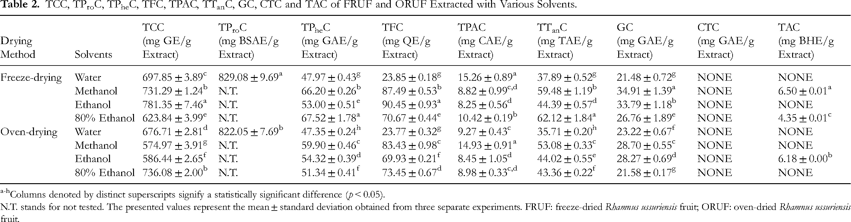
The present investigation evaluated the TCC in various solvent extracts of FRUF, revealing a range of TCC from 623.84 ± 3.99 to 781.35 ± 7.46 mg of glucose equivalents (GE) per gram of extract. In contrast, the TCC of ORUF was found to lie between 574.97 ± 3.91 and 736.08 ± 2.00 mg GE/g extract, as detailed in Table 2. Notable differences were observed among the different groups, with the ethanol extract of FRUF demonstrating the highest TCC.
TCC, TProC, TPheC, TFC, TPAC, TTanC, GC, CTC and TAC of FRUF and ORUF Extracted with Various Solvents.
Columns denoted by distinct superscripts signify a statistically significant difference (p < 0.05).
N.T. stands for not tested. The presented values represent the mean ± standard deviation obtained from three separate experiments. FRUF: freeze-dried Rhamnus ussuriensis fruit; ORUF: oven-dried Rhamnus ussuriensis fruit.
TProC
According to the research, the water extract of FRUF demonstrates a notable TProC of 829.08 ± 9.69 mg of bovine serum albumin equivalents (BSAE)/g extract. The water extract of ORUF exhibits a slightly lower TProC, yet it still attains a concentration of 822.05 ± 7.69 mg BSAE/g (Table 2).
TPheC
The experimental data in Table 2 indicates that the TPheC of different extracts varies between 47.97 ± 0.43 and 67.52 ± 1.78 mg gallic acid equivalents (GAE)/g extract. Among the tested extracts, the 80% ethanol extract of FRUF exhibited the highest content, followed by the methanol extract of FRUF, while the water extract of ORUF showed the lowest levels.
TFC
The TFC ranges from 23.77 ± 0.32 to 90.45 ± 0.93 mg quercetin equivalents (QE)/g extract (Table 2), among the tested extracts, the ethanol extract of FRUF exhibited the highest content, followed by the methanol extract of FRUF.
TPAC
As shown in Table 2, there is a minor variation in TPAC among the solvent extracts, with a relatively low concentration spanning from 8.25 ± 0.56 to 15.26 ± 0.89 mg caffeic acid equivalents (CAE)/g extract. The water extract of FRUF exhibited the highest content, followed by the water extract of ORUF.
TTanC, GC and CTC
To evaluate the tannin content in FRUF and ORUF extracted via various solvents, we analyzed key parameters such as TTanC, GC, and CTC, as shown in Table 2. The TTanC values ranged from 35.71 ± 0.20 to 62.12 ± 1.84 mg tannic acid equivalents (TAE)/g extract, with the highest TTanC recorded in the 80% ethanol extract of FRUF.
TAC
The experimental results revealed that only three of these extracts contained TAC, and the concentration was very low, ranging from 4.35 ± 0.01 to 6.50 ± 0.01 mg of BHE/g extract (Table 2). The methanol extract of FRUF exhibited the highest content.
In Vitro Antioxidant Capability
DPPH and ABTS
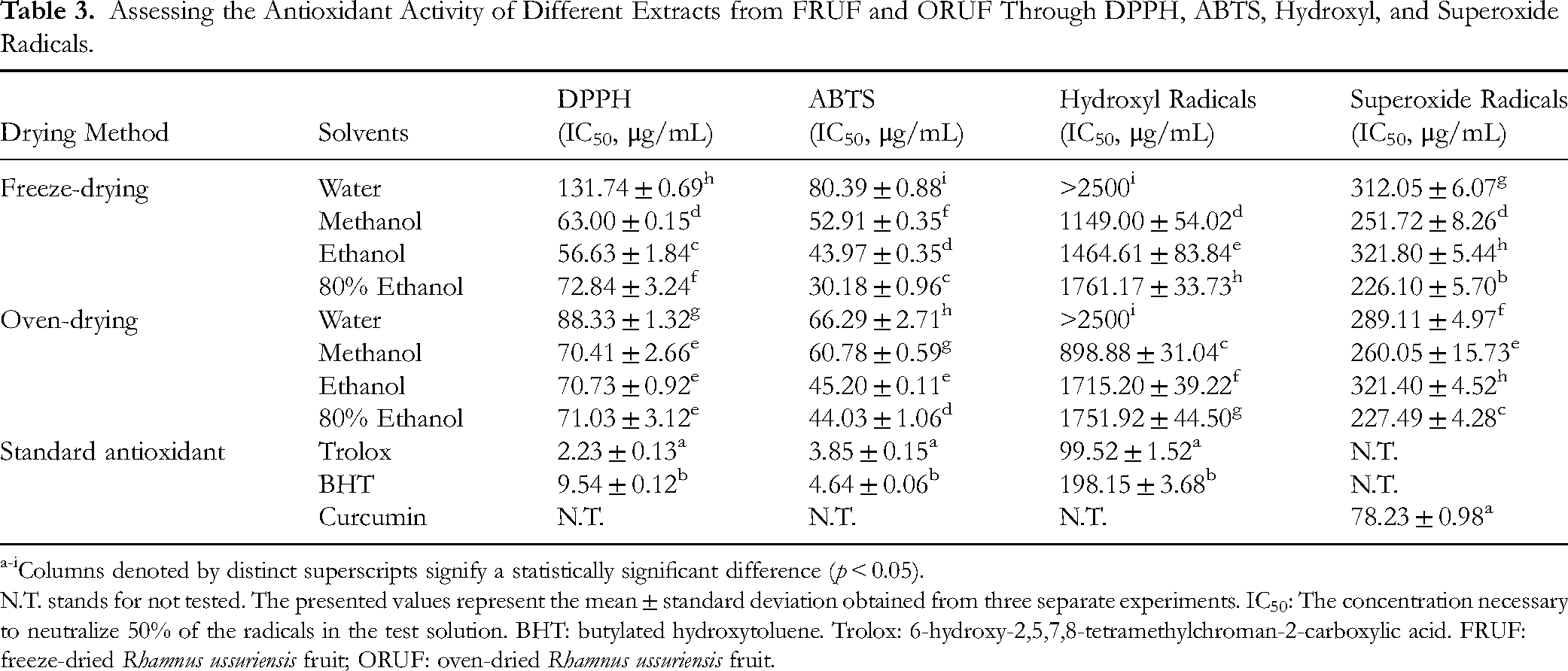
The ethanol extract of FRUF displayed robust scavenging capabilities in the DPPH assay, achieving an IC50 value of 56.63 ± 1.84 μg/mL. Likewise, the 80% ethanol extract exhibited notable scavenging activity in the ABTS assay, yielding an IC50 value of 30.18 ± 0.96 μg/mL (Table 3).
Assessing the Antioxidant Activity of Different Extracts from FRUF and ORUF Through DPPH, ABTS, Hydroxyl, and Superoxide Radicals.
Columns denoted by distinct superscripts signify a statistically significant difference (p < 0.05).
N.T. stands for not tested. The presented values represent the mean ± standard deviation obtained from three separate experiments. IC50: The concentration necessary to neutralize 50% of the radicals in the test solution. BHT: butylated hydroxytoluene. Trolox: 6-hydroxy-2,5,7,8-tetramethylchroman-2-carboxylic acid. FRUF: freeze-dried Rhamnus ussuriensis fruit; ORUF: oven-dried Rhamnus ussuriensis fruit.
Hydroxyl Radicals and Superoxide Radicals
This research indicates that the methanol extract of FRUF and ORUF possesses the greatest scavenging effect on hydroxyl radicals, whereas the 80% ethanol extract of these plants exhibits the highest scavenging activity against superoxide radicals, as detailed in Table 3.
FRAP and CUPRAC
The FRAP assay revealed that all extracts of FRUF and ORUF exhibited moderate ferric reducing ability. Additionally, the CUPRAC assay demonstrated that the 80% ethanol extract of FRUF and ORUF had the best cupric ion reducing ability (Table 4).
Assessing the Antioxidant Properties of various Extracts from FRUF and ORUF Through FRAP, CUPRAC, and Metal ion Chelating.

Columns denoted by distinct superscripts signify a statistically significant difference (p < 0.05).
N.T. stands for not tested. TEAC: trolox equivalent antioxidant capacity. EDTANa2: ethylenediaminetetraacetic acid disodium salt. FRUF: freeze-dried Rhamnus ussuriensis fruit; ORUF: oven-dried Rhamnus ussuriensis fruit.
Metal Ion Chelation
This research has shown that the ethanol extract of FRUF has strong iron chelating properties. Furthermore, the water extract of ORUF exhibits significant copper chelating abilities (see Table 4).
H2O2 and Singlet Oxygen
Consequently, the effective removal of H2O2 is of utmost importance. Experimental results indicate that the ethanol extract of FRUF demonstrates notable H2O2 scavenging capabilities (IC50, 426.05 ± 15.16 μg/mL)(Table 5). Additionally, singlet oxygen, experimental results indicate that the 80% ethanol extract of FRUF exhibits notable singlet oxygen scavenging activity (IC50, 785.30 ± 4.89 μg/mL), rivaling the performance of the positive control, ferulic acid (Table 5).
Assessment of the Antioxidant Properties of various Extracts from FRUF and ORUF Through H2O2, Singlet Oxygen, β-Carotene Decolorization and HClO.

Columns denoted by distinct superscripts signify a statistically significant difference (p < 0.05).
N.T. stands for not tested. BHA: butyl hydroxyanisole. FRUF: freeze-dried Rhamnus ussuriensis fruit; ORUF: oven-dried Rhamnus ussuriensis fruit.
β-Carotene Decolorization
This study found that the ethanol extract of FRUF exhibited comparable antioxidant activity (876.21 ± 22.38) to the positive controls BHT (878.26 ± 30.86; p = 0.982) and BHA (887.37 ± 29.66; p = 0.751), with no statistically significant differences observed. The ethanol and methanol extracts of ORUF showed relatively lower activity (Figure 2 and Table 5).

Alterations in β-Carotene Absorbance Were Observed When Exposed to Various Extracts Derived from FRUF and ORUF. BHT: Butylated Hydroxytoluene; BHA: Butyl Hydroxyanisole. FRUF: Freeze-Dried Rhamnus ussuriensis Fruit; ORUF: Oven-Dried Rhamnus ussuriensis Fruit.
HClO and NO
The HClO scavenging assay revealed that only the ethanol extract of FRUF displayed weak activity, while other extracts showed no detectable effects (Table 5). In contrast, the NO scavenging experiment demonstrated that multiple FRUF and ORUF extracts exhibited significant scavenging capacity. Specifically, the ethanol extract of FRUF exhibited the most significant effect, approaching the efficacy of the positive control, curcumin (Figure 3).

Alterations in Nitric Oxide Absorbance Were Observed When Exposed to various Extracts Derived from FRUF and ORUF. FRUF: Freeze-Dried Rhamnus ussuriensis Fruit; ORUF: Oven-Dried Rhamnus ussuriensis Fruit.
Stability Studies of Ethanol Extract and 80% Ethanol Extract of FRUF
This study evaluated the antioxidant capacity and stability of both the ethanol and 80% ethanol extracts of FRUF through three stability tests. The results of these tests are illustrated in Figures 4-6. These findings revealed that the TPheC and ABTS scavenging potential of the ethanol extract of FRUF maintained a relatively consistent level across varying pH values. In contrast, as the pH level increased, the TPheC content and ABTS scavenging activity of the 80% ethanol extract of FRUF initially rose but subsequently declined, suggesting its instability in both highly acidic and highly alkaline environments. This suggests that the phenolics in this extract may be phenolic glycosides, which are unstable and prone to hydrolysis in strong acid and alkali conditions.

Stability Assessments of the Ethanol Extract and 80% Ethanol Extract of FRUF at Different pH Levels Were Conducted Using TPheC (A) and ABTS (B) Assays. FRUF: Freeze-Dried Rhamnus ussuriensis Fruit.

Thermal Stability Assessments of the Ethanol Extract and 80% Ethanol Extract of FRUF Were Conducted Using TPheC (A) and ABTS (B) Assays. FRUF: Freeze-Dried Rhamnus ussuriensis Fruit.

Stability Assessments of the Ethanol Extract and 80% Ethanol Extract of FRUF in an in Vitro Simulation of the Human Digestive System Were Conducted Using TPheC (A) and ABTS (B) Assays. FRUF: Freeze-Dried Rhamnus ussuriensis Fruit.
Within the initial 30 min of the heating process, notable variations were observed in both the TPheC and ABTS scavenging activity of both the ethanol and 80% ethanol extracts of FRUF. Significantly, the fluctuations were more pronounced in the 80% ethanol extract of FRUF. After this initial period, both parameters stabilized. However, the TPheC of the ethanol extract was consistently higher compared to that of the 80% ethanol extract.
During in vitro digestive system stability tests, both the TPheC of the ethanol extract and that of the 80% ethanol extract of FRUF maintained overall stability. However, the pure ethanol extract exhibited greater stability. It's worth noting that, under gastric conditions, the ABTS scavenging activity of the extracts significantly decreased, but it significantly rebounded under duodenal conditions. The 80% ethanol extract demonstrated a more pronounced fluctuation in activity. While ABTS is generally stable under acidic conditions, excessively acidic environments may compromise its stability, leading to decomposition or alteration that could reduce its efficacy as a radical scavenger.
UHPLC-MS Analysis
The chemical composition of the ethanol extract obtained from freeze-dried R. ussuriensis fruit was characterized by UHPLC-ESI-Q-TOF-MS analysis. By thoroughly comparing molecular and fragment ions with reference materials from peer-reviewed articles (supplemented by detailed analysis methodologies for each peak) and reputable public databases, including the Human Metabolome Database (accessed on October 15, 2024, via https://hmdb.ca/) and MassBank (accessed on October 15, 2024, via https://www.massbank.jp/). Table 6 comprehensively lists fifty-one characterized bioactive compounds, with corresponding structural illustrations provided in Figure 7. The UHPLC-MS analysis in positive-ion mode is displayed in Figure 8, while complete MS and MS/MS spectral data (Figures S1-S90) are available in the Supplemental Material.

Chemical Structures of the Compounds Identified in the Ethanol Extract of Freeze-Dried Rhamnus ussuriensis Fruit.
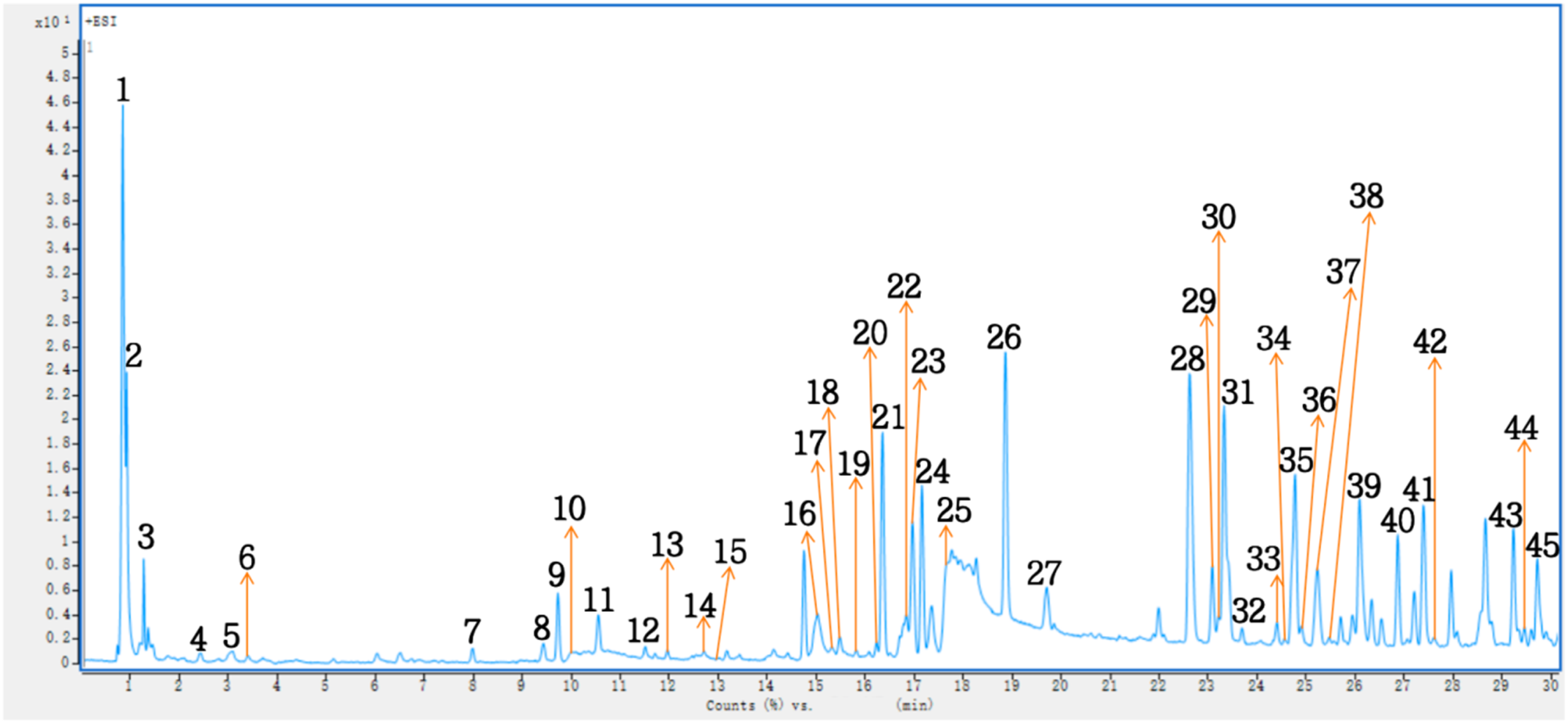
Positive-ion Mode UHPLC-MS Findings of Ethanol Extract of Freeze-dried Rhamnus ussuriensis Fruit.
Identified Compounds in Ethanol Extract of Freeze-Dried Rhamnus ussuriensis Fruit.
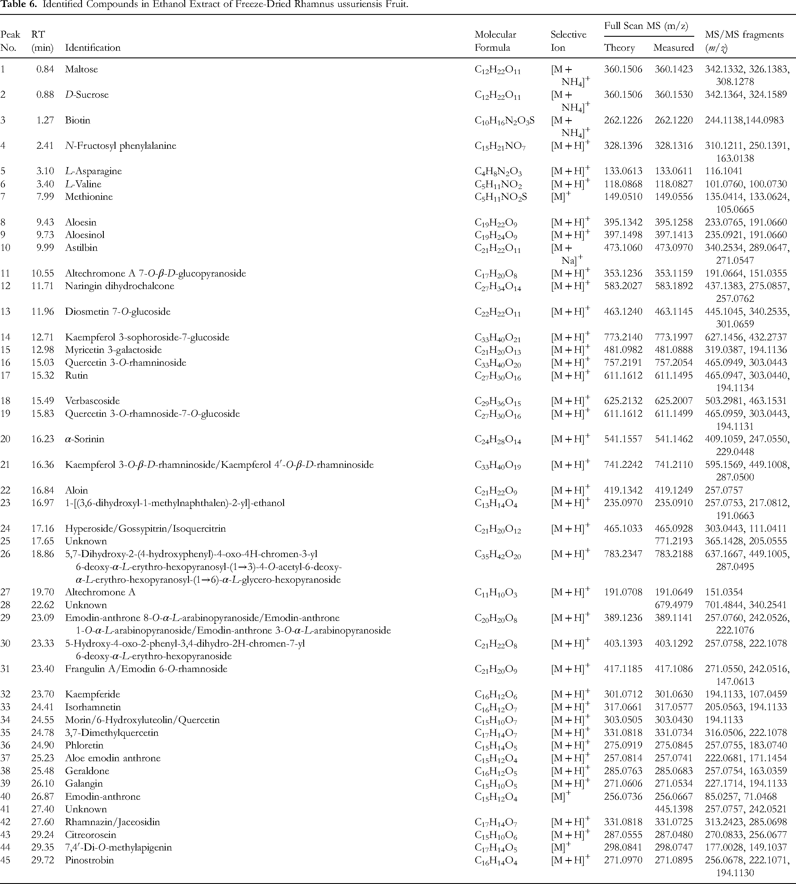
Peak 1 was conclusively identified as maltose by aligning its fragment ions, including m/z 342.1332 for the molecular ion [M]+, 326.1383 for the ion [M−OH + H]+, and 308.1278 for the ion [M−2OH]+, with the reference data provided in. 17 Peak 2, with an m/z of 360.1530, showcased MS2 ions at m/z 342.1364 [M−H2O]+ and 324.1589 [M−2H2O]+, leading to its tentative assignment as D-sucrose. 18 Peak 3, at m/z 262.1220, was recognized as biotin founded on characteristic fragment ions at m/z 244.1138 [M−H]+ and 144.0983 [M−C5H9O2 + H]+. 19 Peak 4, with an m/z of 328.1316, displayed fragment ions at m/z 310.1211 [M−OH]+, 250.1391 [M−C6H5]+, and 163.0138 [C6H11O5]+, suggesting its identification as N-fructosyl phenylalanine. 20 Peak 5, at m/z 133.0611, was recognized as L-asparagine, utilizing the characteristic fragment ion as a foundation, at m/z 116.1041 [M−OH + H]+. 21 Peak 6 exhibited an m/z value of 118.0827 and produced fragment ions with m/z values of 101.0760 and 100.0730, corresponding to the elimination of an −NH2 group and an −OH group, respectively, indicating that peak 6 was L-valine. 22 The major fragment ions of peak 7, at m/z 149.0556, were at m/z 135.0414 [M−CH3 + H]+, 133.0624 [M−OH + H]+, and 105.0665 [M−COOH + H]+, leading to its assignment as methionine. 23 Peak 8 presented a [M + H]+ ion at m/z 395.1258, featuring primary fragment ions at m/z 233.0765 [M−C6H10O5 + H]+ and 191.0660 [M−C6H10O5−C2H2O + H]+, characteristic of aloesin. 24 Peak 9 exhibited a precursor ion [M + H]+ at m/z 397.1413, featuring primary fragment ions of m/z 235.0921 [M−C6H10O5 + H]+ and 191.0660 [M−C6H10O5−C2H4O + H]+, which were indicative of aloesinol, as referenced in. 25 Finally, peak 10, at m/z 473.0970, unveiled MS2 ions at m/z 340.2534 [M−C6H4O2]+, 289.0647 [M−C6H10O5 + H]+, and 271.0547 [M−C6H10O5−OH]+, identifying it as astilbin. 26
The m/z value of Peak 11 was 353.1159, with MS2 ions observed at m/z 191.0664 [M−C6H10O5 + H]+ and 151.0355 [M−C6H10O5−C3H4 + H]+, indicating the presence of altechromone A 7-O-β-D-glucopyranoside. Peak 12, with an m/z value of 583.1892, was determined to be naringin dihydrochalcone. The MS2 spectrum exhibited fragment ions at m/z 437.1383, 275.0857, and 257.0762, which correlated with the losses of −C6H10O4 + H, −C15H12O5 + H, and −C15H12O5−OH, respectively, as reported in. 27 Peak 13 displayed an [M + H]+ ion with an m/z of 463.1145, yielding significant fragment ions at m/z 445.1045 [M−OH]+, 340.2535 [M−C7H7O2 + H]+, and 301.0659 [M−C6H10O5 + H]+, which are indicative of diosmetin 7-O-glucoside, as described in. 28 The mass spectrum for peak 14 revealed an ion with an m/z of 773.1997, and its MS2 spectrum showed fragments at m/z 627.1456 [M−C6H11O5 + NH4]+ and 432.2737 [M−C12H20O10−OH + H]+, which were identified as kaempferol 3-sophoroside-7-glucoside, as stated in. 29 Peak 15, exhibiting an m/z of 481.0888, was determined to be myricetin 3-galactoside, characterized by fragment ions at m/z 319.0387 and 194.1136, which corresponded to the losses of −C6H10O5 + H and −C6H10O5−C6H5O3 + H, respectively, according to. 30 Additionally, peak 16 was identified at an m/z of 757.2054 and characterized as quercetin 3-O-rhamninoside. Its MS2 ions, observed at m/z 465.0949 and 303.0443, suggested the losses of −C12H20O8 + H and −C18H30O13 + H, respectively, as reported in. 31 Peak 17 exhibited an [M + H]+ ion at m/z 611.1495, generating main fragment ions at m/z 465.0947 [M−C6H10O4 + H]+, 303.0440 [M−C12H18O7 + H]+, and 194.1134 [M−C12H20O9−C6H5O2 + H]+, which are characteristic of rutin. 32 Peak 18 was observed at m/z 625.2007 and showed fragment ions at m/z 503.2981 [M−C7H6O2 + H]+ and 463.1531 [M−C9H6O3 + H]+, assigning it as verbascoside. 33 Peak 19 had an m/z value of 611.1499, with MS2 ions at m/z 465.0959 [M−C6H10O4 + H]+, 303.0443 [M−C12H20O9 + H]+, and 194.1131 [M−C12H20O9−C6H5O2 + H]+, based on literature, 34 the compound was provisionally identified as quercetin 3-O-rhamnoside-7-O-glucoside. Lastly, peak 20, with an m/z of 541.1462, displayed MS2 ions at m/z 409.1059 [M−C5H8O4 + H]+, 247.0550 [M−C11H18O9 + H]+, and 229.0448 [M−C11H18O9−OH]+, and was tentatively characterized as α-sorinin. 8
Peak 21, with an m/z of 741.2110, was tentatively discovered as either kaempferol 3-O-β-D-rhamninoside 35 or kaempferol 4′-O-β-D-rhamninoside. 36 The main MS2 ions at m/z 595.1569, 449.1008, and 287.0500 correspond to the loss of −C6H10O4 + H, −C12H20O8 + H, and −C18H30O13 + H, respectively. Peak 22, exhibiting an m/z value of 419.1249, was recognized as aloin, featuring a prominent fragment at m/z 257.0757, which resulted from the loss of −C6H10O5 + H, as referenced in. 37 Peak 23 displayed a precursor ion [M + H]+ with an m/z of 235.0910 and yielded significant fragment ions, including m/z 257.0753 [M + Na]+, 217.0812 [M−OH]+, and 191.0663 [M−C2H4O + H]+, all indicative of 1-[(3,6-dihydroxyl-1-methylnaphthalen)-2-yl]-ethanol. Peak 24, with an m/z of 465.0928, was observed as hyperoside, 38 gossypitrin, 39 or isoquercitrin 40 based on MS2 ions at m/z 303.0443 [M−C6H10O5 + H]+ and 111.0411 [M−C15H14O10 + H]+. The mass spectrum for peak 25 exhibited an ion at m/z 771.2193, accompanied by fragments at m/z 365.1428 and 205.0555 in its MS2 spectrum. Peak 26, displaying an m/z of 783.2188, was identified as the specific compound 5,7-dihydroxy-2-(4-hydroxyphenyl)-4-oxo-4H-chromen-3-yl 6-deoxy-α-L-erythro- hexopyranosyl-(1→3)-4-O-acetyl-6-deoxy-α-L-erythro-hexopyranosyl-(1→6)-α-L-glycero-hexopyranoside, based on characteristic fragment ions such as m/z 637.1667 [M−C6H10O4 + H]+, 449.1005 [M−C14H22O9 + H]+, and 287.0495 [M−C20H32O14 + H]+. Peak 27, with an m/z of 191.0649, was recognized as altechromone A, distinguished by the fragment ion at m/z 151.0354 [M−C3H4 + H]+, as reported in. 41 The mass spectrum of peak 28 revealed an ion at m/z 679.4979, along with fragments at m/z 701.4844 and 340.2541 in its MS2 spectrum. Peak 29 showed an [M + H]+ ion at m/z 389.1141 and yielded significant fragment ions, including m/z 257.0760 [M−C5H8O4 + H]+, 242.0526 [M−C5H8O4−CH3 + H]+, and 222.1076 [M−C5H8O4−2OH]+, indicative of either emodin-anthrone 8-O-α-L-arabinopyranoside, emodin-anthrone 1-O-α-L-arabinopyranoside, or emodin-anthrone 3-O-α-L-arabinopyranoside. Lastly, peak 30, exhibiting an m/z of 403.1292, was identified as 5-hydroxy-4-oxo-2-phenyl-3,4-dihydro-2H-chromen-7-yl 6-deoxy-α-L-erythro- hexopyranoside, based on fragment ions at m/z 257.0758 and 222.1078, resulting from the elimination of −C6H10O4 + H and −C6H11O5−OH, respectively.
Peak 31, which appeared at m/z 417.1086, showed MS2 ions at m/z 271.0550 [M−C6H10O4 + H]+, 242.0516 [M−C6H10O4−CO + H]+, and 147.0613 [C6H10O4 + H]+. It was identified as either frangulin A 42 or emodin 6-O-rhamnoside 43 based on the literature. Peak 32, which appeared at m/z 301.0630, was identified as kaempferide. Its MS2 spectrum featured ions at m/z 194.1133 and 107.0459, indicative of the loss of −C7H7O + H and −C9H5O5 fragments, respectively, as referenced in. 42 Peak 33, with a [M + H]+ ion at m/z 317.0577, was suggested to be isorhamnetin, as its MS2 ions at m/z 205.0563 [M−C6H7O2]+ and 194.1133 [M−C7H7O2 + H]+ matched the literature.8 Peak 34 displayed a [M + H]+ ion at m/z 303.0430, accompanied by a significant fragment ion at m/z 194.1133, which is characteristic of morin, 6-hydroxyluteolin, or quercetin, as indicated by the literature.8,44 Peak 35, located at m/z 331.0734, exhibited fragment ions at m/z 316.0506 and 222.1078, suggesting the loss of −CH3 + H and −C6H5O2 + H, respectively, and was identified as 3,7-dimethylquercetin. 44 Peak 36 was recognized as phloretin at m/z 275.0845, featuring characteristic fragment ions at m/z 257.0755 and 183.0740, which correspond to the loss of −OH and −C6H4O + H, respectively, as reported in. 45 Peak 37, with an m/z of 257.0741, displayed MS2 ions at m/z 222.0681 and 171.1454, indicative of the loss of −2OH and −C4H5O2, respectively, and was tentatively identified as aloe emodin anthrone based on the available literature. 46 Peak 38 exhibited a [M + H]+ ion at m/z 285.0683 and yielded significant fragment ions at m/z 257.0754 and 163.0359, which are indicative of geraldone, as referenced in. 44 The precursor ion [M + H]+ for peak 39 appeared at m/z 271.0534, accompanied by main fragment ions at m/z 227.1714 and 194.1133, corresponding to the loss of −CO−O + H and −C6H5 + H, respectively, and was identified as galangin. 44 Peak 40 showed a [M]+ ion at m/z 256.0667, producing primary fragment ions at m/z 85.0257 and 71.0468, which are characteristic of emodin-anthrone, as indicated in. 42 Peak 41 displayed an ion at m/z 445.1398, with MS2 fragments at m/z 257.0757 and 242.0521. Peak 42, having an m/z value of 331.0725, displayed fragment ions at m/z 313.2423 and 285.0698, which correspond to the loss of −OH and −OH−CO, respectively. Based on the literature, this peak was tentatively identified as either rhamnazin 8 or jaceosidin. 44 Peak 43 exhibited a [M + H]+ ion at m/z 287.0480, accompanied by significant fragment ions at m/z 270.0833 and 256.0677, indicative of the loss of −OH + H and −CH2OH + H, respectively. These fragment ions are characteristic of citreorosein, as reported in. 43 Peak 44 appeared at m/z 298.0747 with MS2 ions at m/z 177.0028 [M−C7H6O2 + H]+ and 149.1037 [M−C8H6O3 + H]+, and was identified as 7,4′-di-O-methylapigenin. 47 Finally, the mass spectrum of peak 45 displayed an ion with an m/z value of 271.0895. The MS2 fragments observed at m/z 256.0678, 222.1071, and 194.1130 correspond to the loss of −CH3, −OH−OCH3, and −C6H5 + H, respectively. Based on this analysis, the peak was identified as pinostrobin, as referenced in. 48
Oxidative Stability Studies of Oils
The research indicates that introducing two doses of the FRUF ethanol extract into EVOO significantly decreased K270 values, outperforming BHA and TBHQ (Figure 9). However, for K232 values, peroxide levels, and acid values, the impact of these doses of the extract was minimal. Yet, it still demonstrated superior performance with significantly lower acidity compared to TBHQ (p < 0.05)(Figures 9–10). In CPSO, the inclusion of the two experimental doses of the extract led to improved K232 and K270 values compared to BHA and TBHQ (p < 0.05), along with a significant reduction in peroxide and acid values comparable to the effects of BHA and TBHQ (p < 0.05)(Figures 9–10).

Alterations in K232 and K270 Concentrations Were Observed in EVOO (A,C) and CPSO (B,D) Supplemented with BHA and TBHQ, as Well as Varying Amounts of the Ethanol Extract Derived from FRUF at 160 °C. BHA: Butyl Hydroxyanisole; TBHQ: Tertiary Butylhydroquinone; FRUF: Freeze-Dried Rhamnus ussuriensis Fruit.

Alterations in the Levels of Acidity Values and Peroxide Values were Observed in EVOO (A,C) and CPSO (B,D) Supplemented with BHA and TBHQ, as Well as Varying Amounts of the Ethanol Extract Derived from FRUF at 160 °C. BHA: Butyl Hydroxyanisole; TBHQ: Tertiary Butylhydroquinone; FRUF: Freeze-Dried Rhamnus ussuriensis Fruit.
Oral Acute Toxicity Study
An evaluation of the acute oral toxicity of the ethanol extract of FRUF was carried out using mice. In this study, a single dose of 2000 mg/kg of the extract was administered orally to twenty mice. Fortunately, all mice remained unaffected for 24 h, suggesting that the ethanol extract of FRUF lacks highly toxic components and poses no notable acute toxicity risk.
Hepatoprotective Activity
The outcomes indicated that both high and low doses of the ethanol extract evidently lowered the viscera index in rats compared to GII (p < 0.001, refer to Figure 11). Remarkably, in relation to their hepatoprotective effects, both doses of the ethanol extract exhibited comparable efficacy to silymarin(p > 0.05), hinting at the potential therapeutic advantages of the ethanol extract of FRUF in mitigating liver damage.

The Results of Treating Rats with Liver Injury Using the Ethanol Extract of FRUF on Their Hepatic Viscera Index are Presented, with Values Indicated as the Mean ± SEM (n = 8). GI: Control Group, GII: D-GalN Group, GIII: D-GalN + FRUF150 Group, GIV: D-GalN + FRUF300 Group, GV: D-GalN + SMN Group. FRUF: Freeze-Dried Rhamnus ussuriensis Fruit; D-GalN: D-Galactosamine; SMN: Silymarin. Significantly Different from the Control Group at $$$ p < 0.001. Significantly Different from the D-GalN Group at *** p < 0.001. Significantly Different from the D-GalN + SMN Group at # p < 0.05 and ### p < 0.001.
A research found that rats in GII had significantly lower albumin levels compared to those in GI (p < 0.001, as shown in Figure 12). After 24 h, the administration of silymarin and both high and low doses of the ethanol extract resulted in a notable elevation of albumin levels in GII (p < 0.001, Figure 12A). It is worth mentioning that the high-dose extract was more potent in boosting albumin levels compared to the low-dose extract, with its effect being nearly comparable to silymarin(p > 0.05).

Impact of the Ethanol Extract of FRUF on Serum ALB (A) and γ-GT (B) Concentrations in Rats with Liver Injury. Data are Presented as Mean ± SEM (n = 8). Effects of the Ethanol Extract of FRUF on Serum ALB (A) and γ-GT (B) in Rats with Liver Injury. Values are Expressed as the Mean ± Standard Error of the Mean (n = 8). GI: Control Group, GII: D-GalN Group, GIII: D-GalN + FRUF150 Group, GIV: D-GalN + FRUF300 Group, GV: D-GalN + SMN Group. FRUF: Freeze-Dried Rhamnus ussuriensis Fruit; D-GalN: D-galactosamine; SMN: Silymarin; ALB: Albumin; γ-GT: γ-Glutamyl Transpeptidase. Significantly Different from the Control Group at $$$ p < 0.001. Significantly Different from the D-GalN Group at *** p < 0.001. Significantly Different from the D-GalN + SMN Group at ### p < 0.001.
At the same time, rat serum levels of ALT, AST, and γ-GT enzymes, predominantly located in liver cells, were assayed. Damage to liver cells leads to the release of these enzymes into the bloodstream, thereby elevating their serum concentrations. The experimental findings reveal that, in comparison to GII, the groups receiving the ethanol extract exhibited a marked decrease in serum γ-GT levels (p < 0.001, as illustrated in Figure 12B). Specifically, the γ-GT levels in GIII and GIV decreased by 70.1% and 66.5%, respectively, compared to GII (p < 0.001).
The research outcomes unequivocally demonstrate that both liver tissue and serum ALT and AST levels in the model group were significantly elevated compared to the control group (p < 0.001, Figure 13), validating the successful establishment of an acute liver damage rat model. Within liver tissue, compared to GII, administration of the ethanol extract in the treatment groups led to a considerable reduction in the activities of crucial liver enzymes, namely ALT and AST (p < 0.001, Figure 13). Significantly, serum ALT levels decreased by 39.1% in GIII and 43.16% in GIV, respectively, while the hepatic ALT levels in these groups decreased by 25.09% and 45.63%, respectively. Similarly, the serum AST levels in GIII and GIV dropped by 20.35% and 72.15%, respectively, and the hepatic AST levels decreased by 43.22% and 56.79%, respectively.

Impact of the Ethanol Extract of FRUF on Serum ALT (A), Liver Tissue ALT (B), serum AST (C), and Liver Tissue AST (D) Levels in Rats with Liver Injury. Data are Presented as Mean ± SEM (n = 8). Effects of the Ethanol Extract of FRUF on Serum ALT (A), Hepatic ALT (B), Serum AST (C) and Hepatic AST (D) in Rats with Liver Injury. Values are Expressed as the Mean ± Standard Error of the Mean (n = 8). GI: Control Group, GII: D-GalN Group, GIII: D-GalN + FRUF150 Group, GIV: D-GalN + FRUF300 Group, GV: D-GalN + SMN Group. FRUF: Freeze-Dried Rhamnus ussuriensis Fruit; D-GalN: D-Galactosamine; SMN: Silymarin; ALT: Alanine Aminotransferase; AST: Aspartate Aminotransferase. Significantly Different from the Control Group at $$$ p < 0.001. Significantly Different from the D-GalN Group at *** p < 0.001. Significantly Different from the D-GalN + SMN Group at ### p < 0.001.
After D-galactosamine administration, GII rats exhibited a significant decrease in CAT levels in both liver and serum (p < 0.001, Figure 14). Conversely, rats treated with silymarin and two doses of ethanol extract showed significantly increased CAT levels in the liver compared to GII (p < 0.001, Figures 14A-B).

The Influence of the Ethanol Extract of FRUF on Serum CAT Levels (A), Liver CAT Levels (B), Serum MDA Concentrations (C), and Liver MDA Levels (D) in Rats with Induced Liver Injury is Presented. Data Are Presented as Mean ± SEM (n = 8). Effects of the Ethanol Extract of FRUF on Serum CAT (A), Hepatic CAT (B), Serum MDA (C) and Hepatic MDA (D) in Rats with Liver Injury. Values are Expressed as the Mean ± Standard Error of the Mean (n = 8). GI: Control Group, GII: D-GalN Group, GIII: D-GalN + FRUF150 Group, GIV: D-GalN + FRUF300 Group, GV: D-GalN + SMN Group. FRUF: Freeze-dried Rhamnus ussuriensis Fruit; D-GalN: D-galactosamine; SMN: Silymarin; CAT: Catalase; MDA: Malondialdehyde. Significantly Different from the Control Group at $$ p < 0.01 and $$$ p < 0.001. Significantly Different from the D-GalN Group at *** p < 0.001. Significantly Different from the D-GalN + SMN Group at ## p < 0.01 and ### p < 0.001.
After D-galactosamine treatment, GII rats showed a marked increase in MDA levels in both liver and serum compared to GI (p < 0.001, Figure 14). Significantly, administering two doses of ethanol extract to GII rats noticeably reduced the liver injury-induced elevation of MDA levels in both liver tissue and serum (p < 0.001, Figures 14C-D). The inhibitory effect was more evident with a higher dose. These observations suggest that the ethanol extract of FRUF could mitigate oxidative stress induced by D-galactosamine injection in rats to a certain degree. On the basis of these experimental results, it is conclusively demonstrated that the ethanol extract of FRUF has a pre-emptive protective effect against liver damage in rats by elevating CAT and albumin levels while decreasing AST, ALT, γ-GT, and MDA levels. Moreover, the high-dose extract exhibited greater efficacy than the low-dose one, and its effectiveness rivaled that of silymarin.
The histopathological findings are illustrated in Figure 15. The control group exhibits a clear liver tissue structure, characterized by a normal cellular arrangement and absence of inflammatory cell infiltration in the vicinity of the portal vein (depicted in Figure 15A). Conversely, in GII, the liver tissue structure undergoes disruption, marked by the disappearance of hepatic cords and the presence of single-cell necrosis (no-tailed arrow) (Figure 15B). Additionally, there is a notable presence of inflammatory cells (long-tailed arrow), which becomes apparent under closer inspection using a 400x microscope, more prominent infiltration and necrosis of inflammatory cells were observed in GII (Figure 15C). It is worth noting that the low-dose group demonstrated considerable improvement in liver cell integrity, showing a relatively reduced count of inflammatory and necrotic cells in comparison to GII (as illustrated in Figure 15D). When compared to the GIII, the high-dose group demonstrated even better results, with reduced inflammation and necrotic cells (Figure 15E). It is worth mentioning that the GV showed the most pronounced therapeutic effect, with a substantial decrease in inflammation and necrotic cells, along with a relatively preserved liver cell structure (Figure 15F).

Histological Examination of Liver Tissue Sections from Various Groups at 200× Magnification was Conducted. The Single-cell Necrosis is Indicated by a No-tailed Arrow, While Inflammatory Cells are Marked by a Long-tailed Arrow. Histological Examination of Liver Sections in Different Groups (200× Magnification). (A): Control Group (200× Magnification); (B): D-GalN Group (200× Magnification); (C): D-GalN Group (400× Magnification); (D): D-GalN + FRUF150 Group (200× Magnification); (E): D-GalN + FRUF300 Group (200× Magnification); (F): D-GalN + SMN Group (200× Magnification). FRUF: Freeze-Dried Rhamnus ussuriensis Fruit; D-GalN: D-Galactosamine; SMN: Silymarin.
Discussions
Qualitative Phytochemical Analysis
However, the experimental results for alkaloids were inconsistent, yielding both negative and positive outcomes. Meanwhile, the Bertrad's reagent tests for both samples also showed inconsistent results, potentially due to the low alkaloid content in the samples. The precise content will require further determination through quantitative analysis. Regarding the four flavonoids tested in the two samples, three were positive and one was negative. This could be attributed to two factors: firstly, the presence of dark-colored components in the fruit samples, which hindered observation; secondly, the low flavonoid content in the sample combined with the low sensitivity of the experiment, making it challenging to detect positive results. However, given the three positive results, it is unlikely that the flavonoid content in the sample is low. Overall, while the different drying methods produced some variability in detection rate, the core profile of active ingredients remained largely consistent.
Extraction Yields
Upon analyzing different plant species, we observed a clear trend: extracts derived from polar solvents have often exhibited antioxidant properties.15,49-50 Consequently, this study employed four solvents—water, ethanol, 80% ethanol, and methanol—to extract FRUF and ORUF. The aim of the extraction process was to assess the content and antioxidant capabilities of various active compounds present in the two dried samples. Upon completion of the extraction, vacuum distillation was utilized to guarantee complete evaporation of all remaining solvents. Rigorous measures were taken to ensure thorough solvent removal, preventing any adverse effects on these experimental outcomes or their practical applications.
Quantitative Phytochemical Analysis
TCC
It is worth mentioning that the TCC in the water extract was relatively lower, potentially due to a decrease in the plant's carbohydrate content and a corresponding increase in glycoside content. Although water is highly effective as a solvent for isolating highly polar carbohydrates such as oligosaccharides and polysaccharides, glycoside compounds, including phenolic glycosides and flavonoid glycosides, may not dissolve readily in water because of their lower polarity. In contrast, organic solvents such as methanol or ethanol, which possess better solubility properties, could more effectively extract a broader spectrum of carbohydrate-related compounds, encompassing glycosides.
TProC
Plant-based protein provides a wide range of health advantages and constitutes a highly nutritious, easily digestible, and well-absorbed source for the human body. It demonstrates various health-promoting biological activities, including anti-inflammatory, antibacterial, antidiabetic, and antioxidant properties. 51 A study was conducted utilizing the method prescribed by AOAC International to determine the protein content in Rhamnus pompana fruit, which was found to be 50 mg/g. 52 This indicates that two drying methods have minimal impact on the protein content, suggesting that the protein present in the plant's fruit is resistant to heat and air. Furthermore, this high protein content not only supplies essential nutrition to the body but also facilitates the conversion of protein into energy, which is vital for maintaining human immune system function.
TPheC
Phenolics and their derivatives are naturally occurring compounds found abundantly in medicinal plants, vegetables, and fruits, and constitute the primary antioxidants in the human diet. Numerous epidemiological studies have revealed their anti-cancer properties, 53 emphasizing their significant impact on health, especially for managing diabetes and cardiovascular diseases. 54 An experiment utilizing 80% methanol to extract different parts of Rhamnus pallasii subsp. Sintenisii demonstrated that the fruit had the highest TPheC, surpassing that of the leaf, bark, and root. 8 Additionally, a report indicates that the TPheC of fresh stem barks and stems of Rhamnus prinoides extracted with 60% ethanol was also high. 12 These findings align with the results of this study.
TFC
Flavonoids, prevalent in grains, fruits, vegetables, and medicinal plants, serve as quintessential representatives of polyphenolic compounds and play pivotal roles across diverse biological processes, possess multiple biological activities, and are vital in preventing metabolic diseases. 55 Studies have shown that 70% ethanol extracts from the leaves, stems, and fruits of 13 species of Rhamnaceae plants contain flavonoids. 10 Upon analyzing the results of this experiment, it was also discovered that various extracts of R. ussuriensis fruit contain flavonoids. The experiment indicated that R. ussuriensis fruit contains a significant amount of flavonoids, which provides direction for component analysis and identification.
TPAC
Phenolic acids are a type of organic acid that exhibit various biological activities and health benefits, widely found in nature. They exhibit a range of beneficial properties, including antibacterial, antioxidant, and anti-inflammatory effects. 56 The experimental results further revealed that R. ussuriensis fruit extracts contain moderate levels of phenolic acids.
TTanC, GC and CTC
Tannins constitute a widespread and diverse class of polyphenolic compounds that are abundant in plant material, are well-known for their antioxidant and antibacterial characteristics.57-58 Broadly speaking, these compounds could be categorized into two structural types: condensed tannins and hydrolyzable tannins, with gallotannin being a prominent example of the second type. It is noteworthy that both FRUF and ORUF extracts were abundant in GC, while CTC was undetectable in all four extracts of both FRUF and ORUF. These observations provide significant information for qualitative analyses. A previous study has reported that the water extract of freeze-dried Rhamnus lycoides is high in tannins, 59 which aligns with the findings of this study.
TAC
Alkaloids, nitrogen-containing compounds prevalent in plants, are renowned for their potent anti-inflammatory, anti-cancer, and detoxifying capabilities. 60 According to some literature, alkaloids have been detected in all extracts of Rhamnus triquetra. 61 This study employed spectrophotometry to measure the TAC of four distinct extracts derived from FRUF and ORUF. The observed inconsistencies in qualitative analysis are explained by this finding, which confirms that the low alkaloid content accounts for these discrepancies. Additionally, the results reveal that, despite their presence, alkaloids are present in relatively low concentrations and do not dominate the plant's composition. This experimental evidence underscores the minimal safety risk posed by the low alkaloid levels.
This study provides valuable insights into R. ussuriensis as a abundant source of bioactive compounds, encompassing proteins, carbohydrates, glycosides, phenolics, tannins, and flavonoids, each exhibiting diverse pharmacological and physiological attributes. Significantly, research has revealed that the overall content of these active ingredients in extracts from FRUF is greater than that in extracts from ORUF. To fully exploit the advantages of these compounds, it is imperative to conduct further research to pinpoint the distinct components within the phenolic and flavonoid categories and elucidate their modes of action. Such endeavors hold the promise of substantially advancing the comprehension of the therapeutic prospects of R. ussuriensis.
In Vitro Antioxidant Capability
DPPH and ABTS
Currently, the most widely used and effective method for evaluating antioxidant activity involves assessing its ability to scavenge free radicals. A widely used method for assessing antioxidant activity involves examining stable free radicals like DPPH and ABTS, which can be determined using spectrophotometry and demonstrated by changes in color. 62 DPPH, being fat-soluble, and ABTS, being water-soluble, serve as excellent tools for screening and assessing natural antioxidants.63-64 Significantly, among the Rhamnus species tested, the stem of Rhamnus prinoides, the leaf of Rhamnus lycioides, and the fruit of Rhamnus pallasii showed the most potent DPPH scavenging capabilities.8,12-13 Additionally, the ability of thirteen species of Rhamnus plants to scavenge ABTS radicals from their leaves, stems, and fruits was evaluated and showed promising results, 10 which are consistent with the experimental results.
Hydroxyl Radicals and Superoxide Radicals
Reactive oxygen species (ROS) are crucial in regulating various physiological functions of organisms. Oxidative stress arises when the production of ROS exceeds the capacity of the cellular defense system, serving as a fundamental principle in the pathophysiology of numerous diseases. 65 Within the body, hydroxyl radicals are a common type of ROS that can cause significant damage to biomolecules like proteins and nucleic acids. 66 Superoxide anion radicals, which are single electron reduction products of oxygen molecules, are another type of ROS produced in the body. 67 Both types of free radicals are closely associated with various inflammatory diseases, including chronic inflammation and cancer.65-66 Therefore, scavenging these two types of free radicals is essential for preventing these diseases.
The water extracts from both drying methods exhibited relatively high IC50 values (>2500 µg/mL), indicating limited hydroxyl radical scavenging capacity in vitro. However, these results still possess pharmacological relevance, as many plant-derived antioxidants (such as high-molecular-weight polyphenols) show lower activity in cell-free assays but exhibit enhanced effects in vivo through metabolite formation or synergistic interactions. 68 This experimental evidence is consistent with prior studies, which emphasized the radical scavenging capability of isotorachrysone derived from Rhamnus nakaharai. 69
FRAP and CUPRAC
Assessing the antioxidant potential of a sample is crucial in the search for therapeutic agents, often entailing the evaluation of its capacity to reduce copper and iron ions. Two prevalent methods for this evaluation are the FRAP and CUPRAC assays. 70 The FRAP assay operates in acidic conditions, while the CUPRAC assay operates under neutral conditions, which mirror the physiological environment more closely. Both methods are renowned for their economy and simplicity. 71 A study has shown that two flavonoids found in Rhamnus alaternus, kaempferol 3-O-β-isorhamninoside and rhamnocitrin 3-O-β-isorhamninoside, possess the ability to reduce both ferric and cupric ions. 72
Metal Ion Chelation
Assessing a sample's capacity to chelate metal ions is essential for gaining insights into its antioxidant potential. 73 The existence of iron and copper ions could accelerate the Fenton reaction, thereby elevating oxidative challenge within living systems. By chelating these metal ions, their reactivity is decreased and their harmful effects are negated. 74 The results highlight the encouraging prospects of R. ussuriensis as a natural chelating agent source. The 6-methoxysorigenin isolated from Rhamnus nakaharai demonstrates superior iron ion chelating ability compared to the positive control trolox. 75
H2O2 and Singlet Oxygen
H2O2 plays a crucial role in the aging process of plants, functioning as a signal that accelerates aging in various species. It accomplishes this by inhibiting the activity of antioxidant enzymes, which leads to an excessive buildup of ROS, causing oxidative stress and cellular damage. 76 In humans, the buildup of H2O2 is closely associated with the advancement of diseases like Parkinson's, Alzheimer's and cerebrovascular accidents. 77 Furthermore, two flavonoids found in Rhamnus alaternus, rhamnocitrin 3-O-β-isorhamninoside and kaempferol 3-O-β-isorhamninoside safeguard cellular DNA from H2O2-induced damage by scavenging this ROS. 72
Additionally, singlet oxygen, another type of ROS, plays a pivotal role in disease initiation, progression, and the aging process. 78 The results support further targeted experiments on singlet oxygen scavenging particularly tailored for the plant genus.
β-Carotene Decolorization
The β-carotene decolorization assay stands as a well-established and frequently employed approach to evaluate the antioxidant capacity of various components and extracts. This method utilizes spectrophotometry to quantify alterations in β-carotene concentration, acting as a proxy for the antioxidant potential of the tested sample. 79 Antioxidants have the ability to delay the rate of β-carotene oxidation, thereby slowing its bleaching process. By observing the decrease in β-carotene absorption over time, one can ascertain the strength of the antioxidants present. The methanol extract of Rhamnus alaternus leaves showed an inhibition rate of β-carotene that was half that of BHT, 80 and the weaker results may be attributed to the use of leaves instead of fruits. The findings suggest that R. ussuriensis holds promise as a natural antioxidant source with the ability to hinder oxidative processes.
HClO and NO
HClO, a weakly acidic yet highly oxidizing disinfectant, functions as a swiftly reactive molecule produced by the human immune system in reaction to the detection of pathogens or potentially damaging stimuli. This property renders HClO highly effective in disinfection and sterilization. However, in certain contexts, it may also be linked to oxidative processes within organisms. 81 NO serves as a vital signaling molecule within biological cells, overseeing a wide range of cellular functions and engaging in the physiological mechanisms of the cardiovascular and immune systems, while also adjusting biological signals. However, high levels of NO can cause oxidative stress, leading to various human diseases. 82 Among the results of inhibiting NO production from extracts of thirteen species of Rhamnus plants, seven extracts showed an inhibition rate exceeding 50%. 10 The results indicate that R. ussuriensis could possess natural conponents capable of assisting in the regulation of NO levels within the body.
The use of an oven for sample drying presents both significant advantages and notable limitations that must be acknowledged. One of its primary benefits is its ability to efficiently and rapidly eliminate moisture from samples, particularly suited for batch processing, which drastically enhances work efficiency. Additionally, oven equipment is relatively straightforward, user-friendly, and cost-effective. The control over temperature and time parameters is straightforward, leading to a highly repeatable and consistent drying process, making it an excellent choice for standardized operations in both laboratory and industrial settings. A key disadvantage of oven drying arises when the sample contains antioxidant components such as flavonoids and phenolics, as excessively high temperatures can degrade these active ingredients, leading to loss of biological activity and nutritional value. 83 A report confirm that drying temperature significantly impacts plant components—for instance, the same water extract dried at 60 °C yields 118 more degradation-derived compounds than at 40 °C, 84 highlighting the sensitivity of thermolabile phytochemicals to thermal processing. To optimize the balance between drying efficiency and thermolabile component protection, the drying temperature was maintained at 50 °C based on literature evidence 84 and empirical optimization. This temperature regime was selected to simultaneously ensure: 1) effective moisture removal, 2) maximal retention of antioxidant constituents, and 3) preservation of functional properties.
In recent years, freeze-drying technology has emerged as a promising alternative with numerous advantages. Firstly, since it operates under vacuum conditions, it can safeguard easily oxidizable components and maintain their original chemical structure. 85 Secondly, the dried sample is lightweight, porous, and sponge-like, enabling it to quickly and fully dissolve upon rehydration, almost instantaneously restoring its original shape. This characteristic is advantageous for subsequent chemical analysis and applications. Finally, freeze-drying can remove over 95% of moisture, allowing the dried product to be stored for extended periods without deterioration, making it suitable for long-term storage and transportation. Despite its benefits, freeze-drying also has its disadvantages. Firstly, the equipment required for freeze-drying is complex and consumes a significant amount of energy, leading to relatively high costs. Secondly, when dealing with a large number of samples, the freeze-drying process can be time-consuming, potentially impacting experimental efficiency.
Given these considerations, this experimental design incorporated two drying methods: freeze-drying and oven-drying at 50 °C. This dual methodology offers an intriguing and significant investigation into how various drying methods influence the chemical composition and antioxidant properties of R. ussuriensis fruit. Through comparative analysis of these two methodologies, this study seeks to identify the optimal drying protocol for maximal preservation of functional and nutritional properties in the sample matrix. Although both drying methods achieved visually dry, stable-weight samples, the absence of moisture content measurements limits mechanistic interpretation of how residual moisture may influence ingredient stability. Future studies should incorporate these parameters to refine drying protocols.
Our previous extraction studies employed eight solvents, including four non-polar solvents: ethyl ether, ethyl acetate, dichloromethane, and n-hexane.49-50 The evaluation of activity mainly involved two areas: antioxidant research and enzyme inhibition research. After a thorough analysis of several plant species, a distinct trend emerged: extracts using polar solvents frequently exhibited antioxidant capacity, whereas non-polar solvent extracts predominantly showed enzyme inhibitory activity. As a result, the research direction split into two distinct yet complementary paths: one focused on extracting polar solvent extracts to investigate their antioxidant properties, and the other on extracting non-polar solvent extracts to explore their enzyme inhibitory potential. This strategic division underpins the rationale of our ongoing research efforts.
The quantified antioxidant compounds—TPheC, TFC, and TTanC—showed strong correlations with antioxidant activity: 1) DPPH/ABTS scavenging: The ethanol extract of FRUF exhibited the highest TPheC and TFC, corresponding to its superior DPPH and ABTS radical scavenging capacities, indicating that phenolics and flavonoids are key contributors to free radical neutralization. 2) FRAP/CUPRAC assays: The 80% ethanol extract's high TTanC aligned with its significant reducing power, suggesting tannins play a role in electron transfer mechanisms. 3) metal chelation: The ethanol extract's effective iron chelation correlated with its flavonoid content, consistent with the known metal-chelating properties of flavonoid glycosides such as rutin and quercetin derivatives. These findings demonstrate that the antioxidant efficacy of R. ussuriensis extracts arises from the synergistic interaction of phenolics, flavonoids, and tannins, with freeze-drying better preserving these bioactive compounds.
The extensive experiments yielded two crucial findings that greatly enhance the knowledge of the antioxidant characteristics of R. ussuriensis fruit. Firstly, notable variations in the content of active compounds and antioxidant potential were noted among the different extracts obtained from FRUF and ORUF. Significantly, both types of extracts exhibited robust antioxidant capacity, yet the overall effectiveness of the freeze-dried extracts was superior. This suggests that the freeze-drying method is a more favorable choice for studying the antioxidant properties of R. ussuriensis fruit, as it may preserve a higher concentration of active compounds and enhance their antioxidant activity. Secondly, this study highlighted the significance of the extraction solvent in influencing the quality and efficacy of the resultant extracts. These results indicated that, overall, extracts obtained using ethanol or 80% ethanol were more effective than those obtained using methanol or water. The experimental evidence can be explained by the greater solubility of antioxidant constituents in ethanol and 80% ethanol, which allows for more efficient extraction of these beneficial components. Consequently, we selected the ethanol extract and 80% ethanol extract of FRUF as the primary targets for further research.
This research has not only provided valuable insights into the antioxidant properties of R. ussuriensis fruit but also identified the optimal extraction methods and solvents for obtaining the most effective extracts. These findings will guide the future research endeavors and contribute to the development of new antioxidant-rich products derived from R. ussuriensis fruit.
Stability Studies of Ethanol Extract and 80% Ethanol Extract of FRUF
Based on these findings, the TPheC of the ethanol extract of FRUF is higher, and its active ingredients are more stable, which is expected to preserve its efficacy across various physiological conditions. These findings establish a basis for the storage, processing, and potential utilization of these extracts in pharmaceuticals and foods, leading to the selection of the ethanol extract for further in-depth investigation.
UHPLC-MS Analysis
The meticulous chemical profiling of the ethanol extract provides valuable insights into its diverse compound composition. Remarkably, many of the identified compounds have previously been isolated from the Rhamnus genus and are recognized for their antioxidant capabilities. These findings underscore the significant therapeutic capacity of R. ussuriensis fruit, with these compounds functioning as a cornerstone for future research endeavors. By identifying the specific antioxidants within the extract, the study establishes a foundation for focused therapeutic innovations and progress in medical applications, setting the stage for future advancements.
Based on the UHPLC-MS findings and antioxidant assays, the structural features of key compounds were closely linked to their radical scavenging capacity. Flavonoid glycosides, such as quercetin derivatives and kaempferol glycosides, exhibited strong DPPH and ABTS scavenging activities due to their phenolic hydroxyl groups and conjugated π-electron systems. The catechol (3′,4′-dihydroxy) groups in quercetin derivatives enhanced hydrogen donation and electron delocalization, 86 while glycosylation at the 3-position (eg, rutin, hyperoside) stabilized the flavonoid radical intermediate, prolonging antioxidant activity. 87 Phenolic acids and anthraquinones, including aloesin and emodin-anthrone, showed moderate activity in metal chelation and H2O2 scavenging, as their carbonyl and hydroxyl groups facilitated redox reactions, with the anthraquinone core in emodin derivatives enabling resonance stabilization of radicals. 88 Significantly, the methoxy group in kaempferide reduced activity compared to quercetin analogs, underscoring the importance of free hydroxyl groups for radical neutralization. Overall, the structure–activity relationships revealed that hydroxylation pattern, glycosylation, and conjugation were critical determinants of antioxidant efficacy.
Oxidative Stability Studies of Oils
When oil is subjected to high temperatures, its chemical stability undergoes severe deterioration, resulting in a substantial acceleration of the oxidation process. In this process, high temperatures stimulate unsaturated bonds in oil molecules to generate free radicals, which trigger chain reactions and lead to the formation of polymers. This not only alters the fundamental properties of the oil but may also generate various compounds harmful to human health, such as carbonyl compounds and epoxy groups, which possess differing levels of toxicity and pose various health risks. 89 Consequently, adding antioxidants to oils can effectively enhance their stability. Nevertheless, employing synthetic antioxidants involves specific hazards.90-91 As a result, the food industry has a pressing demand for natural antioxidants. The peroxide and acid values are commonly used to gauge the initial oxidation level of oils. Furthermore, the K232 and K270 indices are widely adopted to evaluate the primary and secondary oxidation stages of oils, respectively. 92 These metrics are crucial for monitoring oil oxidation, thereby safeguarding the safety and quality of fried foods.
These findings suggest that the ethanol extract of FRUF is suitable for use in CPSO, which contains polyunsaturated fatty acids with multiple double bonds that can lead to instability. However, it is not suitable for EVOO, which is a representative of monounsaturated fatty acids. Further research is necessary to consider its use as an oil stabilizer.
Oral Acute Toxicity Study
According to a prior study, it was announced that Rhamnus davurica exhibits low or no toxicity to human liver cells (L-O2). 93 Additionally, the median lethal dose of the water extract of freeze-dried Rhamnus lycoides in mice was found to be 2900 ± 202 mg/kg, confirming its extremely low toxicity. 59 Nevertheless, the safety of R. ussuriensis fruit cannot be solely guaranteed by these findings, necessitating additional research, encompassing preclinical and clinical trials, to ascertain its safety and effectiveness.
Hepatoprotective Activity
The liver, which is crucial for many biological processes, is prone to injuries that can lead to liver diseases. D-galactosamine, a hepatotoxic compound, is often used to model liver injury and screen for hepatoprotective drugs. 94 However, little research has been conducted on the potential benefits of R. ussuriensis fruit for liver diseases. To address this, a rat study was conducted using D-galactosamine-induced liver injury to evaluate the hepatoprotective effects of FRUF ethanol extract. A range of biochemical indicators, such as the viscera index, activities of liver enzymes (including ALT, AST, and γ-GT), CAT activity, MDA levels, and albumin levels, were measured to quantify the extent of acute liver damage in the rats.
Serum albumin levels serve as a crucial indicator of liver function. As liver function declines, the synthesis of albumin decreases, leading to reduced levels of serum albumin. 95 Elevated ALT and AST levels serve as indicators of liver damage. 96 The decrease in liver enzyme levels implies a mitigation of liver damage and inflammation. 97 These findings are akin to those observed with silymarin treatment, with the high-dose extract exhibiting a more pronounced effect.
D-Galactosamine not only induces liver damage but also enhances ROS generation in the body while reducing antioxidant efficacy. This, in turn, leads to an elevation in MDA levels and a reduction in CAT activity, as reported in. 98 These findings affirm that the ethanol extract of FRUF possesses a pre-emptive protective effect against D-galactosamine-induced liver damage, and this protective action is dose-dependent.
These results indicate its therapeutic potential for liver-related diseases. A notable study limitation involves the incomplete assessment of key antioxidant enzymes, including superoxide dismutase, peroxidase, and glutathione peroxidase. These enzymatic biomarkers will be prioritized in subsequent investigations to comprehensively evaluate the antioxidant defense system.
The ethanol extract of FRUF exhibits dual antioxidant efficacy, demonstrated through both hepatoprotective effects in biological systems and stabilization of CPSO in lipid matrices. In vitro and in vivo studies reveal its capacity to mitigate D-galactosamine induced liver injury in rats by reducing serum hepatic markers (ALT, AST, γ-GT) and enhancing antioxidant defenses through elevated CAT activity and decreased MDA levels. Parallel experiments with CPSO demonstrate superior inhibition of lipid oxidation (reduced K232 and K270 values) compared to synthetic antioxidants. These effects are unified by the extract's high flavonoid and phenolic content, particularly quercetin derivatives and kaempferol glycosides identified via UHPLC-MS. These compounds employ hydrogen donation and electron delocalization mechanisms to neutralize ROS, as evidenced by strong radical scavenging activity (low IC50 in DPPH/ABTS assays). The antioxidant action manifests simultaneously in biological and food systems: preserving hepatocyte integrity (histopathological confirmation of reduced necrosis/inflammation) and delaying lipid peroxidation in oils. This mechanistic synergy highlights the extract's potential as a multifunctional natural antioxidant for both therapeutic applications in oxidative liver disorders and food preservation technologies.
Conclusions
This study systematically evaluated the effects of different drying methods (freeze-drying vs oven-drying) on the chemical composition and antioxidant properties of R. ussuriensis fruit. The results demonstrated that freeze-drying better preserved active ingredients and maintained higher antioxidant activity compared to oven-drying. While both ethanol and 80% ethanol extracts showed similar phytochemical profiles and antioxidant capacity, stability tests revealed superior stability of the ethanol extract. UHPLC-MS analysis identified fifty-one compounds in this extract, including twenty-six flavonoids and seventeen phenolics, which may contribute to its observed antioxidant effects. In vitro testing showed the extract's ability to enhance oxidative stability in sunflower seed oil. Acute toxicity evaluation revealed no adverse effects at the tested doses. In a rat model of D-galactosamine-induced liver injury, the ethanol extract significantly improved both histopathological markers and biochemical parameters.
Supplemental Material
sj-doc-1-npx-10.1177_1934578X251349455 - Supplemental material for Screening the Extracts from Two Drying Methods of Rhamnus ussuriensis J.J. Vassil.: Assessment According to Active Ingredient Content, Antioxidant Potency, and Hepatoprotective Effects in Rats
Supplemental material, sj-doc-1-npx-10.1177_1934578X251349455 for Screening the Extracts from Two Drying Methods of Rhamnus ussuriensis J.J. Vassil.: Assessment According to Active Ingredient Content, Antioxidant Potency, and Hepatoprotective Effects in Rats by Hao Pang, Yang Han, Shuang Sun, Zheng Xing, Li Li, Guangqing Xia, Junyi Zhu, Jing Han and Hao Zang in Natural Product Communications
Footnotes
Abbreviations
Acknowledgements
We thank professor Junlin Yu, from Tonghua Normal University, collected and identified plants for us.

Funding
The authors disclosed receipt of the following financial support for the research, authorship, and/or publication of this article: This work was supported by the Science and Technology Development Plan Project of Jilin Province, China [grant number 20250601025RC]; and the independently selected topic project for basic scientific research of the Education Department of Liaoning Province, China [grant number LJ212410163034].
Declaration of Conflicting Interests
The authors declared no potential conflicts of interest with respect to the research, authorship, and/or publication of this article.
Supplemental Material
Supplemental material for this article is available online.
References
Supplementary Material
Please find the following supplemental material available below.
For Open Access articles published under a Creative Commons License, all supplemental material carries the same license as the article it is associated with.
For non-Open Access articles published, all supplemental material carries a non-exclusive license, and permission requests for re-use of supplemental material or any part of supplemental material shall be sent directly to the copyright owner as specified in the copyright notice associated with the article.
