
Abstract
Introduction
Hyperglycemia is one of the leading risk factors for developing metabolic syndrome. 1 Metabolic syndrome is a health problem characterized by various symptoms, including insulin resistance, high blood sugar, hypertension, dyslipidemia, inflammation, and abdominal obesity that raise the risk of diabetes mellitus (DM) and cardiovascular disease. 2 The leading cause of mortality and morbidity in both T1DM and T2DM is the negative impact created by hyperglycemia on the vascular system. DM is a medical condition that potentially results in high rates of mortality due to late-onset organ failure. 3 Until now, no treatment has been effective for curing DM; however, treatment modalities like lifestyle modifications, treatment of obesity, oral hypoglycemic agents, and agents that decrease insulin resistance are still the recommended first-line treatment, especially for obese patients. 4 Empagliflozin (EMPA) is a sodium-glucose cotransporter-2 (SGLT-2) inhibitor that works as an antidiabetic and has antihypertensive and cardiovascular protective function approved for treating T2DM. 5
Herbal medicines are known for their potential impact on improving and maintaining human health and are used to treat various disorders. 6 Cardamom is a spice made from plant seeds belonging to the ginger family (Zingiberaceae), and its scientific name is Elettaria cardamomum. 7 Cardamom is a good source of volatile oils, fixed oils, phenolic acids and sterols. 8 The pharmacologically active substances of cardamom have shown broad-spectrum activities, including antihypertensive, antioxidant, lipid-modifying, anti-inflammatory, hepatoprotective, anti-obesity, and antidiabetic effects. 9 It was found that cardamom supplementation enhanced antioxidant enzyme production and activity in DM and decreased oxidative stress, inflammatory factors, and lipid profile, improving serum insulin and blood glucose. 10 Another study also stated that green cardamom supplementation for eight weeks in pre-diabetic women lowered serum total cholesterol (TC), low-density lipoprotein (LDL-C), and insulin resistance but not blood pressure and glycemic indices. 11 Additionally, a randomized placebo-controlled clinical trial showed that green cardamom supplementation improved glycemic control, lipid profile and oxidative stress in patients with T2DM. 12
Nanoparticle (NP) systems loaded with cardamom essential oil (CEO)-lipid carriers (CEO-NLC) present a promising delivery system of drug therapy that can improve its performance and rise above its limitations. 13 This study was critical because it aimed to determine the effect of CEO-NLC as a blood glucose-lowering, weight-reducing, anti-inflammatory, and antioxidant agent while observing its impact on the liver, pancreas, and kidneys.
Materials and Methods
Preparation of CEO-NLC
CEO-NLC were prepared using the low-energy nanoemulsification approach in conjunction with high-shear homogenization and sonication at the Biochemistry Laboratory, College of Medicine, University of Sulaimani, Sulaimaniyah, Iraq. Briefly, the CEO was dissolved in olive oil, and the mixture was added to heated cocoa butter (solid lipid). Then, the lipid phase dropwise was homogenized at high shear (Heidolph, Nuremberg, Germany) for 45 min at 20000 rpm, and the warmed aqueous surfactant (Tween-80) was added to it. After that, sonication was performed ten times at one-minute intervals using an ultrasonic processor (Sonics, Vibracell, USA). The ultrasonic processor was constructed to perform at 0.5 cycles/second at 70% amplitude (200 W, 24 kHz). These procedures allowed the suspension's temperature to be maintained at 40 ± 5 °C. Before the creation of heated oil in water nanoemulsions, it was set in a refrigerator to undergo cooling to 4 °C, leading to the lipid phase to recrystallize, causing the formation of the NLC. The assessment of the weight ratio of CEO to olive oil in the formula revealed it to be 1:10, taking into consideration that 330 mg of cocoa butter, 500 mg of Tween-80, and 25 mL of purified water were the remaining variables.
Characterization of CEO-NLC
Through the employment of Dynamic Light Scattering (DLS) and the zeta sizer (Malvern Instruments, UK), CEO-NLCs’ particle size (z-average size), zeta potential, and polydispersity index (PDI) were assessed. Using scanning electron microscopy (KYKY-EM3200), the surface morphology of acquired NLC was investigated. DSC thermal analyzer (LINSEIS, DSC model P10) and X-ray diffraction (XRD) analysis facilitated thermal analysis. 14
Animals, Diabetes Induction, and Study Protocol
A total of 24 male Sprague Dawley rats, aged 8–10 weeks and weighing between 200–250 g, were used and divided randomly into four groups of six animals each after acclimatization for one week.15,16 Negative control (NC, G1) and disease control (DC, G2) received normal saline, the treatment group received CEO-NLC (600 mg/kg), and the standard treatment group received EMPA (10 mg/kg; G4). 17 To induce DM intraperitoneally, rats from groups G2, G3, and G4 were injected with 50 mg/kg Streptozotocin (STZ) (Sigma, St. Louis, MO, USA) solution dissolved in 0.1 mM citrate buffer (pH 4.5). Then, rats were allowed free access to 10% glucose water to enhance hyperglycemia and fed with a high-fat diet (43% carbohydrate, 17% protein, and 40% ghee). Three days later, a random blood glucose (RBG) test was performed on groups 2, 3, and 4 through a tail vein puncture to see if they had diabetes. The test was repeated on the seventh day after the induction to confirm their diabetic status. RBG values of ≥300 mg/dL were considered as diabetes. Consequently, animals received the treatments daily/orally for four consecutive weeks using a gavage tube. On the zero, seventh and 28th days of the experiment, RBG was done. An assessment of the body weight of the rats was also performed on the days zero, seventh, 14th, 21st and 28th. On the day 29th of the experiment, the rats were sacrificed using intraperitoneal injection of a mixture (0.2 mL) of xylazine (13 mg/kg) and ketamine (87 mg/kg). Then, 5.0–7.0 mL of blood was collected immediately from the caudal vena cava for further analysis.
Serological Analysis
To assess the inflammatory status in each animal group, the serum proinflammatory mediators, including TNF-α (Cat. No. E0764 Ra), IL-6 (Cat. No. E0135 Ra), and IL-10 (Cat. No. E0108Ra), were investigated using an ELISA kit (Bioassay Technology Laboratory, UK) in compliance with the manufacturer's instructions.
Biochemical Analysis
C-Reactive Protein (CRP), total antioxidant capacity (T-AOC), liver function test, including total serum bilirubin (TSB), alkaline phosphatase (ALP), glutamic-pyruvic transaminase (GPT), and glutamic oxaloacetic transaminase (GOT), α-amylase, magnesium and kidney function test (Urea and Creatinine) were also investigated.
Histopathological Analysis
The liver, kidney and pancreas were collected directly after animal excising for histopathological analysis and detection of any changes. The tissues were obtained, cleaned with cold saline, and preserved with 10% formalin. The samples were paraffin blocked, and through the utilization of a microtome, they were cut into 3.0 µm slices. The slices were stained and immobilized on glass slides using hematoxylin and eosin (H & E). The examination was performed using a binocular light microscope, and micrographs had been acquired.
Statistical Analysis
The data was analyzed using Statistical Package for Social Science (IBM, Chicago, USA, Version 27). One-way analysis of variance (ANOVA) was used to determine the correlation between different groups, while a post hoc test was used to determine the correlation between each group and the diabetic control group. The results were revealed statistically significant when the p < 0.05.
Results
Effect of CEO-NLC on Blood Glucose Level
Significant decrease in blood glucose level achieved in CEO-NLC group and EMPA group versus DC (350 mg/dL in CEO-NLC, 375 mg/dL in EMPA vs 400 mg/dL in DC group) (p < 0.001) in day 7. Then, CEO-NLC continued to decrease more significantly (p < 0.001) on day 28 as it was 250 mg/dL, while EMPA became 300 mg/dL (p = 0.02) (Figure 1).

Effect of cardamom essential oil lipid carrier nanoparticle (CEO-NLC) on blood glucose level of studied rats. Data was analyzed using ANOVA (post hoc) test. Values with (*) are significantly different from the diabetic control using Tukey test (*p < 0.05), (**p < 0.01), (***p < 0.001), and (****p < 0.0001).
CEO-NLC Effect on Body Weight
A significant decrease in body weight was observed in the CEO-NLC group gradually throughout the treatment days with p = 0.014 on day 14, p = 0.007 on day 21, and p = 0.004 on day 28, but not on day 7 (p = 0.09). Whereas the EMPA group had less steady weight loss (Figure 2).
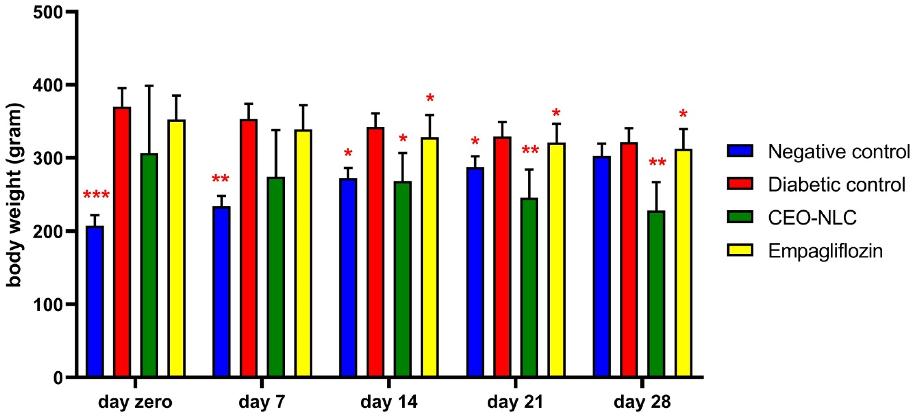
The cardamom essential oil lipid carrier nanoparticle (CEO-NLC) effect on body weight of studied rats. Data was analyzed using ANOVA (post hoc) test. Values with (*) are significantly different from the diabetic control using Tukey test (*p < 0.05), (**p < 0.01), (***p < 0.001), and (****p < 0.0001).
CEO-NLC Effect on Inflammatory Mediators and Antioxidants
An increase in the IL-6, TNF-α, and CRP with a decrease in IL-10 was seen in the G1 and G2 groups. In contrast, lower levels of IL-6, TNF-α, and CRP and higher levels of IL-10 were seen in CEO-NLC/EMPA groups, with an increased T-AOC (Figure 3).

Effect of CEO-NLC on the serum Interleukin (IL)-6 (p < 0.003), Tumor Necrosis Factor (TNF)-α (p < 0.001), IL-10 (p < 0.000), C-Reactive Protein (CRP) (p < 0.001) and Total Antioxidant Capacity (p < 0.000) levels in streptozotocin induced diabetic rats. Data was analyzed using ANOVA (post hoc) test. Values with (*) are significantly different from the diabetic control using Tukey test (*p < 0.05), (**p < 0.01), (***p < 0.001), and (****p < 0.0001).
Effect of CEO-NLC on the serum Levels of Enzymes in Liver and Pancreas
The DC group demonstrated an increase in AST, ALT, ALP, and α-amylase levels. On the other hand, the CEO-NLC group showed lower levels of these liver enzymes. However, the TSB levels did not show any significant changes. Magnesium levels were elevated, which indicates that magnesium plays an essential role in glycolysis (Figures 4 and 5).

Effect of CEO-NLC on the serum levels of alkaline phosphatase (ALP) (p < 0.046), glutamic-pyruvic transaminase (GPT) (p < 0.000), and glutamic oxaloacetic transaminase (GOT) (p < 0.006) in streptozotocin induced diabetes mellitus in rat model. Data was analyzed using ANOVA (post hoc) test. Values with (*) are significantly different from the diabetic control using Tukey test (*p < 0.05), (**p < 0.01), (***p < 0.001), and (****p < 0.0001).

Effect of CEO-NLC on the serum levels of α-amylase (p < 0.021), magnesium (p < 0.010) and total serum bilirubin (TSB) (p < 0.73) in streptozotocin induced diabetic rats. Data was analyzed using ANOVA (post hoc) test. Values with (*) are significantly different from the diabetic control using Tukey test (*p < 0.05) and (**p < 0.01).
CEO-NLC Effect on Kidney Function Tests
After the CEO-NLC administration, serum urea and creatinine were significantly lowered compared to the DC, indicating nephroprotective properties (Figure 6).

CEO-NLC effect on the serum levels of urea (p < 0.009) and creatinine (p < 0.022) in streptozotocin induced diabetic rats. Data was analyzed using ANOVA (post hoc) test. Values with (*) are significantly different from the diabetic control using Tukey test (*p < 0.05) and (**p < 0.01).
Histopathological Analysis
Liver
In NC, the liver revealed a central vein surrounded by hepatocytes and typical sinusoidal capillaries, Kupffer cells can be evident, while in the DC group, the liver revealed a central vein surrounded by hepatocytes with fatty changes manifested by the presence of numerous lipid vacuoles in their cytoplasm, that pushing nucleus to the periphery of the cell. Some necrotic hepatocytes manifested by pyknotic nuclei are present throughout the section. Regarding treating the group with CEO-NLC, better modifications were seen than EMPA because the standard histological structure of the liver is preserved. In contrast, in the EMPA group, Kupffer cell hyperplasia and deposition of homogenous eosinophilic material along the sinusoids within the spaces suggested a mild degree of amyloidosis (Figure 7).

(A) Liver of control negative rats revealed central vein (C) surrounded by hepatocytes (H) and typical sinusoidal capillaries (S), and Kupffer cells (K) can be evident in the given section. (B) Liver of diabetic control rats revealed central vein (C) surrounded by hepatocytes with fatty changes (F) manifested by presence of numerous lipid vacuole in their cytoplasm, that pushing nucleus to periphery of the cell. There are some necrotic hepatocytes manifested by pyknotic nuclei (P). (C) Liver of treated group with nanoparticles shows normal histological structure of liver is preserved, central vein (C) surrounded by hepatocytes (H) and typical sinusoidal capillaries (S) with presence of Kupffer cells (K). (D) Liver of Empagliflozin group shows central veins (C) surrounded by hepatocytes (H), Kupffer cell hyperplasia (HK), and deposition of homogenous eosinophilic material along the sinusoids within the spaces (CA) that suggested mild degree of amyloidosis. H&E staining, 40×.
Pancreas
In NC, pancreatic tissue composed of lobules separated by interlobular stroma that contain islets of Langerhans with its exocrine duct, surrounded by coroner acinar glands, while in the DC group, pancreas revealed degenerated islet of Langerhans, manifested by severe cellular reduction, fatty degeneration, most of the tissue occupied by acinar cells, with infiltration of inflammatory cells. In the CEO-NLC group, the pancreas revealed a regenerated islet of Langerhans surrounded by acinar gland cells. In contrast, in the EMPA group, the pancreas still revealed a degenerated islet of Langerhans, manifested by severe cellular reduction and fatty degeneration with infiltration of inflammatory cells (Figure 8).

(A) Pancreatic tissue of negative control rat shows lobules separated by interlobular stroma (S) that contain islets of Langerhans (I) with its exocrine duct (D), surrounded by coroner acinar glands (A). (B) Pancreas tissue of diabetic control group reveals degenerated islet of Langerhans (DI), manifested by severe cellular reduction, fatty degeneration (F), and most of tissue occupied by acinar cells (A), with infiltration of inflammatory cells (IN). (C) Pancreas of treated animals with nanoparticles reveals regenerated islet of Langerhans (R), surrounded by acinar gland cells (A), and ducts of exocrine glands (D) with connective tissue septa (S). (D) Pancreas tissues of Empagliflozin group reveals degenerated islet of Langerhans (DI), manifested by severe cellular reduction, fatty degeneration (F), most of tissue occupied by acinar cells (A), with infiltration of inflammatory cells. H & E staining, 40×.
Kidneys
In the NC group, renal tissue is observed where the cortex contains glomeruli. These glomeruli comprise a network of capillaries surrounded by Bowman's capsule and proximal and distal convoluted tubules. However, in the DC group, the renal cortex showed nodular glomerulosclerosis, which is characterized by the presence of hyaline nodules within the lobules of glomeruli. The glomerular capillaries surrounding the nodules have thickened walls, and there is also the presence of blood vessel atheroma. Additionally, glycogen deposition appears as cytoplasmic vacuoles in the epithelial cell lining of proximal convoluting tubules. About the CEO-NLC group, nodular glomerulosclerosis, hyaline nodules within the lobules of glomeruli surrounded peripherally by glomerular capillaries with thickened walls in proximal and distal convoluted tubules and throughout the section were seen with no substantial pathological changes. These lesions are better than the EMPA group in which the renal cortex revealed some degree of glomerulosclerosis, hyaline nodules within the lobules of glomeruli, surrounded peripherally by glomerular capillaries with thickened walls. Also, there is blood vessel atheroma in the proximal convoluting tubules’ epithelial cell lining, and glycogen deposition appears as a cytoplasmic vacuole (Figure 9).

(A) Kidney tissue of negative control group shows renal cortex that contains glomeruli (G), network of capillaries (C) surrounded by Bowman's capsule (B), proximal convoluted tubules (P) stained density pink with brush border, and distal convoluted tubules (D) pale in color. (B) Kidney tissues of diabetic control group shows renal cortex with nodular glomerulosclerosis, manifested by hyaline nodules within the lobules of glomeruli surrounded peripherally by glomerular capillaries with thickened walls. There is blood vessel atheroma (A), and in epithelial cell lining of proximal convoluting tubules there is glycogen deposition appearing as cytoplasmic vacuole (G). Nodular glomerulosclerosis also called as Kimmelstiel Wilson (KW) lesions or interpapillary glomerulosclerosis. Tare specific for type 1 diabetes (juvenile onset diabetes). (C) Kidney tissues of treated animals with nanoparticles reveals some degree of glomerulosclerosis, hyaline nodules (ND) within the lobules of glomeruli, surrounded peripherally by glomerular capillaries with thickened walls. There is blood vessel atheroma (A), in epithelial cell lining of proximal convoluting tubules there is glycogen deposition appearing as cytoplasmic vacuole (G). (D) Renal cortex of Empagliflozin group reveals nodular glomerulosclerosis, hyaline nodules within the lobules of glomeruli surrounded peripherally by glomerular capillaries with thickened walls, in proximal (P) and distal (D) convoluted tubules. There are no significant pathological changes throughout the section. H&E staining, 40×.
Discussion
The routine use of spices in our food significantly impacts our behavior, physical development, and overall health. It is interesting that the applications of phytochemical constituents from medical plants to managing disorders related to general health problems such as inflammation, loss of body weight, and cosmetic uses are attracting attention worldwide. Considering the importance of the spices, the present study investigated the effect of CEO-NLC on body weight and blood glucose levels in the diabetic-induced rat model. This study showed that compared to EMPA, on days 14 and 28, CEO-NLC supplementation significantly decreases blood glucose and body weight. These findings were similar to studies conducted on spices’ uses, and in vitro studies reported that cardamom effectively decreases inflammatory markers and lowers blood glucose in diabetic patients.18,19 In 2014, one day, we supported the cardamom effect on reducing the glycemic level in rats with no effect on weight loss, w, which contradicts these results on the positive impact of CEO-NLC for weight loss. 20
IL-10 has been proven to have several anti-inflammatory and protective features that lower cardiovascular risk. 21 At the same time, IL-6 and TNF-α are vital proinflammatory factors promptly and transiently produced in response to infections and tissue injuries.22,23 There is a significant decrease in the levels of the inflammatory markers IL-6, TNF-α, and CRP, with an increase of IL-10 and T-AOC but with enhanced results in the CEO-NLC group than the antidiabetic drug, proving the anti-inflammatory and protective function of the cardamom. The randomized control trial by Zahedi et al showed that cardamom supplementation improved serum levels of adhesion molecules and reduced some inflammatory biomarkers in T2DM patients. 24
Two separate animal studies have shown that supplementing with cardamom can significantly decrease weight gain, as well as lower levels of the liver enzymes AST and ALT.25,26 This study observed that the administration of CEO-NLC leads to a significant decrease in liver enzymes. However, EMPA treatment had no significant effect on liver function improvement and reported the highest level for all liver enzymes compared to other groups. Similarly, Böhm et al 2023 stated that beneficial effects were not observed on liver tests after treatment with EMPA. 27
A study conducted by Feng et al has shown that high levels of magnesium in the body have anti-inflammatory and organ preservation functions. On the other hand, hypomagnesemia can hinder the proper glucose transport by the type 4 glucose transporter. This can cause an increase in insulin resistance. Additionally, hypomagnesemia can affect lipid metabolism, trigger oxidative stress, and impair the antioxidant system of endothelial cells. As a result, it plays a significant role in the onset and progression of DM and its associated macrovascular and microvascular complications. 28 This study demonstrates that CEO-NLC can effectively increase the level of magnesium. This confirmed the anti-inflammatory and glycemic-lowering effects of the CEO-NLC.
An increase in creatinine and urea confirms kidney damage, as they are end-products of protein metabolism that must be excreted by the kidneys. 29 There was a significant decrease in urea and creatinine levels in the CEO-NLC group in contrast to the DC and EMPA groups. Thus, it proved the kidney protection effect of cardamom oil. This result was confirmed by a study that observed significant protection of the kidneys of rats from the structural and functional changes associated with gentamicin by co-administering it with cardamom. 30 Collectively, this study confirms that CEO-NLC reduces obesity and mitigates DM in rats fed with high sugar/high fat diet through variable parameters (Figure 10). The current study had limitations, such as small animal houses and facility shortages. Therefore, further studies using more advanced laboratory tests are required to corroborate gene expression.

Illustrating the mechanistic understanding of how CEO-NLC exerts its effects.
Conclusions
The CEO-NLC have been found to positively impact reducing blood sugar levels, aiding weight loss, diminishing inflammation and oxidative stress, and enhancing the liver and kidney function in rats with induced diabetes. This study proved their potential organ preservation function.
Footnotes
Acknowledgements
The author acknowledges the technical assistance of the Histopathology Department and Biochemistry Laboratory staff at Smart Health Tower, Sulaimaniyah, Iraq.
Data Availability
The datasets used and/or analyzed during the current study are all provided in the manuscript.
Declaration of Conflicting Interests
The author declared no potential conflicts of interest with respect to the research, authorship, and/or publication of this article.
Funding
The author received no financial support for the research, authorship, and/or publication of this article.
Informed Consent
Not applicable.
Ethical Approval
The ethical handling, utilization, and animal euthanasia procedures used in this investigation were certified by the ethics committee of the College of Pharmacy, University of Sulaimani, Iraq. The ethical permission number for this study is PH94–23, which was on April 10, 2023. Measures were taken to minimize pain and discomfort, and experiments followed the ARRIVE Guidelines for animal ethics.
Human and Animal Rights
Measures were taken to minimize pain and discomfort, and experiments followed the ARRIVE Guidelines for animal ethics guidelines and protect animal rights.