
Abstract
Introduction
Heavy metals are environmental contaminants, which are common and are caused by industrial pollution and human activities. From a global perspective, lead (Pb), a xenobiotic, continues to pose a hazard to human health.1,2 Pb exposure occurs by eating, inhalation, cutaneous absorption, and transplacental absorption.3,4 Globally, Pb exposure remains a significant public health concern, particularly in low and middle income countries. According to the World Health Organization, 5 Pb exposure contributes to approximately 540 000 mortality every year, with the highest number in sub-Saharan Africa due to environmental pollution from mining, industrial activities, and use of Pb-containing products. Around one million people die annually from lead-related disorders, which account for 3% of chronic renal diseases, 4.6% of cardiovascular disease, and 30% of the world's cases of idiopathic cognitive impairment.6,7
According to Halliwell, 8 lead exposure has detrimental effects on human health, particularly on the nervous, cardiovascular, and reproductive systems. It affects various biological processes, including enzyme inhibition, oxidative stress, and disruption of calcium homeostasis. Neurotoxic effects of Pb are thought to be mediated through oxidative stress by generating reactive oxygen species, leading to cellular damage, lipid peroxidation, inflammation, and disruption of neurotransmitter function (Figure 1). ROS has the ability to damage several macromolecules such as proteins, DNA, and lipids of the cells in the body. Studies have shown that the levels of ROS were found to be increased in neurons of people with neurodegenerative diseases, resulting in mitochondrial dysfunction and the secretion of redox metals that react with oxygen that ultimately resulted in neuronal death.9–11
Wyss-Coray and Mucke 12 reported that neurodegenerative diseases can lead to chronic inflammation because of the activation of the microglia. Even though inducing factors of inflammation differ among neurodegenerative diseases, chronic inflammation is mainly stimulated in brain cells through common processes.13,14 Once prolong activation of neuroinflammation takes place, proinflammatory mediators are secreted due to the chronic activation of microglia, and the ROS level is elevated, which might be injurious to neurons. 15 Additionally, Pb can also interfere with calcium signaling, impair synaptic function, and damage neuronal structures, leading to cognitive deficits, behavioral problems, and developmental delays in children. 16 The central nervous system is susceptible to Pb poisoning. Pb exposure has been associated with cognitive deficits, executive function changes, inappropriate social behavior, and disturbances in fine motor control.3,17
Musa sapientum is a member of the Musaceae family and has been shown to have significant antiulcer, antidiabetic, antiinflammatory, and antioxidant characteristics.18–20 M sapientum peels contain a variety of antioxidant compounds, 21 and extracts from the peels of different banana species can be used to treat diseases caused by the generation of free radicals. M sapientum peels are a rich source of phytochemical molecules. Bioactive compounds such as polyphenols, carotenoids, and flavonoids have been shown to have antioxidants and neuroprotective properties. 22 , 23 This study aims to investigate the cognitive and antioxidative potentials of ripe M sapientum peels on lead acetate-induced neurotoxicity in female wistar rats. The study considered female animals because approximately two-thirds of AD patients are females, and studies have shown that women have higher prevalence of AD.24,25
Materials and Methods
Drugs and Chemicals
Donepezil and lead acetate were purchased from Sigma-Aldrich Cooperation and administered orally via oral gavage. All reagents used in this study were of analytical grade.
Animals
Thirty mature female wistar rats at 6 months old, weighing between 120 and 160 g were collected from the animal house of Physiology Department at Alex Ekwueme Federal University. The animals were kept in a normal environment, given 2 weeks to acclimatize, and then placed into six groups at random. The animals were kept in standard laboratory settings with a 12 h light/dark cycle, relative humidity of 50 ± 5%, and room temperature (23 ± 2 °C). The animals were allowed free access to feed (raw chow, Vital feeds Nigeria Ltd, Jos) and water ad libitum. The National Institutes of Health's rules for the care and use of laboratory animals were meticulously adhered to when handling the animals, 27 and the ethical approval number AEFUNAI 2024/00135 for this experiment was acquired from Alex Ekwueme Federal University Ndufu Alike, Ebonyi State, Faculty of Basic Medical Sciences Ethical Approval Committee.
Extraction of Plant Material
The peels of M sapientum were obtained from a local market in Ikwo, Local Government Area, Ebonyi State. It was identified and authenticated by a Taxonomist in the Department of Botany and Ecological studies, University of Port Harcourt, Nigeria. The voucher specimen was deposited in the department with the herbarium number:
Gas Chromatography–Mass Spectrometry (GC–MS) Analysis
Analysis of M sapientum peels was done using GC–MS as reported previously by Puraikalan. 15 The study used GC–MS analysis, Perkin Elmer GC Claurus 500 system with an Elite 5-MS fused silica capillary column made up of 5% Diphenyl and 95% Dimethyl poly siloxane and Turbo Mass. Also, the study employed 70 eV of the ionization energy. Helium gas was utilized as a carrier at a constant flow rate of 1 mL/min. Two microliters of the sample was injected into the column. Initially the oven temperature was 110 °C, and later it was elevated to 200 °C and eventually raised to 280 °C. The total time for running was estimated to be 40 min.
Experimental Design
Thirty (30) adult female wistar rats (6 months old) were randomly divided into six groups containing five animals each, and they received daily oral administration of ethanol extract ripe M sapientum and donepezil for 3 weeks (21 days) via oral gavage. The grouping was as follows:
Group 1 (Normal control): rats received 10 mL/kg of distilled water daily for 21 days. Group 2: rats received 100 mg/kg lead acetate for 21 days. Group 3: rats were concomitantly treated with 100 mg/kg lead acetate and 2.5 mg/kg of donepezil for 21 days. Group 4: rats were concomitantly treated with 100 mg/kg lead acetate and 100 mg/kg of ethanol extract of M sapientum for 21 days. Group 5: rats were concomitantly treated with 100 mg/kg lead acetate and 200 mg/kg of ethanol extract of M sapientum for 21 days. Group 6: rats were concomitantly treated with 100 mg/kg lead acetate and 400 mg/kg of ethanol extract of M sapientum for 21 days.
After 21 days of oral administration the rats were subjected to neurobehavioral experiment Object recognition task and T-Maze Simple Alternation Test. The brains were meticulously removed from the skull, weighed, and homogenized. Phosphate buffer 0.1 M, pH 7.4 at 4 °C was used to prepare 10% (w/v) tissue homogenate. The homogenates were centrifuged for 10 min at 3000 rpm to separate the supernatants, which were then used to conduct biochemical tests on acetylcholinesterase (AChE), malondialdehyde (MDA) and superoxide dismutase (SOD) activities. A histological study of the hippocampus region of the brain was also carried out.
Behavioral Tests
Novel Object Recognition Test (NORT)
NORT was carried out in a black open-field box (45 cm × 45 cm × 50 cm) as previously described. 28 `All testing occurs during the dark-phase (the active phase from 6 pm to 8 pm) of the light cycle. Testing is conducted under dim white-light illumination (about 150 lx). The testing arena was clean with cotton wool and methylated spirit and allowed to dry before use. All animals had an adaptation period of 5 min without objects. After 5 min, the subjects were returned to their home cage. After adaptation, the animals were placed in the center of the box with two identical objects and permitted to search for 3 min. The time spent by the mice exploring each object was determined (defined as the acquisition trial). After 24 h, the animals were permitted to explore the objects, including the original object used in the training session and a novel object for 3 min. The time spent by the mice exploring the novel and the familiar objects was measured (defined as the test trial).
T-Maze Simple Alternation Test
T-mazes are used to study how rodents function with memory. 29 The T-maze is made up of the base, the left arm, and the right arm with a length of about 50 and 10 cm width. The different tasks, such as left-right discrimination and forced alternation, are mainly used with rodents to test the reference and working memory. The natural tendency of rats in a T-maze, however, is to alternate their choice of goal arm. Alternation reflects the motivation of the animal to explore its environment and locate the presence of resources such as food, water, mates, or shelter. Animals do not need to be deprived of such resources to show alternation behavior; in this case it is called ‘spontaneous alternation’. The maze was set so that the central partition was in place. The animal was placed in the start area and allowed it to choose a goal arm. After each alternation the animals were returned to the maze for another alternation test. After 30 s, each animal was return to the maze for another alternation test. Each animal was subjected to five pairs of different trials, making a total of 10 trials.
With each pair of trial, the animal was determined to have passed or failed. Any animal that repeats a particular alternation (picked the same arm) in a pair of trial was considered to have failed the trial, while the animal that picked the alternate arm in a single pair of trial was considered to have passed the trial. At the end of the trial, the percentage (%) alternation was calculated for each animal.
Percentage (%) alternation was calculated thus:
No. of phases with good alternation/Total No. of phases × 100 (%).
Biochemical Analysis
The rats were anesthetized with pentobarbital sodium (30 mg/kg i.p.), and the brain was excised and homogenized with external ice-cold saline bath. The homogenates were centrifuged at 3000 rpm for 30 min at 4°C, and the supernatant was collected and stored at −80°C before the assay. The Acetyl cholinesterase (AChE) activities of the brain were measured using the method described by Ellman. 30 SOD activity was determined as previously described 31 by spectrophotometry at 420 nm.
Histopathological Analysis
The granule cells were examined in the histological section. The hippocampus (dentate gyrus) was perfusion-fixed in 0.1 M phosphate buffer with 4% paraformaldehyde. The fixative was applied to the brains afterward and left overnight at 48°C. The brains were stained with hematoxylin–eosin and embedded in paraffin. At 400 magnification, lesions were examined under a microscope. 32
Statistical Analysis
Graph Pad prism (version 8) software was used for the statistical analysis at a probability level of p < 0.05. One-way analysis of variance (ANOVA) and Tukey multiple comparison post hoc tests were carried out and were presented as mean ± standard error of mean (SEM).
Results
GC–MS Analysis of Bioactive Components of Musa sapientum Peel
The phytochemical compounds found in Musa sapientum extract from GC–MS instruments are listed in Table 1. The bioactive compounds are represented by the various peaks. The peaks were 12, representing a total of 12 bioactive compounds. The table shows the retention times of 12 bioactive compounds, molecular formula, molecular weight, peak and areas. The retention time ranged from 4.50 (4H-Pyran-4-one,2,3-dihydro-5-dihydroxy-6-methyl) to 38.49 (9,19-cyclolanostan-3-ol, acetate, (3β)). The 9,19-Cyclolanostan-3-ol, acetate, (3β) has the highest retention time, while Sucrose has the highest peak area 344 (%).
GC–MS Analysis of Bioactive Components of Banana Peel Powder.

Evaluation of Novel Object Recognition Test
Total Exploratory Time (Identical Object)
The total exploratory time of rats when exploring the two identical objects is reported in Figure 2. The result shows that there was a significant (p < 0.05) reduction in the total exploratory time of the lead acetate group and a significant (p < 0.05) increase in Donepezil-treated group when compared with the control group. Comparing the ethanol extract groups with the control, there was a significant (p < 0.05) increase in the ethanol extract middle- and high-dose-treated groups in a dose-dependent manner.

Summary of lead acetate neurotoxicity showing increased acetylcholinesterase (AChE) activity, decreased acetylcholine (Ach) activity, increased malondialdehyde (MDA) level and lipid peroxidation, disruption of antioxidant system and decreased superoxide dismutase activity (SOD) in the CNS. 26

Effect of Musa sapientum on total exploratory time for the two familiar objects. Data presented as mean ± SEM; (n = 5); *control; #, Pb; $, donepezil; (p < 0.05).
Total Exploratory Time (Familiar and Novel)
The total exploratory time of rats when exploring the familiar and novel objects is reported in Figure 3. The result shows that there was a significant (p < 0.05) reduction in the total exploratory time of the lead acetate group and a significant (p < 0.05) increase in the donepezil-treated group when compared with the control group. Comparing the ethanol extract groups with the control, there was a significant (p < 0.05) increase in the ethanol extract high-dose-treated groups.
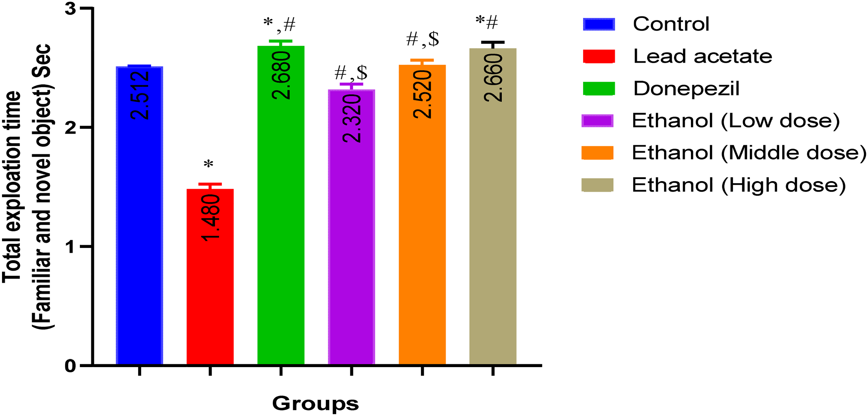
Effect of Musa sapientum on total exploratory time for the two familiar and novel objects. Data presented as mean ± SEM; (n = 5); *control; #, Pb; $, donepezil; (p < 0.05).
Absolute Discrimination Time
The absolute discrimination time of rats during the novel object task is reported in Figure 4. The result shows that there was a significant (p < 0.05) reduction in the absolute discrimination time of lead acetate group and a significant (p < 0.05) increase in donepezil-treated group when compared with the control group. Comparing the ethanol extract groups with the control, there was a significant (p < 0.05) increase in the ethanol extract low- and high-dose-treated groups. Comparing the ethanol extract groups with the lead acetate group, there was a significant (p < 0.05) increase in the ethanol extract low- and high-dose-treated groups.

Effect of Musa sapientum on absolute discrimination measures. Data presented as mean ± SEM; (n = 5); *control; # Pb; (p < 0.05).
Discrimination Index
The discrimination index of rats during the novel object task is reported in Figure 5. The result shows that there was a significant (p < 0.05) decrease in the discrimination index of the lead acetate group compared with the control group and a significant (p < 0.05) increase in donepezil-treated group when compared with the control group. Comparing the ethanol extract groups with the control, there was a significant (p < 0.05) increase in the ethanol extract low- and high-dose groups. Also, there was a significant (p < 0.05) increase in the donepezil-treated group, as well as a significant (p < 0.05) increase in the ethanol extract-treated groups in a dose-dependent manner when compared with the lead acetate-treated group.

Effect of Musa sapientum on discrimination index. Data presented as mean ± SEM; (n = 5); *control; #, Pb; (p < 0.05).
T-Maze Simple Alternation Test
The percentage alternation in the simple alternation Test is reported in Figure 6. The result shows that there was a significant (p < 0.05) decrease in the percentage alternation of the lead acetate group compared with the control group. Comparing the ethanol extract groups with the lead acetate group, there was a significant (p < 0.05) increase in the ethanol extract low-, middle-, and high-dose groups in a dose-dependent manner.

Effect of Musa sapientum on percentage alternation. Data presented as mean ± SEM; (n = 5); *control; #, Pb; (p < 0.05).
Effects of Ethanol Extract of Ripe Banana (Musa sapientum) Peels on Acetylcholinesterase (AChE) Activity
The effects of ethanol extract of ripe banana (Musa sapientum) peels in AChE activity is represented in Figure 7. The result shows that there was a significant (p < 0.05) increase in lead acetate- and donepezil-treated groups when compared with the control group. Comparing the ethanol extract treated group with the control group there was a significant (p < 0.05) decrease in ethanol high dose. Also, there was a significant (p < 0.05) reduction in the ethanol extract treated groups in a dose dependent manner when compared with Lead acetate treated group.

Effect of Musa sapientum on acetylcholinesterase activity (AChE). (A) Superoxide dismutase activity (SOD); (B) malondialdehyde activity (MDA); (C) data presented as mean ± SEM; (n = 5); *control; #, Pb; (p < 0.05).
Effects of Ethanol Extract of Ripe Banana (Musa sapientum) Peels on Superoxide Dismutase (SOD) Activity
The effects of ethanol extract of ripe banana (M sapientum) peels in SOD activity is represented in Figure 7. The result shows that there was a significant (p < 0.05) decrease in lead acetate- and donepezil-treated groups when compared with the control group. Comparing the ethanol extract-treated group with the control group, there was a significant (p < 0 .05) increase in ethanol middle and high doses. Also, there was a significant (p < 0.05) increase in the ethanol extract-treated groups in a dose-dependent manner and also a significant (p < 0.05) increase in the donepezil-treated group when compared with the lead acetate-treated group.
Effects of Ethanol Extract of Ripe Banana (Musa sapientum) Peels on Malondehyde Aldehyde Activity
The effects of ethanol extract of ripe banana (M sapientum) peels in Malondehyde aldehyde is represented in Figure 7. The result shows that there was a significant (p < 0.05) increase in the lead acetate- and donepezil-treated groups and a significant (p < 0.05) decrease in the ethanol extract high dose when compared with the control group. Also, there is a significant (p < 0.05) decrease in the donepezil-treated group and ethanol extract high dose when compared with the lead acetate group.
Histology Staining
Figure 8 shows the histology staining of the hippocampus. Photomicrograph of the hippocampus of the control group shows normal and active granular cells (GC) (×400) (H/E) (a); photomicrograph of the hippocampus of the group treated with lead acetate shows severe degeneration, severe vacuolation (V) and granular cell atrophy (A) (×400) (H/E) (b); photomicrograph of the hippocampus of the group treated with lead acetate and donepezil shows mild regeneration with vacuolation (V) and pyknotic (P) granular cell (×400) (H/E) (c); photomicrograph of the hippocampus of the group treated with lead acetate and 100 mg/kg EMS shows mild regeneration with moderate vacuolation (V) and granular cell atrophy (A) (×400) (H/E) (d); photomicrograph of the hippocampus of the group treated with lead acetate and 200 mg/kg EMS shows mild regeneration with moderate vacuolation (V) and granular cell atrophy (A) (×400) (H/E) (e); photomicrograph of the hippocampus of the group treated with lead acetate and 400 mg/kg EMS shows mild regeneration with moderate vacuolation (V) and granular cell atrophy (A) (×400) (H/E) (f).

Photomicrograph of the hippocampus (×400) (H/E). H&E, hematoxylin and eosin; V, vacuolation; P, pyknotic; GC, granular cells.
Discussion
Lead exposure has continued to pose a hazard to human health.1,33 This study explored the potential of M sapientum to protect against lead acetate-induced neuronal damage in male wistar rats. It has been revealed that the brain is a potential target for lead toxicity.34,35 Lead can go deep into the blood brain barrier, accumulate and distort the functions of the brain causing several neurological impairments such as Alzheimer's disease and Parkinsonism.36,37 The bioactive compounds as shown in this present study are consistent with those revealed in a work done by Puraikalan. 15 A study exposed the bioactive compounds and phytochemical aspect of the most popular banana specie from a local organic farm in Thailand. It was revealed that acetic acid, formic acid, 1,2-benzenediol,3-methyl-, and 4-hydroxy-2-methylacetophone were the phytochemicals found in banana peels and exhibited antioxidant and antimicrobial activities. The study suggests the possibility of applying banana peels to supplement animal and aquatic feed to improve growth.30,38 Studies have also shown that n-hexadecanoic acid and hexadecanoic acid, ethyl ester showed anticancerous, antioxidative, hypocholesterolemic, and antipsychotic properties. 39 Puraikalan 15 revealed that banana peels had a significant amount of total phenols, flavonoids, and tannin. It has been suggested that antioxidants and phenolic ingredients in several medicinal plants can penetrate the blood brain barrier and chelate heavy metals.40,41 Several animal and human studies have shown the involvement of oxidative damage due to the generation of reactive oxygen species (ROS) in neurotoxicity associated with lead exposure. 42 This study reports a change in the antioxidant status of hippocampal tissues following lead acetate exposure; this was indicated by an elevation in the level of MDA and AChE activities and a decrease in SOD content. Studies have shown that lead enhances the production of reactive oxygen species (ROS), which attack cellular compartments and promote the peroxidation of cell membrane lipids, disrupting the integrity and function of the cell membrane and resulting in cell death. 43
Exposure to lead acetate caused the rats to develop memory deficits. Rats exposed to lead acetate for 21 days spent less time exploring new objects when compared with rats in the control, suggesting an impaired recognition memory. Similarly, rats exposed to lead acetate for 21 days had a reduced percentage alternation in the T-maze simple alternation task when compared with rats in the control, suggesting a deficit in spatial memory. Similar results on the neurotoxicity of lead acetate have been reported by Yousef et al 44 that Coenzyme Q10 (CoQ10) has beneficial effects against PbAc-induced neuronal damage through its antioxidant, antiinflammatory, antiapoptotic, and neuromodulatory activities. Gurer and Ercal 45 reported that Lead acetate is a white crystalline chemical compound that has a sweet taste and is found under the earth's crust. Lightfoot and Yeager 46 suggested that the contamination of air, water, soil, food by paints, disposable materials of factories such as batteries and leaded gasoline is the principal reason for lead poisoning. Campbell et al 47 revealed that water is a vital source of lead poisoning, especially due to the leaking of lead from water pipes. Flora et al 48 reported that lead acetate induces experimental hepatic injury in rats via the induction of oxidative stress following an imbalance between free radical generation and the antioxidant defense system. This oxidative stress results in the generation of reactive oxygen species (ROS), leading to serious damage to different biomolecules. Atawodi et al 49 suggested that lead generates free radicals that damage the vital organs, including the liver, by reducing the activities of antioxidant enzymes and increasing lipid peroxidation. Merill et al 50 revealed that lead toxicity results in neuropsychiatric disorders ranging from headache, difficulty concentrating, and delayed motor nerve conduction, as well as to delayed reaction times and irritability. Henretig et al 51 suggested that exposure to higher levels of lead causes encephalopathy, characterized by swelling of the brain tissue associated with delirium, coma, and seizures. Patrick et al 52 reported that chronic lead toxicity results in short-term memory loss, nausea, depression, loss of coordination, numbness and tingling in the extremities, and abdominal pain.
Treatment with M sapientum significantly elevated the level of SOD and reduced the elevated level of MDA in hippocampal tissue following lead acetate exposure. A previous study suggests that M sapientum stem extract has the potential to prevent the neuronal oxidative injury pretreatment with MSSE at 100 mg/kg, which decreased the MDA levels significantly and appeared to restore the reduced GSH level in the brain tissues of PTZ-kindled mice. Similarly, Samad et al 53 show that banana pulp and peel via their antioxidant potential increase activities of CAT, GSH, and SOD in the brain.
Histological sections of the hippocampus in the group treated with lead acetate showed severe degeneration, vacuolation (V), and severe granular cell atrophy (A). This observation could explain the reason behind the deficit in the hippocampus including memory impairment and cognitive deficit. The present finding is in tandem with previous reports. Khaled et al 32 had observed neuronal damage in hippocampus in which there was neurodegeneration in the CA1 and CA3 regions. In this region of the brain, there were scar formation, demyelination, and neuronal atrophy which could result from even low exposure to lead. However, administration of M sapientum peels was able to reduce the impairment caused by lead acetate, and this could be due to the fact that the extract has the ability to chelate lead in the brain. This finding is consistent with the findings of previous studies.54,55
Acetylcholine is considered as one of the important neurotransmitter implicated in memory function. One of the major markers for cholinergic function is the determination of acetylcholinestrase (AChE) activity. 56 In this study, banana peel extract increased reference memory and also increased the activities of antioxidant enzyme in the brain which may be attributed to the decreased AChE activity in the rats. This could be a result of the phytochemical present in banana peels which could potentially enhance synaptic plasticity 57 which in turn affects the brain neurotransmitter levels. Samad et al 53 suggested that phytochemicals present in banana pulp and peel via their potential antioxidants improve the memory by increasing the AChE activity to regulate brain neurotransmitter levels.
Limitation of the Study
This study was able to unravel the beneficial role of ripe M sapientum peels in protecting the brain against neurotoxicity via antioxidant. The study was limited to certain biochemical biomarkers, neurobehavioral experiments, and histological analysis of the hippocampus and the use of female animal models. More molecular studies should be done pertaining the signaling pathway or gene expression in order to unravel the mechanism of action of ripe M sapientum extract. Docking studies could also be done on M sapientum plant extract to ascertain specific bioactive molecules that cause its neuroprotective effect.
Conclusion
In conclusion, the study suggests that ripe M sapientum peels via their antioxidant mechanism enhanced cognitive functions and may be beneficial in mitigating neurotoxicity as well as preventing neurodegenerative disorders. The findings of the present study bring to the fore the intrinsic neuroprotective potential of ripe M sapientum peels against lead-induced brain insult and may be formulated after several other tests as a pharmaceutical agent that will help in the treatment of several neurodegenerative diseases.
Footnotes
Acknowledgments
The authors are grateful to the academic and technical staff of Physiology Department, Faculty of Basic Medical Sciences, Alex Ekwueme Federal University, Ndufu Alike, Nigeria for their technical support.
Author's Contributions
The study was conceived and designed by Uduak Anthony Inwang; Azibuike Raphael Nwaji and Uduak Anthony Inwang performed the experiment. The original draft was written by Uduak Anthony Inwang, Ekementeabasi Aniebo Umoh, and Onwe Uchewa. Inwang Uduak analyzed and Ezekiel E. Ben interpreted the data.
Declaration of Conflicting Interests
The authors declared no potential conflicts of interest with respect to the research, authorship, and/or publication of this article.
Funding
The authors received no financial support for the research, authorship, and/or publication of this article.
Statement of Animal Right
All the experimental procedures involving rats were done in accordance to the National Institutes of Health's rules for the care and use of laboratory animals. The ethical approval for this investigation was provided by the Faculty of Basic Medical sciences ethical approval committee, Alex Ekwueme Federal University Ndufu Alike, Nigeria.
Informed Consent
There are no human subjects in this article; therefore, informed consent is not applicable.