
Abstract
Objectives
Caralluma speciosa (N.E. Br.) N.E. Br. (Asclepiadaceae) and other succulent plants of the genus Caralluma have mainly been known for their appetite suppressing and weight loss properties, due to the presence of pregnane compounds. This study aimed to investigate chemical constituents and evaluate antimicrobial and antioxidant activities of stems of C speciosa.
Methods
The chloroform and methanol stems extracts were subjected to silica gel chromatographic separation. The structures of isolated compounds were established by FT-IR, 1D-NMR, and GC-MS instruments, and in comparison with reported data. The antimicrobial and antioxidant activities were performed using agar disc diffusion and DPPH assaying methods.
Results
The phytochemical investigation afforded 5 compounds, 3 pregnane derivatives (
Conclusion
All the extracts and isolated compounds
Keywords
Introduction
Caralluma speciosa (N.E. Br.) N.E. Br. (Asclepiadaceae) is a succulent perennial herb which grows in Acacia woodland and bush land. It is found in Ethiopia, Sudan, Somalia, Uganda, Kenya, and Tanzania. 1 The genus Caralluma is composed of nearly 260 species which have been reported to be used mainly as food sources. In India, for example, fruits and young shoots of C edulis are consumed as a vegetable. 2 The plant species belonging to this genus are mainly consumed for the purpose of antiobesity and appetite suppression. For instance, C adscendens fimbriata is used as an appetite suppressant in India. 3 The appetite suppressing and weight loss properties of Caralluma species are believed due to the presence of their major phytoconstituents, pregnane glycosides (C21 steroidal compounds). Pregnane glycosides, previously reported from the succulent African plant, Hoodia species, are confirmed for their appetite suppressing and antiobesity property. 4 Caralluma species have also been reported for their ethnobotanical uses to cure human ailments such as malaria, inflammation, hyperglycemia, ulcers, and cancer. The fresh stems of C attenuate and Caralluma stalagmifera, for example, are used against diabetes and migraine. 5 Caralluma tuberculata and Caralluma fimbriata are also used to cure rheumatism, diabetes, leprosy, paralysis, joint pains, fever, inflammation, and pyretic. 2 In Ethiopia, the stems and sap of C speciosa are used for the treatment of skin cyst and tumor, gangrene, poison, swollen body, wound, and itching skin. 6
Many pregnane glycosides and extracts of various species of the genus Caralluma have also been reported for their biological activities. For instance, pregnane glycosides isolated from C penicillata, C tuberculata, and C russelliana showed antitrypanosomal and antiplasmodial potency. 7 Six compounds isolated from C tuberculata were tested for their antimalarial and antitrypanosomal activities. 8 The ethanolic extract of C tuberculata was also found as protector of gastric mucosa. In addition, C arabica was reported for its anti-inflammatory, antihyperglycemic, antinociceptive, gastric mucosa protective, and antiulcer properties. 9 C umbellata was also evaluated for anti-inflammatory and antinociceptive activity. Caralluma fimbriata was another species studied for its antifungal and anthelmintic activities.2,10 According to the biological activity study reported by Jitwasinkul and Charoensuksai, 11 the aqueous ethanolic extract of fresh C speciosa showed cytotoxic, antiproliferative, and pro-apoptotic activities against head-and-neck (HN22) and colon (HT29) cancer cell lines.
The chemical constituents reported from the genus Caralluma are mainly dominated by pregnane glycosides. Besides, megastigmane glycosides and few flavones have also been isolated from some species of the genus. 12 For instance, 4 acylated pregnane glycosides were identified from the aerial parts of C russelliana. 13 Five steroidal glycosides (stalagmosides) and 2 steroidal glycosides (indicosides) were reported from whole parts of C stalagmifera and C indica, respectively. 14 In addition, 5 steroidal glycosides and 27 pregnane glycosides were reported from the whole part of C dalzielii. 15 Moreover, 18 fatty acids, 4 hydrocarbons and a β-sitosterol were reported from C edulis. 16 Seventy-four volatile compounds and several fatty acids were also identified from stems and fruits of C europaea. 17 However, to the best of our knowledge, no research works were reported on the chemical constituents, and antimicrobial and antioxidant activities of the stems of C speciosa. Hence, this research work aimed to investigate the chemical constituents and evaluate the antimicrobial and antioxidant activities of the stems of C speciosa. The isolated compounds, reported herein, were also subjected to computational study to predict their drug-likeness, ADME, and toxicity properties using the SwissADME and ProTox-II property explorer software.
Results and Discussion
Structural Identification of Isolated Compounds 1-5
Previously reported phytochemical studies on the genus Caralluma have revealed that members of this genus have been dominated by the C21 steroidal-like compounds, known as pregnanes.
4
Such classes of compounds exhibit in common fused tetracyclic skeletal structure with mainly 2 methyl groups at C-10 and C-13 and 2 extended carbon-chain at C-17 position. Pregnane compounds differ from each other by the presence or absence of different sugar units and other aglycone esterified groups (such as acetyl, benzoyl, and tigloyl) at different positions of the basic skeleton.
18
In the present investigation, 3 pregnane derivatives (

Structures of compounds
From the 1H, 13C, and DEPT-135 NMR spectra (Supplemental Figures S1, S2, and S3), aliphatic proton and carbon signals (including methyl group) were observed at δH 0.88 to 2.04 and δC 14.6 to 34.3 ppm, respectively. Briefly, in the 1H spectrum, a multiplet methine signal at δH 5.80 (1H, H-2) and a doublet of doublet methylene signal at δH 4.96 (2H, dd, H-1, J = 16.4, 11.1 Hz) were appeared to indicate the presence of an olefinic bond. The proton spectrum also displayed 3 aliphatic methylene multiplet signals, integrated for 2, 4, and 42 protons, respectively, at δH 2.06 (2H, H-3), 1.38 (4H, H-4/26), and 1.25 (42H, H-5 to H-25) and a triplet methyl signal at δH 0.88 (3H, H-27, J = 12.87 Hz). The existence of the olefinic bond was also evidenced by the appearance of methine (=CH-) and methylene (=CH2) carbon signals at δC 139.8 (C-2) and 114.5 (C-1), respectively, in the 13C and DEPT-135 spectra. The 13C and DEPT-135 spectra also showed 6 aliphatic carbon signals, which attributed to 25 carbons, at δC 34.3 (C-3), 32.4 (C-25), 29.9 (C-5 and C-6), 30.2 (C-4 and C-7 to C-24), 23.2 (C-26), and 14.6 (C-27). The absence of any quaternary carbon signal in the DEPT-135 spectrum also confirmed that compound
In the 1H NMR spectrum (Supplemental Figure S7), 2 doublet of doublet signals were observed at δH 7.53 (2H, dd, J = 4.45, 4.87 Hz) and 7.70 (2H, dd, J = 4.45, 4.87) due to the protons of symmetrical ortho disubstituted aromatic ester with an AA’BB’ spin system. The spectrum also showed an intense multiplet proton signal at the chemical shift range of δH 1.25 to 1.36, integrated for a total of 16 protons, indicated the presence of 8 methylene (-CH2-) protons in at similar chemical environment. In addition, aliphatic proton signal was observed at δH 4.21 (4H, br t, J = 12.38 Hz) for 2 set of oxygenated methylenes neighboring with a multiplet signal at δH 2.33 (2H) of methine (-CH-) protons. The 1H spectrum also showed a doublet of doublet signal at δH 0.89 (6H, dd, J = 9.83, 12.04) due to 2 symmetric methyl protons adjacent to 2 nonequivalent methylene protons; and a triplet signal at δH 0.78 (6H, t, J = 8.56) for 2 symmetric methyl protons next to 2 equivalent methylene protons, each.
In the 13C spectrum (Supplemental Figure S8), 3 aromatic carbon signals at δC 128.6, 130.7 and 132.2 along with an ester carbonyl carbon signal at δC 167.6 were showed which were due to the presence of 2 symmetrically esterified benzoic aromatic groups; and this was supported by the DEPT-135 spectrum (Supplemental Figure S9) in which 2 quaternary carbon signals (δC 132.2 and 167.5) and a signal at δC 67.5 (C-1/1’) of 2 symmetric oxygenated methylene carbons were revealed. The DEPT-135 spectrum also indicated the presence of an intense methine carbon signal at δC 38.5 (C-2/2’) together with 2 consecutive methyl carbon signals at δC 10.8 and 13.9 ascribed to 2 equivalent methyl groups, each. The obtained NMR spectral data is presented in Supplemental Table S2.
The IR spectrum (Supplemental Figure S10) showed an absorption peak of the ester carbonyl group (C = O) of the phthalate unit at 1734cm−1 together with the C-O and aromatic C-H stretching at wavenumbers of 1274 and 731 cm−1, respectively. It also displayed an intense peak at 2918 to 2855 cm−1 which represents the C-H stretching of the long chain alkyl groups. From the GC/MS spectrum (Supplemental Figures S11 and S12), a molecular ion peak at m/z 390 in the EI-MS and major peak with retention time of 37.031 min in the GC were appeared which is compatible with the molecular formula of C24H38O4 (calculated MW, 390). A base peak at m/z 149.0 (calcd. 149.0 for C8H5O3) was also obtained, including 4 relatively intense product ions at m/z 167.0 (C8H7O4, calcd. MW, 167), 57.1 (C4H9, calcd. MW, 57), 71.0 (C5H11, calcd. MW, 71), and 279.0 (C16H23O4, calcd. MW, 279). Based on the above spectral information and reported data,
16
the structure of compound
It is observed that the 1H and 13C NMR spectra (Supplemental Figures S13 and S14) contained many complicated aliphatic proton and carbon signals at δH 0.88 to 2.02 and δC 10.5 to 55.7 ppm, respectively. This showed that compound
From the 13C spectrum, those 3 signals shown at δC 73.8, 86.5, and 70.4 were attributed to 3 oxygenated carbon signals (C-12, C-14, and C-20, respectively) of the pregnane part. Besides, the 3 consecutive signals observed at δC 10.5, 12.5, and 19.2 were because of the presence of 3 methyl carbons (C-18, C-19, and C-21, respectively) of the pregnane unit. Furthermore, 2 close signals at δC 129.2 and 129.6, 127.9 and 128.3, and 132.3 and 132.8 were the ortho (C-3’/7’), metha (C-4’/6’), and para (C-5’) carbons of the 2 benzoyl groups attached at C-12 and C-20, respectively. The presence of the 2 ester groups of the benzoyl moieties (at C-12 and C-20) was also witnessed by the presence of 2 ester carbonyl carbon signals at δC 165.8 (C-12) and 166.4 (C-20) in the 13C spectrum. In the DEPT-135 spectrum, 7 quaternary signals were confirmed; 3 signals at δC 35.2, 53.2, and 86.5 due to the C-10, C-13, and C-14 (oxygenated with OH) of the pregnane unit; 2 signals at δC 130.4 and 130.5 belong to C-2’ of the 2 benzoyl groups; and the remaining 2 signals at δC 165.8 and 166.4 due to the 2 carbonyl carbons of the benzoyl ester groups (Supplemental Table S3). The overall 13C and DEPT-135 data of the aglycone part of compound
The 1H spectrum also showed many multiplet signals of oxymethinic protons in the chemical shift range of 3.53 to 4.98 ppm. This indicated the presence of sugar moieties within the pregnane unit. The number of consecutively connected saccharide units can be recognized by the number of anomeric carbon signals at around δC 90 to 110 and anomeric proton signals at δH 4 to 5 ppm.
18
In our case, the presence of 2 intense carbon signals at δC 95.3 (C-1’’) and 99.1(C-1’’’) in the 13C spectrum together with 2 proton signals at δH 4.98 (1H, br d, H-1’’, J = 10.52 Hz) and 4.90 (1H, br d, H-1’’’, J = 6.89 Hz) in the 1H spectrum inferred that there were 2 anomeric carbons and associated protons which belong to 2 interlinked sugar moieties. In addition, 2 proximate carbon signals at δC 57.0 (C-7’’) and 57.7 (C-7’’’) along with 2 intense singlet proton signals at δH 3.53 (3H, s, H-7’’) and 3.54 (3H, s, H-7’’’) were observed which ascribed to 2 methoxy (MeO-) groups. This revealed that the 2 sugar units were cymarose-I (C-1’’) and cymarose-II (C-1’’’).
18
Besides, the presence of 2 methyl groups at δC 17.9 and 18.1 (C-6’’ and C-6’’’, respectively) and at δH 1.32 (6H, br s, H-6’’ and H-6’’’) in combination with the methylene carbon signal (δC 36.1, C-5’’ and C-5’’’) and multiplet proton signal (δH 1.79, 4H, m, H-5’’ and H-5’’’) was additional evidence to the 2 sugar moieties to be cymarose-I and II.
18
In reference to reported data for similar compound,
18
the disaccharide units were attached to the pregnane unit at C-3 (δC 72.2). The IR spectrum (Supplemental Figure S16) also provided supportive information by showing major bands at 3420, 2918 to 2848, 1713, and 710 cm−1 of the hydroxyl, alkyl, carbonyl, and aromatic groups, respectively. Based on the spectroscopic data (Supplemental Tables S3 and S4) and comparison with reported data,18–20 the structure of compound
The acquired 1H, 13C, and DEPT-135 spectra (Supplemental Figures S17, S18, and S19) confirmed the presence of a pregnane aglycone nucleus as in compound
But, herein, it is very important to note that the attachment of the benzoyl ester moiety outside the pregnane aglycone nucleus (C-22) of compound
The 1H, 13C, and DEPT-135 spectral feature (Supplemental Figures S23, S24, and S25) of compound
Bacteriological Assay
The antibacterial activity results of 5 crude extracts (n-hexane, chloroform, 1:1 of chloroform: methanol, methanol, and ethanol) and 5 isolated compounds

DPPH radical scavenging percentage (%) versus different concentrations of Caralluma speciosa stems extracts and ascorbic acid. HE, n-hexane extract; CE, chloroform extract; CME, 1:1 of chloroform: methanol extract; ME, methanol extract; EE, ethanol extract; AA, ascorbic acid.
As can be indicated in Supplemental Table S8, the best diameter of inhibition zone (8.65 ± 1.63 mm) against Escherichia coli was recorded by chloroform and then followed by methanol (7.09 ± 0.88 mm) extracts at the maximum concentration (100 mg/mL). Whereas the n-hexane, chloroform: methanol (1:1), and ethanol extracts (EEs) did not show any activity against E coli at all tested concentrations. However, the mean difference between the extracts was not significant (P > .05). All tested extracts were found with no activity against the methicillin-resistant Staphylococcus aureus bacterium at all concentrations, except the EE which tried to show a significantly different (P < .05) slight inhibitory effect (7.55 ± 0.25 mm) at the higher concentration (100 mg/mL). On the other hand, the Gram-negative Pseudomonas aeruginosa bacterium showed a total resistant against all evaluated extracts at all concentrations.
All the isolated compounds
Antifungal Activity Assay
The chloroform: methanol (1:1) extract exhibited prominent inhibitory potential against Candida albicans at all evaluated concentrations; except for the minimum concentration (12.5 mg/mL) which showed zero diameter of inhibition zone. This extract recorded the inhibitory values of 9.00 ± 1.00 mm for 25 mg/mL, 14.67 ± 0.58 mm for 50 mg/mL, and 17.17 ± 1.04 mm for 100 mg/mL. The mentioned extract in general was found as active as the standard antifungal drug ketoconazole 0.01 mg/disc (17.67 ± 2.52 mm) mainly at the maximum concentration.
Examination of Antioxidant Activity
The crude extracts and isolated compounds
DPPH Free Radical Scavenging Activity Assay
The obtained results (Supplemental Tables S10 and S11, and Figures 2 and 3) revealed that each tested analyte showed a directly correlated (dose-dependent) activity, that is, the DPPH radical scavenging activity percentage was getting increased as the concentration increased. Ethanol extract showed a better DPPH radical scavenging percentage (58.092 ± 0.00) followed by chloroform extract (CE, 51.303 ± 0.04) in comparison to AA (95.342 ± 0.02) at the highest concentration of 500 µg/mL (Supplemental Table S10 and Figure 2). The inhibitory activity of the tested extracts against the DPPH radicals was generally observed negligible with higher IC50 values in reference to the standard AA (5.62 µg/mL).

DPPH radical scavenging percentage (%) versus different concentrations (µg/mL) of isolated compounds
Among the isolated compounds, compound
Potassium ferric reducing antioxidant power assay
The obtained PFRAP results of tested extracts and isolated compounds
Drug-Likeness, ADME, and Toxicity Properties of Isolated Compounds 1-5
The obtained in silico drug-likeness, ADME, and toxicity properties of the isolated compounds
In this study, both the in vitro and in vivo anticancer activity of the studied C speciosa stems were not included to support the claimed traditional medicinal use against tumor. Besides, the experimental cytotoxicity effect of the isolated compounds was not studied herein based on the obtained computational study result. Furthermore, the plant extracts and isolated compounds were evaluated against few microbial strains and oxidants. All these points could be considered as the limitations of the present study.
Conclusions
The phytochemical investigation resulted in isolation of 3 pregnane derivatives (
Experimental
Plant Material
The stems of C speciosa (Figure 4) were collected during October 2022 from the prehistoric Harla town and its surroundings, Eastern Ethiopia, located at a distance of 515 km from the capital Addis Ababa. The area is found with the geographical coordinates of 9°27′ and 9°39′N latitude, and 41°38′ and 42°20′E longitude at elevation of 950 to 2260 m above sea level. The mean annual temperature and rainfall are about 22.8 °C and 1000 mm, respectively. 6 The taxonomy of the plant was identified and a voucher specimen (voucher number, AHU111) was preserved in the herbarium of Haramaya University. Collected stems (2 kg) were washed with tap water, dried in shade at room temperature for 2 weeks, and ground into powder with electrical blender. Powdered samples were stored in a glass container and kept at 4 °C refrigerator for further experimental activities.
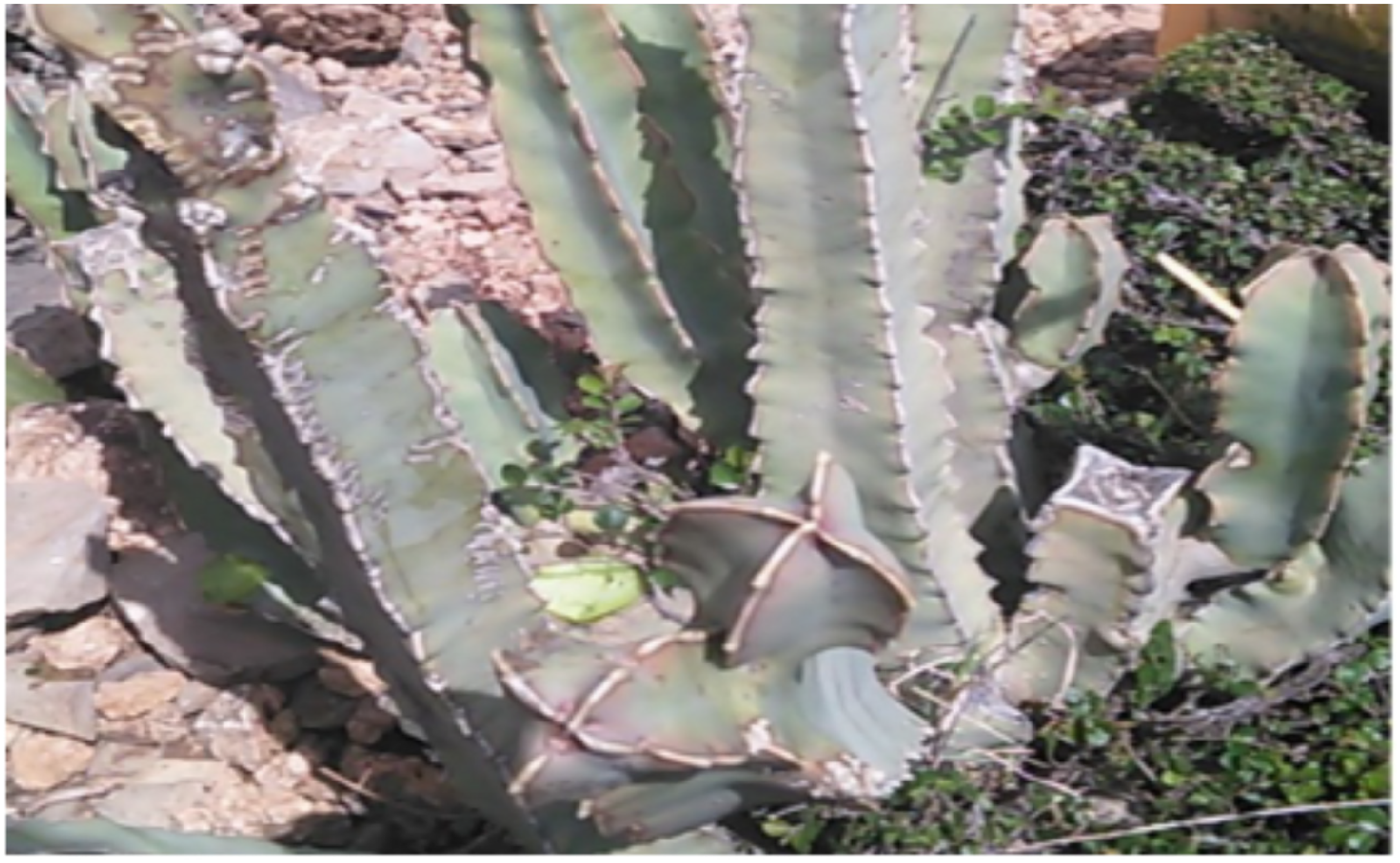
Picture of Caralluma speciosa (photo by Tsegu K., October, 2022).
Chemicals and Instruments
Various chemicals such as the TLC detecting agents (iodine vapor, vanillin/MeOH/H2SO4), analytical grade organic solvents (methanol, ethanol, n-hexane, ethyl acetate, chloroform, and dichloromethane), inorganic reagents (ferric chloride, potassium ferricyanide, and 0.2 M potassium phosphate buffer), culture media (MHA and PDA), and others (trichloroacetic acid, DMSO, chloramphenicol standard, ketoconazole standard, 230-400 mesh size normal silica gel, AA, acetic acid, and DPPH free radical) were used. Silica gel 60 F254 (Merck) precoated aluminum TLC sheet, glass column, electrical laboratory blender, suction filtration apparatus, Petri plates, orbital shaker (Hy-5A, Movel Scientific Instrument Co. Ltd), rotary evaporator (Rotary vacuum, Jainsons), incubator (Binder B28), autoclave (tuttnauer 3150EL), UV-lamp cabinet (UVP Chromato-Vue C-70G, Analytik Jena), UV-Vis spectrophotometer (Cecil CE4001 UV/VIS), Spectrum 65 FT-IR (PerkinElmer, 4000-400 cm−1), GC-MS spectrometry (7890B and 5977A, Agilent Technologies), and 1H (400 MHz) and 13C (100 MHz) NMR spectroscopy were employed.
Extraction and Chromatographic Separation
Coarse powder of C speciosa stems (500 g) was extracted with n-hexane (3×, 2.5 L) via shaking on orbital shaker for 24 h. The extract solution was then filtered by suction filtration, and solvent was evaporated by rotary evaporator resulted in yellow n-hexane extract (6 g). The remained n-hexane residue was consecutively extracted with chloroform, chloroform: methanol (1:1), ethanol, and methanol using the same extraction steps which afforded the corresponding yield of 19, 18.9, 8, and 10 g. The TLC profiles of all the extracts were examined using the solvent systems: n-hexane/DCM/AcOH (1:1:0.1), DCM/EtOAc/AcOH (1:1:0.1), and EtOAc/MeOH/AcOH (3:1:0.1); and the visualizing techniques: UV-lamp (254 and 365 nm) followed by iodine vapor. Thereafter, the chloroform and methanol extracts were fractionated over column chromatography (CC) normal silica gel as follows.
The chloroform extract (15 g) was reconstituted in chloroform (100 mL), adsorbed on silica gel (20 g), and dried using rotary evaporator. Adsorbed and concentrated sample was then applied onto CC silica gel (200 g). Three hundred twenty-five fractions were collected using the eluents n-hexane/DCM/EtOAc/acetone with gradient of polarity. Among the fractions, Fr.1-25 (eluted with n-hexane), Fr.88-118 (eluted with DCM), and Fr.204-211 (eluted with DCM/EtOAc, 1:1) yielded compounds
The methanol extract (9 g) was dissolved in methanol (25 mL), adsorbed on silica gel (10 g), and fractionated over CC silica gel (150 g) using DCM/EtOAc/MeOH as eluents in increasing ratio of polarity. As a result, 167 fractions were collected, out of which Fr.74-83 (eluted with DCM/EtOAc, 1:4) furnished compound
Evaluation of In Vitro Antimicrobial Activity
Bacteriological assay
The antibacterial activity of all extracts and isolated compounds
Antifungal activity assay
The chloroform: methanol (1:1) extract was assessed for its potential antifungal activity against C albicans ATCC10231 using the disc diffusion method mentioned above. A PDA medium was prepared as per the instruction of the manufacturer. The PDA-containing plate was then inoculated with C albicans suspension by streaking with a sterilized swab. Similar to the antibacterial activity experiment, 4 concentrations (12.5, 25, 50, and 100 mg/mL) were prepared in DMSO and 100 µL amount of each concentration was loaded onto sterile Whatman No. 1 filter paper disc (6 mm). Thereafter, the impregnated discs were placed onto the surface of the inoculated agar plates using sterile forceps. Commercial Ketoconazole/Tilt disc (0.01 mg/disc) was used as a standard drug. Then the PDA plates were sealed with Para film and incubated at 27 °C for 3 to 5 days. After incubation, the diameters of the zone of inhibition around each disc were measured using caliper (in mm). Experiments were conducted in duplicate, and results were expressed in mean and standard deviation.
Examination of antioxidant activity
The antioxidative effect of the extracts and isolated compounds
DPPH free radical scavenging activity assay
The DPPH free radical trapping power of the extracts and isolated compounds
Potassium ferric reducing antioxidant power assay
In the present study, ferric ion reducing power of the extracts and isolated compounds
General information of the isolated compounds 1-5
Heptacos-1-ene (
Di-(2-ethylhexyl) phthalate (
12β,20-O-dibenzoly-14β-hydroxy-5α, 6 dihydropregane-3-O-β-cymaropyranosyl-(1-4)-β-cymaropyranoside (
Hexadecahydro-10, 13-dimethyl-17-vinyl-1H cyclopenta[a]phenanthren-12-yl) methyl benzoate (
12β,20-O-dibenzoly-14β-acetyl-5α,6-dihydropregane-3-O-β-thevetopyranosyl-(1-4)-β-cymaropyranosyl-(1-4)-β-cymaropyranoside (
Statistical Data Analysis
The mean and standard deviation of antimicrobial and antioxidant activities were performed by descriptive statistics of SPSS version 20 software. The mean difference between the extracts was analyzed by one-way analysis of variance (ANOVA) using the Tukey HSD test and was considered as significant at 0.05 level.
Supplemental Material
sj-docx-1-npx-10.1177_1934578X231220110 - Supplemental material for In Silico Pharmacokinetics Properties and In Vitro Bioactivities of Pregnane Derivatives and Other Compounds From Stems of Caralluma speciosa
Supplemental material, sj-docx-1-npx-10.1177_1934578X231220110 for In Silico Pharmacokinetics Properties and In Vitro Bioactivities of Pregnane Derivatives and Other Compounds From Stems of Caralluma speciosa by Tsegu Kiros, Seid Mohammed, Aman Dekebo and Yadessa Melaku in Natural Product Communications
Footnotes
Acknowledgments
The authors are thankful to Dr Mesfin Getachew, Addis Ababa Science and Technology University, and Dr Yonas Chebude, Addis Ababa University, Ethiopia, for conducting the NMR and FT-IR spectral analysis, respectively. The authors are also grateful to Dr Anteneh Belayneh, Haramaya University, Ethiopia, for the botanical identification of studied plant.
Authors’ Note
1D-NMR, IR, and GC-MS spectra and in silico pharmacokinetics properties of compounds
Declaration of Conflicting Interests
The author(s) declared no potential conflicts of interest with respect to the research, authorship, and/or publication of this article.
Funding
The author(s) disclosed receipt of the following financial support for the research, authorship, and/or publication of this article: This work was supported by Adama Science and Technology University under Grant (grant number ASTU/AS-R/003/2020); and the World Academy of Sciences (TWAS) and the United Nations Educational, Scientific and Cultural Organization (UNESCO) under the TWAS Research Grant (grant number RGA No. 20-274 RG/CHE/AF/AC_G – FR3240314163).
Supplemental Material
Supplemental material for this article is available online.
References
Supplementary Material
Please find the following supplemental material available below.
For Open Access articles published under a Creative Commons License, all supplemental material carries the same license as the article it is associated with.
For non-Open Access articles published, all supplemental material carries a non-exclusive license, and permission requests for re-use of supplemental material or any part of supplemental material shall be sent directly to the copyright owner as specified in the copyright notice associated with the article.