
Abstract
Background
Malaria caused by Plasmodium falciparum with an average prevalence of 75% in Nigeria, can be treated. Chemical-based medications, one of the methods used to treat it, frequently causes hematological abnormalities in the blood tissues. However, the ethnobotanical and scientific usage of Mangifera indica for the management of malaria in Nigeria necessitated this study on the antiplasmodial evaluation, the hematological profiling of Plasmodium berghei-infected mice treated with Mangifera indica's herbal formulation as well as the isolation and characterization of bioactive compounds from the plants.
Method
The plant's coarse leaves (3 kg) and stem bark (3.2 g) were weighed, divided into groups of leaves (250.71 g), stem bark (509.34 g), leaves/stem bark (1:1) (101.24/101.24 g), leaves/stem bark (1:2) (164.39/328.78 g), and leaves/stem bark (2:1) (218.50/109.25 g), macerated in 3 L of ethanol for 72 h, filtered, and concentrated. For each extract, the following weights and yields were recorded: 31.3 g leaves (15.2%), 43.8 g stem bark (8.7%), 34.8 g leaves/stem bark (1:1) (17.2%), 29.2 g leaves/stem bark (1:2) (5.92%), and 26.1 g leaves/stem bark (2:1) (7.96%). According to established methods, the herbal formulation was employed for hematological testing, GC-MS analysis, and in vivo antiplasmodial evaluation based on standard protocols.
Results
The leaves: Stem bark (1:2) extract (the best therapeutic response) of 98.92% was better than ACT (98.63%). GCMS analysis revealed predominantly mangiferin and esters of linoleic acids that could have enhanced erythropoiesis, mitigated infections, as well as boosted platelet aggregation in P berghei-infected mice as observed from the hematological assay.
Conclusion
Therefore, the extract of leaves: stem bark (1:2) had the best antiplasmodial therapeutic response and hematoprotective activity in P berghei-infected mice.
Keywords
Background
Malaria is a mosquito-borne disease that affects humans and other animals. 1 With a projected 68 million cases and 194 000 deaths from the disease in 2021, malaria is a significant public health concern in Nigeria, according to the World Health Organization. 2 Nigeria has the highest global malaria burden, accounting for about 27% of all cases worldwide. Anywhere in the country is susceptible to transmission at any time of year. 2 Additionally, malaria is caused by Plasmodium falciparum and the parasite causes hematological changes like anemia, thrombocytopenia, leukocytosis, monocytosis, eosinophilia, and neutrophilia (a mild to moderate atypical lymphocytosis). 3 Anemia has been reported as the second in the list of hematological abnormalities in malaria infections according to Elizabeth et al. 3 These hematological aberrations associated with malaria infection vary depending on the following factors: Malaria immunity, demographic factors, level of malaria endemicity, background, and hemoglobinopathy. 4 There are some complexities and uncertainties on the pathophysiological processes that induce the hematological changes in malaria; however, the correct understanding and diagnosis of hematological alterations in malaria patients guide the clinician in the effective therapeutic interventions to avert major complications. 4 For over two decades, World Health Organization has proposed hyperparasitemia as a feature in severe P falciparum malaria. Some researchers have reported a relationship between parasite density and the severity of malaria infection. 5 Additionally, high parasitemia results in anemia due to excessive hemolysis of parasitized erythrocytes. 5 Most patients have presented leukocytosis and thrombocytopenia, where high parasitemia led to the mobilization of leucocytes and platelets (PLTs) destruction resulting in decreased PLT but increased white blood cell (WBC) counts. 5
Over the years, phytomedicines have been reported as an alternative in the management of malaria and its complications in some countries. Mangifera indica (mango) is a pharmacologically rich and diverse plant with some of its reported pharmacological potentials as antimalarial, 6 antioxidant, and anti-inflammatory 7 activities; moreover, it contains bioactive compound (mangiferin) also reported to have antioxidant 8 and antimalarial 9 activities. Many of the secondary metabolites with antiplasmodial activities isolated from the plant parts may possibly have some hematological implications in animal models.
It is worthy of note that to avert the hematological alterations associated with malaria infections, people have used several treatment regimens including chemical-based medications and ethnobotanicals. 10 Despite the efforts exerted to provide effective anti-malarial drugs, patients still develop hematological derangements due to the drugs’ metabolites or parasite pathogenicity; therefore, this research was undertaken to validate the antiplasmodial efficacy of Mangifera indica's herbal formulation, unravel its hematological profile in Plasmodium berghei-infected, as well as characterize the chemical constituents in the plant that could attenuate and ameliorate malaria pathogenicity and blood parameters derangement. The findings have unraveled that mangiferin and esters of linoleic acids from the plant's herbal formulation are responsible for the observed antiplasmodial therapeutic responses and attenuation of the various hematological derangement from parasite pathogenicity in P berghei-infected mice.
Methods
Ethics Approval
This study was approved by the Cross River State Educational Research Board, Cross River State, Nigeria.
Statement of Animal Rights
All the experimental procedures involving mice were conducted in accordance with the International Animal Care guidelines of Arthur Jarvis University, Nigeria, and were approved by the Ethics Committee Faculty of Natural and Applied Science.
Statement of Informed Consent
There are no human subjects in this article and informed consent is not applicable.
Processing of Mangifera indica's Parts and the Preparation of Herbal Formulation
The plant parts were washed with distilled water and sun dried; with mortar and pestle, pulverization yielded coarse particles of the plant parts. The coarse particles were divided into groups consisting of leaves (250.7 g), stem bark (509.34 g), leaves/stem bark (1:1) (101.24/101.24 g), leaves/stem bark (1:2) (164.39/328.78 g), and leaves/stem bark (2:1) (218.50/109.25 g). Each group was macerated in ethanol (3L), shaken occasionally within 72 h, filtered with sieve cloth and Whatman filter paper, and concentrated with the aid of a rotary evaporator (at 40 °C) to get the various ethanol extracts. The weights and % yields for each extract were: 31.3 g leaves (15.2%), 43.8 g stem bark (8.7%), 34.8 g leaves/stem bark (1:1) (17.2%), 29.2 g leaves/stem bark (1:2) (5.92%), and 26.1 g leaves/stem bark (2:1) (7.96%). The herbal formulation was preserved in a refrigerator for 3 days and used for the study. 6
Acquisition, Handling of Mice, and Ethical Approval
The Animal House located in the Faculty of Science, Arthur Jarvis University, Cross River State, Nigeria, provided 55 Albino Wistar mice of (13-27 g) that were used in the study. The NIH guidelines for handling and caring for experimental animals (NIH publication No. 8023; revised 1978) were judiciously implemented. The mice were given the freedom to a standard pellet diet, water, and kept at ambient temperature. The approval (approval number: AJUFNAS/0036) for this study's experimental protocol was given by the Animal Ethics Committee of our Faculty and compliance was judiciously monitored.
Determination of Acute Toxicity
Lethal dose (LD50) and effective doses (ED50) were geometrically calculated by using the method reported by Lorke 11 which involved phases I and II. The rats were observed for 24 h while the number of deaths was recorded in order to establish the medial dosage of the herbal formulation of the plant as well as the one that might induce toxicity in a short term in mice. After administering the herbal formulation (50-2000 mg/kg) through the peritoneum, toxicity symptoms such as gasping, slowed breathing, palpitations, and death could be seen within 24 h.
Parasites and Inoculum Preparation
National Institute of Medical Research (NIMR), Nigeria provided the donor mice infected with chloroquine-sensitive P berghei (NK-65) strain used for the study. An inoculum of P berghei-infected erythrocytes with 20% minimal peripheral parasitemia was obtained through cardiac puncture of infected mice and emptied into an anticoagulant-coated tube. The parasite's percentage in the blood was evaluated by counting the number of P berghei-infected erythrocytes against the entire erythrocytes. The concentration of the blood cells was measured, and the final inoculum, which corresponds to the typical dose for inoculating a mouse, included 1 × 10⁷ P berghei-infected erythrocytes in 0.2 mL of normal saline. 12
Drug Formulation and Administration to Mice
Exactly 100 mL of distilled water was used in the dissolution of tablets of ACT (560 mg) and administered at dosage (8 mg/kg body weight of mice) as well as 20 mL of distilled water enhanced the dissolution of tablets of artesunate (100 mg) and administered at dosage (5 mg/kg body weight of mice), both used as a positive control in the study.
In Vivo Antiplasmodial Determination
A method reported by Asanga et al 12 was used for the evaluation of in vivo antimalarial property of the herbal formulation. To assess the schizonticidal activity of the plant during established infection, 55 mice were inoculated intraperitoneally with a standard inoculum of 1 × 10⁷ P berghei parasitized red blood cells (RBCs) on the first day (D0). After 72 h, the mice were randomized into 11 groups of 5 mice each. Mice in group 1 received distilled water (10 mL/kg) and served as the negative control group. Groups 2 and 3 (positive control groups) received ACT (5 mg/kg/day) and Artesunate (5 mg/kg). Moreover, groups 4 to 11were administered with the herbal formulation (300 mg/kg). The drugs and herbal formulation were orally administered to the animals once daily for 5 days. Tail blood samples from each mouse were collected daily for 3 days, stained with Giemsa's stain, thick and thin films prepared were used to monitor the level of parasitaemia. Parasite density was estimated by counting the number of plasmodium parasites against 200 or 500 WBCs and expressing the resultant number of parasites/µL by blood assuming a WBC count of 8000 per µL of blood according to the relative value method. 12 Parasite density was deduced by using the formula: No. of parasites counted X 8000/WBC Counts = Parasites/µL
The mean survival time (MST) of each treatment group was determined over a period of 30 days (D0-D29) as MST = sum of survival time for each group/ No. of mice in that group.
Percentage (%) growth inhibition was calculated as:

Examination Procedure
Each slide was prepared by measuring blood (2 μL) for the thin film as well as 6 μL for the thick film according to a report by Owusu-Agyei et al, 13 whereas their parasite densities and % growth inhibition of P berghei were calculated and expressed as earlier reported by Asanga et al. 12
Gas Chromatography-Mass Spectroscopy
The Mangifera indica's herbal formulation were weighed (10 mg), dissolved in DMSO, and subjected to GC-MS analysis according to the method described by Asanga et al. 6 Two µL (split ratio 10:1; split flow 12 mL/min) was injected into an Agilent system consisting of a model 7890N gas chromatograph and a model mass detector Triple Quad 7000A in EI model at 70 eV (m/z range 40-600 amu; Agilent Technologies, Santa Clara, California, USA. GC column was an HP-5 ms fused silica capillary with a 5% phenyl-methyl polysiloxane stationary phase (30 m × 250 μm × 0.25 μm). The carrier gas was helium with a column head pressure of 9.7853 psi and flow rate of 1.2 mL/min. Inlet temperature was 250 °C and mass selective detector temperature was 250 °C. The GC oven temperature programing used was as follows: 50 °C initial temperature was held for 10 min; increased at 6 °C/min to 190 °C for 20 min; increased 7 °C/min to 290 °C for 30 min. Identification of compounds was achieved based on their retention indices and by comparison of their mass spectral fragmentation patterns with the National Institute of Standards and Technology (NIST) database/ChemStation data system.
Collection of Blood and Hematological Parameters Profiling
After the sixth-day administration, each treatment group contributed five mice that were anesthetized under chloroform based on animals’ ethical committee protocol, adequately sacrificed; then, 1 mL of blood was collected from each mouse through the cardiac puncture, emptied into EDTA bottle for hematological analysis using automated hematology analyzer system Kx 21, (SYSMEX; Corporation, Japan). The principle adopted by this instrument is based on densitometry and differential blood cell indices and automatically quantifies their values and makes a deduction based on the average blood cells evaluated in the samples considered. 10
Statistical Analysis
GraphPad Prism version 9.0 (GraphPad Software, Incorporated, San Diego, CA, USA) was used to analyze the statistics and expressed as mean ± SEM. The significant differences within and between the different groups (leaves/stem bark (1:2), ACT, stem bark + artesunate, leaves/stem bark (1:1) + artesunate, leaves + artesunate, stem bark, artesunate, leaves/stem bark (1:1), leaves/stem bark (2:1), leaves, negative control) were analyzed using one-way ANOVA; then, their means were compared through the use of Turkey's HSD post hoc test. The differences at a confidence level of 95% were considered to be statistically significant.
Results
Antiplasmodial Assay of the Herbal Formulation
Table 1 shows the schizonticidal activities of Mangifera indica's herbal formulation as compared with the negative control, ACT, and artesunate groups to establish their pharmacological activities. Parasites’ percentage growth inhibition on day 5 (D5), in decreasing order, were as follows: leaves/stem bark (1:2) (98.92%) > ACT (98.63%) > stem bark + artesunate (97.08%) > leaves/stem bark (1:1) (91.10%) + artesunate (95.41%) > leaves + artesunate (95.24%) > stem bark (95.20%) > artesunate (92.34%) > leaves/stem bark (1:1) (91.10%) > leaves/stem bark (2:1) (86.04%) > leaves (81.02%) > negative control (−136%).
In Vivo Curative Antiplasmodial Test (n = 5).

a* significant decrease; P < .05 versus control; a** implies significant increases P < .05 versus control.
GCMS Analysis of the Ethanol Extracts of Mangifera indica
Results from GC-MS spectra showed the presence of many compounds with different retention times and percentage abundance (area %), as illustrated in Tables 2 and 3. The mass spectrometer (MS) analyzed the eluted compounds at varying times to characterize their area %, nature, and retention times. The appearance of the peaks at different m/z ratios arose from the fragmentation of large compounds into smaller fragments. The GC-MS analysis of plant extract showed that the most abundant compounds were mangiferin, 9,12-octadecadienoic acid, oleic acid, 9,12-octadecadienal, 9-oxabicyclo (6.1.0) nonane, hexadecadienal, and spirohexane.
GCMS Analysis of Ethanol Extract of Mangifera indica Stem Bark.

GCMS Analysis of Ethanol Extract of Mangifera indica Leaves.

Discussion
Antiplasmodial microscopic assessment focuses on the schizont stage of the parasite being responsible for the clinical symptoms in malaria patients; so, the evaluation of schizonticidal activity of a drug provides insight on its therapeutic efficacy. Consequently, the schizonticidal activity of the herbal formulation of Mangifera indica in this study (Table 1), revealed substantial reductions in the parasite densities and improvements in MST as compared with the negative control. These findings aligned with similar research outcomes by Asanga et al 12 on Nauclea latifolia roots’ extract and fractions (150 mg/kg). However, Kumaratilake et al 14 reported that fatty acids induce direct destruction of malaria parasites, whereas others act through neutrophil priming. In addition, the history of malaria chemotherapy is traceable to natural products from plant sources; however, these studies revealed some of the natural products with already reported pharmacological activities. Maenpuen et al 9 reported the antimalarial activities of mangiferin, whereas Asanga et al 6 reported that linoleic acid esters in Mangifera indica are responsible for the plant's antimalarial activity through the inhibition of fatty acid biosynthesis in the parasite's apicoplast. Therefore, the predominance of mangiferin and esters of linoleic acid (Tables 2 and 3) in this herbal formulation may have aided in the destruction of the parasites. Conversely, a similar finding was reported by Asanga et al 12 on the significant increases in MST of mice; therefore, the prolonged MST for the parasitized mice was due to better parasitemia clearance by the bioactive compounds in the herbal formulation; hence, better schizonticidal activities.
The essence of RBC estimation is for anemia evaluation as well as the investigation of normal erythropoiesis; however, the assay also gives clue on the O2 carrying capacity of the blood. It is worthy of note that Schnell et al 15 reported the optimal range of RBC counts in an adult mouse as 9.23-9.42×10⁶/mm3; however, the result (Table 4) revealed significant elevation (P < .05) in RBC counts in all the mice except for the groups treated with leaves:stem bark (1:1) as compared with the negative control; implying that the herbal formulation enhanced erythropoiesis in mice. This report was in tandem with that earlier reported by Cyril-Olutayo et al 16 and Asanga et al 10 on other plants. In addition, the higher RBC counts for mice treated with leaves/stem bark (1:2) as compared to the artesunate group, suggested enhanced erythropoiesis. Nevertheless, the general low RBC counts in all the mice could have resulted from hemolysis potentiated by the parasites as well as the predominant linoleic acid esters (Tables 2 and 3) as Yuan et al 17 had posited that linoleic acid induces RBC and hemoglobin (Hb) damages via oxidative mechanism.
Effect of the Treatments on Hb, MCH, MCV, HCT, MCV, MCH, MCHC RDW-CV, RDW-SD Levels, and RBC Counts of Parasitized Mice (n = 5).
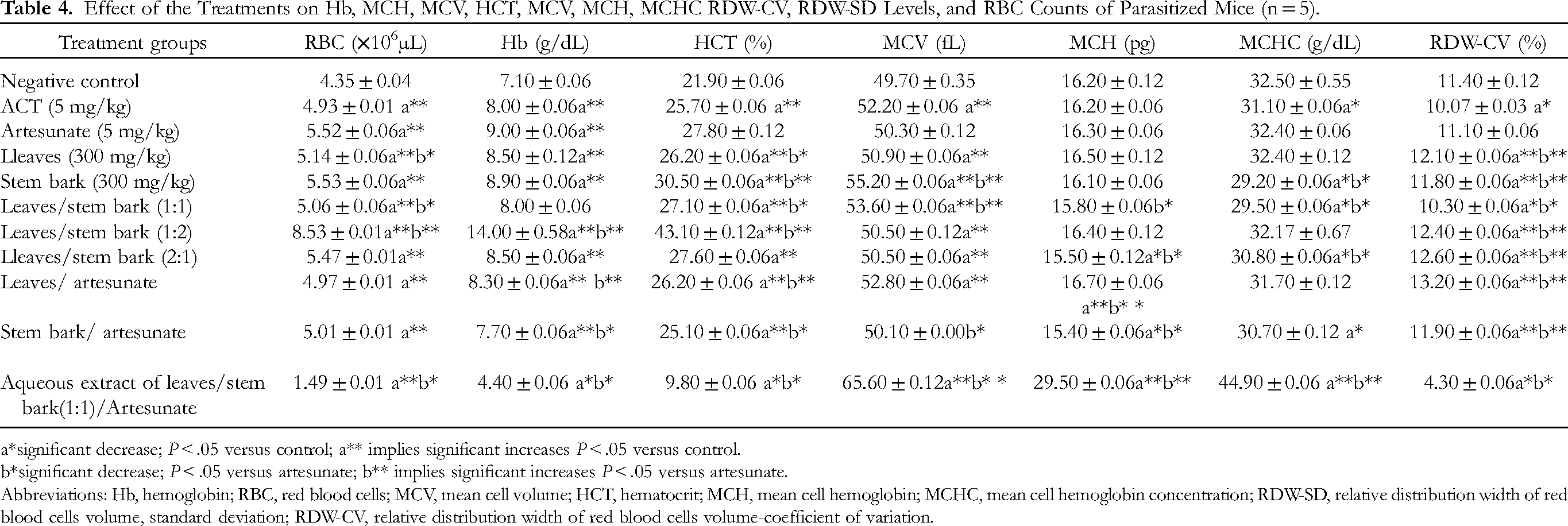
a*significant decrease; P < .05 versus control; a** implies significant increases P < .05 versus control.
b*significant decrease; P < .05 versus artesunate; b** implies significant increases P < .05 versus artesunate.
Abbreviations: Hb, hemoglobin; RBC, red blood cells; MCV, mean cell volume; HCT, hematocrit; MCH, mean cell hemoglobin; MCHC, mean cell hemoglobin concentration; RDW-SD, relative distribution width of red blood cells volume, standard deviation; RDW-CV, relative distribution width of red blood cells volume-coefficient of variation.
Also, Hb as an intracellular protein gives blood its red color and is used for the diagnosis of anemia (blood index <12 g/dL). 18 However, the result (Table 4) revealed significant elevation (P < .05) in the Hb levels in all the mice except those treated with leaves:stem bark (1:1)/Artesunate as compared with the negative control; this was in line with the report by Lathia and Joshi 19 that some plants possess antianemic potentials. More so, the elevation in the Hb levels in mice treated with leaves/Artesunate and leaves:stem bark (1:2) as compared with the ACT, implied better efficacy. On the whole, the herbal formulation and ACT averted the resultant anemia induced by the infiltration of P berghei in RBCs; a shred of clear evidence that they contain antianemic metabolites like mangiferin (Tables 2 and 3) known to have antioxidant activities.
Moreover, hematocrit (HCT) as an index for the diagnosis of anemia, measures the number of relative erythrocytes in a blood sample. Schnell et al 15 reported 45.6%-46.0% as the optimal range of HCT in an adult mouse; however, anemia could result due to destruction, excessive loss, or decreased production of RBCs evidenced by low Hb and HCT. From the result (Table 4), the HCT levels in all the mice significantly increased (P < .05) except for those treated with leaves:stem bark (1:1)/Artesunate as compared with the negative control. This result showed consistency with the report by Momoh et al 20 and Aleksandro et al 21 that drugs and extracts could prevent nonimmune destruction of erythrocytes infected with P berghei in conditions of observed hyperparasitemia. However, the HCT levels in mice that were treated with leaves/artesunate, stem bark, and leaves:stem bark (1:2) were better than ACT; a pointer that these herbal formulations were more potent in the attenuation of anemia induced by the P berghei in mice.
Furthermore, the classification of the size of RBCs into normocytic, macrocytic, or microcytic is of great essence in the diagnosis of malaria. 22 Schnell et al 15 reported that the normal range of mean cell volume (MCV) in a healthy mouse is 48.9-50.0U3; however, the result (Table 4) revealed that all the mice except those treated with the stem bark/artesunate significantly increased (P < .05) their MCV levels as compared with the negative control. This result showed inconsistency with that earlier reported by Momoh et al 20 in a similar study. More so, the MCV levels in all the mice with exception to the negative control group were above the reference range; an indication of a possible macrocytic anemic condition. In addition, mean cell hemoglobin (MCH) as the amount of Hb in the erythrocyte is considered as the difference in the evaluated MCV. Its optimum value in adult mice according to Schnell et al 15 is 14.7-14.8UUG; however, Table 4 revealed significant increases (P < .05) in the MCH levels of mice treated with the leaves/artesunate and leaves:stem bark (1:1)/artesunate as compared with the negative control and ACT, respectively. This result was inconsistent with that reported by Momoh et al 20 in a similar study. Nevertheless, stem bark/artesunate administration caused significant decreases (P < .05) in the MCH levels of mice as compared with the negative control and ACT; this report showed inconsistency with that reported by Mohammed et al 23 in another study.
Unarguably, Table 4 revealed significant increases (P < .05) in mean cell hemoglobin concentration (MCHC) levels in mice treated with leaves:stem bark (1:1)/artesunate as compared with the negative control and ACT. The result showed inconsistency with the reports by Tanko et al 24 and Momoh et al 20 in similar studies. It is worthy of note that MCHC is the mean Hb concentration in 100 mL of erythrocytes and according to Schnell et al, 15 its optimum value in an adult mouse is 29.5%-30.3%. However, the administration of the leaves, stem bark, and leaves:stem bark (1:1) extracts caused significant reductions (P < .05) in the MCHC levels in mice as compared with the negative control and artesunate groups.
In addition, WBC protects the body against antigenic infiltrations and as observed in the negative control group (Table 5), leucocytosis gives an indication of infection. Schnell et al 15 reported the optimal range of leucocytes in an adult mouse as 2.8-5.4 × 10⁹/L; however, Table 5 further revealed significant decreases (P < .05) in WBC counts in all the mice except for those treated with leaves:stem bark (2:1) and stem bark as compared with the negative control. The result was consistent with those reported by some researchers on other plants,10,16,20 that the observed leucopenia may be related to apoptosis from the depletion of lymphocyte subsets. 25 More so, the administration of stem bark/artesunate and leaves:stem bark (1:1)/artesunate that caused significant elevation (P < .05) in the WBC counts in mice as compared with the ACT imply that the leucocytosis could have resulted from the oxidative damages by esters of linoleic acid (Tables 2 and 3) and P berghei in the infected mice. Therefore, leaves/stem bark (1:2) had the best anti-infective activities.
Effect of the Treatment on the WBC and PLT Counts, MON, NEU, EOS, BAS, and MPV Levels of P berghei-Infected Mice (n = 5).

a* significant decrease; P < .05 versus control; a** implies significant increases P < .05 versus control.
b* significant decrease; P < .05 versus artesunate; b** implies significant increases P < .05 versus artesunate. Abbreviations: WBC count, white blood cell; LYM, lymphocytes; BAS, basophils level; NEU, neutrophils level; EOS, eosinophils level; MON, monocytes level; PLT, platelet counts; MPV, mean platelet volume; PDW, platelet distribution width; PCT, plateletcrits; P-LCR, platelet large cell ratio.
Consequently, lymphocytes are leucocytes that defend the body against immunologic reactions and pathogen invasion. According to Schnell et al, 15 its optimal range in an adult mouse is 84% to 87%; however, Table 5 showed that the lymphocyte levels significantly decreased (P < .05) in all the mice as compared with the negative control. So, lymphocytosis was observed in mice treated with distilled water, leaves: stem bark (2:1), and stem bark whereas the other groups of mice revealed lymphocytopenia; these results showed consistency with the report by Asanga et al 10 and Okoroiwu et al 26 on the possible synthesis of antibodies due to parasite infiltration in the mice. Therefore, leaves:stem bark (1:2) and leaves/artesunate revealed better activity than ACT implying that their metabolites responded better in the mobilization lymphocytes to attenuate parasite-induced infection in mice.
Basophils are granulocytes that are mobilized in the blood after the invasion of an allergen or parasites. According to Schnell et al, 15 basophils level in the differential blood count is 0.5% to 1%; however, the results as shown in Table 5 revealed a significant reduction (P < .05) in basophils level in all the mice except for those treated with leaves:stem bark (1:1)/artesunate as compared with the negative control. There was consistency in this result with those reported by some researchers on other plants.23,24 Hence, the plant's herbal formulation has significant antiparasitic activities. Consequently, neutrophils are granulocytes of leucocyte origin, they play roles in phagocytosis and Johnson 27 reported its normal range as 1.77-3.38 × 10³/mm3. Conditions such as inflammatory or metabolic disorders, bacterial infections, and severe trauma results in neutrophilia whereas neutropenia is noticed during the ingestion of rotten foods and viral infection; increased neutrophil removal from the system by macrophages, decreased level of neutrophil production, or parasitic infections like malaria. 28 The result (Table 5) revealed significant elevation (P < .05) in the neutrophils level in all the mice except for those treated with leaves/artesunate as compared with ACT and negative control. This result was inconsistent with other reports on neutropenia as reported by Okoroiwu et al 26 and Chenl and Sendo. 28 Conversely, eosinophils are granulocytes that are found in the blood during parasitic infection. According to Gannong, 29 the differential WBC count is 1%-4%; however, the result as presented in Table 5 revealed no change (P > .05) in eosinophil levels in all the mice; therefore, this result suggested the inability of the herbal formulation and ACT to arrest allergy induced by P berghei infiltration of mice or maybe that the period of administration was limited.
Platelets play critical role in clotting of blood as well as acute phase reaction to inflammation and infection and its normal value in mice is 8.9-12.3 × 10⁵/mm3 according to Schnell et al. 15 However, Table 5 revealed thrombocytosis in all the mice except for those treated with artesunate, leaves:stem bark (2:1), leaves:stem bark (1:2) in which thrombocytopenia was observed as compared with the negative control. Therefore, the high PLT counts noticed in this study were inconsistent with the reports by Igbeneghu and Odaibo, 30 Asanga et al, 10 and Taha et al. 31 Conversely, the thrombocytopenia in the negative control group may have resulted from the intricate produced by malaria antigens thereby leading to segregation of injured PLTs by macrophages in the spleen. 32 In addition, mean platelet volume (MPV) is used in the investigation of the ability of a drug to enhance blood clotting and it reveals the PLTs’ average sizes. So, the results (Table 5) revealed significant elevation (P < .05) in MPV levels in all the mice with the exception to those treated with ACT and leaves:stem bark (1:2) as compared with the negative control. The result showed inconsistency with the report by Yakubu et al; 33 however, it was consistent with the report by Momoh et al. 20 Therefore, the limitation of this study was the nonprofiling of mangiferin, esters of linoleic acids, and other compounds to validate the reproducibility of the attenuation of hematological derangements as well as the observed antiplasmodial therapeutic indices as unraveled through the administration of the herbal formulation in this study. Moreover, limited time and adequate instrumentation were great challenges for this study.
Conclusion
The antiplasmodial therapeutic response of leaves:stem bark (1:2) extract was better than ACT with the predominant compounds in the herbal formulation being esters of linoleic acid and mangiferin. These compounds could have played critical role in the observed erythropoiesis, infection mitigation, and PLT aggregation in P berghei-infected mice. Therefore, the leaves:stem bark (1:2) had the best ability to ameliorate anemia and leukocytosis whereas leaves:stem bark (1:1)/artesunate and leaves/artesunate showed the best activity in averting thrombocytopenia.
Footnotes
Acknowledgments
We appreciate Mr Ndiana, Chemical pathology laboratory and Mr Ukpong, Clinical Pharmacology, University of Uyo Teaching Hospital, Nigeria for their professional guidance on this study.
Authors’ Contributions
The conceptualization, design, and methodology were carried out by EEA. The financial resources were provided by EEA, FEO, and MO. Statistical analysis was done by EAU, HUO, and EEA whereas the drafting and proofreading were done by EEA, OAE, and MOO. The manuscript and the authors’ list have been read and approved by all the authors.
Availability of Data
All datasets generated in this study is available from the corresponding author on request.
Consent for Publication
Not applicable
Declaration of Conflicting Interests
The author(s) declared no potential conflicts of interest with respect to the research, authorship, and/or publication of this article.
Ethics Approval and Consent to Participate
The experimental protocol was strictly followed and approved by the Faculty of natural and Applied Science animal Ethics Committee, Arthur Jarvis University.
Funding
The author(s) received no financial support for the research, authorship, and/or publication of this article.