In a phytochemical investigation of the rhizomes of Kniphofia reflexa, an endemic plant used to treat relapsing fevers in Kejom, northwestern Cameroon, 12 known (1-12) compounds were obtained following chromatographic methods and purification, together with 3 new derivatives (13-15) prepared by acetylation. One-dimensional and 2-dimensional nuclear magnetic resonance spectroscopic studies together with infrared and ultraviolet spectral analyses in association with data found in the literature were used to determine the structure of the compounds. In the in vitro evaluation of compounds 1-9, 12-14, and the crude extract against Plasmodium falciparum chloroquine-sensitive (D6) and chloroquine-resistant (W2) strains, cassiamin C (1) [IC50 0.57 ± 0.54 (D6); 0.78 ± 0.08 (W2)], and crude extract [IC50 1.06 ± 0.22 (D6); 1.08 ± 0.12 (W2)] were highly active against the parasites. Kniphofiarexine (12) was inactive. However, its derivative, kniphofiarexine B (14), was moderately active. In the in vivo studies, the extract suppressed Plasmodium berghei growth, but did not clear completely the parasites.
Malaria is one of the main life-threatening illnesses, despite enormous efforts in research, being one of the main causes of disease and death worldwide.1 In 2020, 90% of malaria cases and deaths occurred in Africa.2 Malaria is still a challenge to humans as the incidence of drug resistance keeps rising, coupled with a lack of therapies that can efficiently target the intricate life cycle of the malaria parasite.3,4 Resistance to artemisinin,5 the most recommended component for malarial combination therapies, is reported in sub-Saharan Africa and other centers of endemism.6 Because of the use of Kniphofia reflexa Hutchinson ex Codd. Asphodelaceae in the treatment of relapsing fevers, supported by literature data on the antiplasmodial activities within classes of natural products like terpenes,7‐10 flavonoids,11‐13 alkaloids,14‐17 quassinoids,7,18 xanthones,19‐22 quinones, and anthraquinones,23‐29 a needed study of this plant was conducted considering its unique endemic nature, coupled to its classification. To the best of our knowledge, no study is reported on the antiplasmodial and antimalarial potential of chemical constituents of K reflexa.
Results and Discussion
Identification of Compounds
Phytochemical study of the rhizome of K reflexa (Asphodelaceae) led to the isolation of 12 previously described compounds (1-12), including 2 anthraquinones (8 and 9), 1 phenyl anthraquinone (10), 4 anthraquinone dimers (1, 3, 4, and 11), 3 naphthalene derivatives (2, 5, and 12), 1 indanone (7), and 1 benzene derivative (6). The acetylation of 12 yielded 3 new derivatives (13-15). The structures of compounds 1-12 were elucidated from their 1-dimensional (1D) and 2-dimensional (2D) nuclear magnetic resonance (NMR) spectral data and that found in the literature to be 2 anthraquinones, chrysophanol (9),30 and helminthosporin (8)31,32; 1 phenyl anthraquinone, knipholone (10)33; 4 anthraquinone dimers, cassiamin C (1),34 microcarpin (3),35 chrysotalunin (4),36 and 10-hydroxy-10,7′-(chrysophanolanthrone)-chrysophanol (11)37; 3 derivatives of naphthalene, 2-acetyl-1,8-dimethoxy-3-methylnaphthalene (2),38 2-acetyl-1-hydroxy-8-methoxy-3-methylnaphthalene (5),37 and kniphofiarexine (12); one indanone39; and a benzene derivative, 2׳,4׳,6׳-trimethoxyacetophenone (6).40 The acetylation of kniphofiarexine (12) yielded 3 new derivatives to which we assign trivial names of kniphofiarexine A (13), kniphofiarexine B (14), and kniphofiarexine C (15) (Figure 1).
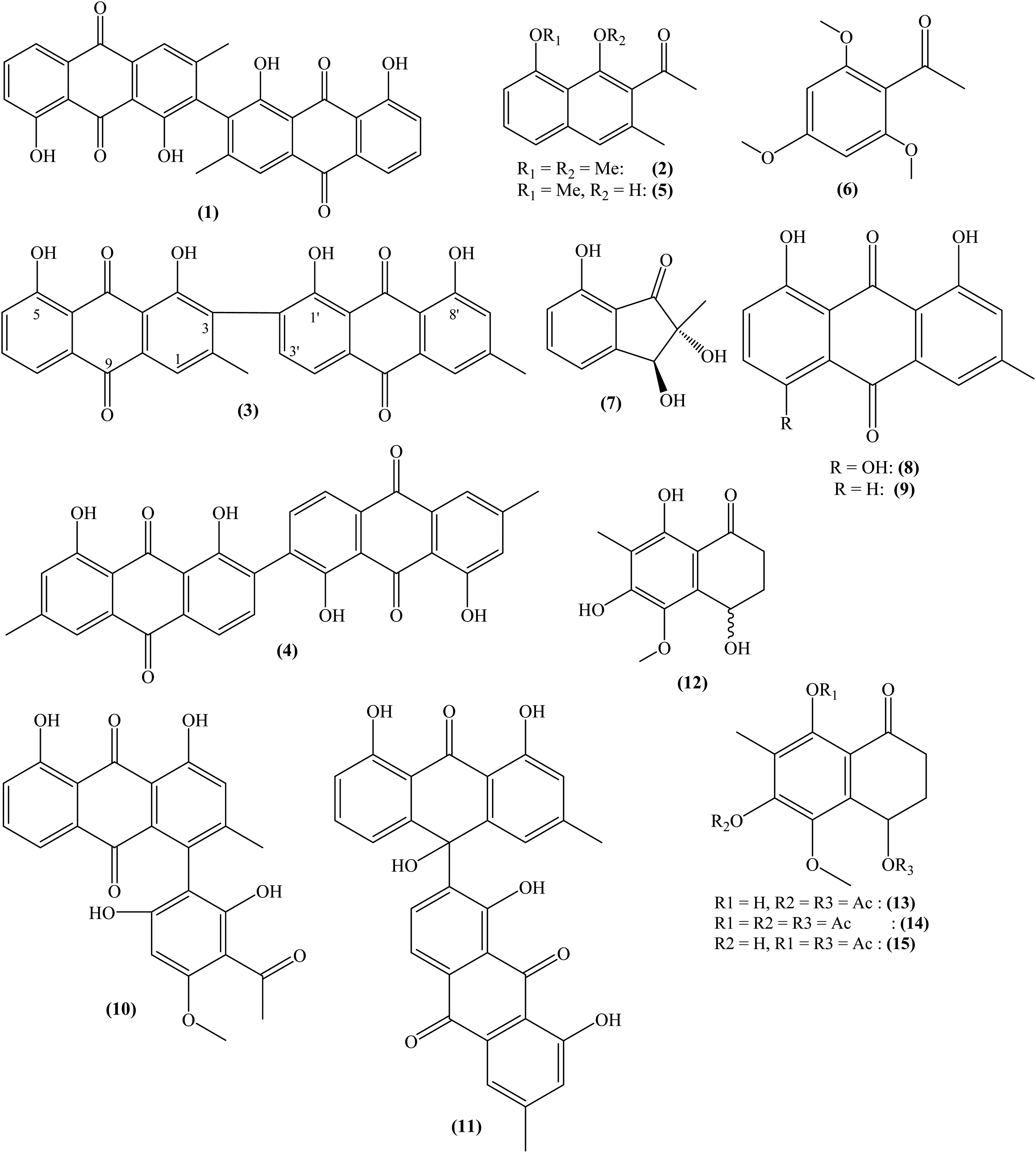
Isolated compounds from rhizomes of Kniphofia reflexa.
Antimalarial Activities
Among the pure compounds and methanol (MeOH) crude extract assessed for in vitro antiplasmodial activity on chloroquine-sensitive (D6) and chloroquine-resistant (W2) strains of Plasmodium falciparum, the extract exhibited the best activity (IC50 1.06 ± 0.22 against D6; 1.08 ± 0.12 against W2). All anthraquinone dimers showed some activity, with cassiamin C (1) (IC50 0.57 ± 0.54 against D6; 0.78 ± 0.08 against W2) having significant antiplasmodial property, while microcarpin (3) and chrysotalunin (4) were only weakly active. Compounds 2, 5, and 7 displayed modest to weak antiplasmodial activities, respectively (Supplemental Table S1). Though kniphofiarexine (12) was inactive (IC50 180.6 ± 6.78 against D6; 45.91 ± 3.76 against W2), its derivative kniphofiarexine B (14) was more potent (IC50 1.55 ± 0.55 against D6; 11.41 ± 1.09 against W2), suggesting that hydroxyl substituents affect activity since complete acetylation greatly improved the antiplasmodial activity. In all tests, quinine (IC50 0.09 ± 0.005 against D6; 0.12 ± 0.03 against W2) was the reference drug (Supplemental Table S2). Compounds 8 and 9 were inactive, whereas their dimers were active. This might be due to the oxidative coupling of anthraquinones, as perceived in compounds 10 and 11. The oxidative coupling has previously been postulated to be a possibility for novel antimalarials presenting significant activities against P falciparum.41
In the in vivo study, at the maximum dose of 5000 mg/kg body weight, animals treated with the MeOH crude extract survived the 14-day observation period, as well as a constant increase in body weight during an acute toxicity test, attesting the safety of K reflexa in the treatment of malaria. On a 4-day study period, the in vivo suppression of Plasmodium berghei by the crude extract showed a significant suppression of 12.7% ± 1.1% of the parasites as opposed to 14.6% ± 2.0% by quinine (Supplemental Table S2 and Figure S1). However, the extract could not satisfactorily clear the parasites on a 5-day curative period as compared to quinine in the curative test (Supplemental Table S3 and Figure S2). The low acute cytotoxicity is indicative of the fact that the suppression or clearance of the parasites is due to the combined activities of contained phytochemicals and not toxicity.
Experimental
General Experimental Procedures
Optical rotations were determined on a JASCO P-2000 polarimeter, while infrared spectra were measured on a JASCO J-810 spectrophotometer. For thin-layer chromatography, silica gel 60F254 and RP-18F254 were used, coated on aluminum sheets and glass, respectively. Merck silica gel 60 and Sephadex LH-20 were used for column chromatography. Preparative high-performance liquid chromatography (HPLC) was performed using SIL D-60-10 and ODS-M-80 columns on LC-90BW and LC908C machines, respectively (Japan Analytical Industry Co. Ltd). A JOEL MS600H-1 mass spectrometer was used to record mass spectrometry (MS), and a MAT95XP recorded high-resolution electron ionization-MS. 1D and 2D NMR spectra were obtained using an Avance AV-500 MHz spectrophotometer, and an Avance AV-400 MHz spectrophotometer from Bruker was used for 13C-NMR spectra.
Plant Material
K reflexa was collected in February 2015 from Lake Bambili, North-West Region of Cameroon (altitude: 2320 m above sea level; latitude: 5°55′28.49′′ and longitude: 10°14′41.03′′) and identified by Mr. Tadjouteu Fulbert, at the National Herbarium of Cameroon in Yaoundé, where a specimen is deposited (collection No. 66930/HNC).
Extraction and Isolation
Rhizomes of K reflexa were airdried and powdered (4.5 kg) prior to extraction with MeOH at ambient temperature for 96 h to obtain 461 g of crude extract. Part of this (450 g) was separated in 500 mL fractions by flash chromatography using an evolving solvent system from n-hexane, through ethyl acetate to MeOH; these were regrouped to get 14 fractions (F.1-14). From the isocratic CC of F.12 (1.4 g), 2 subfractions F.12-1 (62 mg) and F.12-2 (93 mg) were selected. F.12-2 on synchronized Sephadex LH-20 at 50% dichloromethane (DCM)/MeOH and 100% MeOH yielded compound 1 (1.8 mg) after purification by HPLC (isocratic, 100% MeOH). F.3 (650 mg) by CC with increasing polarity of n-hexane/EtOAc yielded 14 subfractions (F.3-1 to F.3-14). F.3-4 (243 mg) by monopolar CC (15% Hex/EtOAc), followed by Sephadex LH-20 (100% MeOH) gave compounds 5 (25 mg) and 2 (80 mg), respectively. F.4 (163 mg) by CC using a gradient of EtOAc/MeOH issued Fr.4-3 (89 mg), which again, on gradient CC, eluting with 5% DCM/MeOH underwent isocratic CC (50% Hex/DCM) to yield compound 8 (3.4 mg). F.5 (1.2 g) using an increasing polarity of n-hexane/EtOAc gave fluorescent orange slate-like crystals of 9 (42 mg) at 18% Hex/EtOAc, and a deep red amorphous solid at 45% Hex/EtOAc, (10, 213 mg). F.5-7 (32 mg) by pTLC gave compound 3 (3 mg). F.5-9 (78 mg) gave 6 (31 mg) by HPLC (40% Hex/EtOAc). Compound 12 (103 mg), eluted with 30% Hex/EtOAc was then purified by HPLC (50% MeOH/H2O), while 7 (19 mg) came out at 35% Hex/EtOAc, and systematically underwent further CC (40% Hex/EtOAc monopolar) and Sephadex LH-20 (50% DCM/MeOH), then pTLC from F.8-6 (437 mg). F.6 (200 mg) issued 11 (9.2 mg) on (isocratic, 25% Hex/EtOAc) CC and HPLC (isocratic, 20% Hex/EtOAc). F.10 (1.8 g) on a solvent gradient (DCM/MeOH) CC, yielded compound 4 (1.8 mg), which was crystallized from 3% DCM/MeOH.
Acetylation of Kniphofiarexine (12)
Compound 12 (15.2 mg) was acetylated42 and 18.0 mg of the product was separated by recycling HPLC to yield 3 new derivatives: kniphofiarexine A (13) (2.4 mg), kniphofiarexine B (14) (1.4 mg), and kniphofiarexine C (15) (1.2 mg) (Supplemental Table S4 and Figures S3-S22).
Antimalaria Tests
The activity of K reflexa was investigated on chloroquine-sensitive (D6) and chloroquine-resistant (W2) strains of P falciparum according to the method of Trager and Jensen, with adaptation.43 Uninfected O+ human red blood cells, supplemented by RPMI-1640 and solutions, obtained from Corning®, were used for in vitro antiplasmodial tests. The solutions used were sterilized using syringe-adapted 0.22 µm filters, and 96 well microtiter plates (Costar®) were used. The concentration inhibiting 50% of the growth of parasites (IC50) was assessed using GraphPad Prism version 7.03. For in vivo antimalarial tests, 6-8-week-old male and female BALB/C mice were used, of body weight 27-32 g, kept at room temperature and with a 12 h light/12 h dark cycle, with sufficient food and water. P berghei strain was offered by BEI-Resources.
In the in vivo antimalarial tests, 5 mg of crude extract was dissolved in 200 µL dimethyl sulfoxide (DMSO; Sigma) and then diluted with purified water to obtain stock solutions. Male and female BALB/C mice (6-8 weeks and 27-32 g) were maintained in the MRABL animal house of the Faculty of Health Sciences, University of Buea at 25 °C with a 12 h light/12 h dark cycle, with enough food and water. All universally agreed guidelines on animal use in laboratory experiments were observed. The protocol was appraised and accepted by the University of Buea Institutional Ethics Review Board for Animal Use. BEI-Resources donated the P berghei. Serial passage of blood from infected mice to noninfected mice was made every week to maintain the parasites.44 To inoculate the parasites, 20% to 30% of hitherto infected P berghei albino mice were used as donors. They were sedated and sacrificed through the thoracic region to expose the heart. Heparinized tubes containing 0.5% trisodium citrate were used to collect blood by cardiac puncture. Blood was diluted with 0.9% saline depending on the parasitemia of the donor.45
In vivo suppressive assessment of P berghei lasted 4 days to measure the schizontocidal potential of tested samples against P berghei-infected BALB/C mice, after a previously described method with necessary alterations.45 Animals were given 400 mg/kg daily enteral dose of the extract dissolved in 100 µL of 2% DMSO/H2O. In total, 10 mg/kg body weight of quinine was administered as a positive control to another group, whereas the negative control animals received only 100 µL of the vehicle. The endurance rate was observed every day for 2 weeks after inoculation. Parasitemia and percentage inhibition were evaluated by Giemsa-stained thin blood smears from the tail of each animal on the 5th day.46 The percentage of parasite growth suppression was projected using the following equation:
where A is the average parasitemia for the negative control group and B is the average parasitemia for the test group.
In vivo curative test (Rane test) was used to establish the curative potential of the samples, following the previously described method.47 Animals in the negative control group received corresponding volumes of 2% DMSO in distilled water (vehicle) daily. Giemsa-stained thin blood films were prepared every day for 5 days to monitor parasitemia.42 Mean endurance times for the groups were established over a period of 2 weeks.
Conclusion
The chemical study of the rhizomes of K reflexa led to the isolation of twelve previously described compounds (1-12), as well as 3 new derivatives (13-15) by acetylation. In vitro evaluation of compounds 1-9, 12-14, and the crude extract against chloroquine-sensitive (D6) and chloroquine-resistant (W2) strains of P falciparum showed that cassiamin C (1) [IC50 0.57 ± 0.54 (D6); 0.78 ± 0.08 (W2)], and crude extract [IC50 1.06 ± 0.22 (D6); 1.08 ± 0.12 (W2)] were highly active against the parasites. Kniphofiarexine (12) was not active, whereas its derivative kniphofiarexine B (14) was moderately active. The antiplasmodial and antimalarial effects were never due to toxic effects but to the activities of the isolated phytochemicals and crude extract. To the best of our awareness, the antiplasmodial and antimalarial potential of the chemical constituents of K reflexa, which supports its traditional use in the treatment of malaria, is reported for the first time in this study.
Supplemental Material
sj-docx-1-npx-10.1177_1934578X221133582 - Supplemental material for In Vitro and in Vivo Evaluation of the Antimalarial Activities of Kniphofia reflexa Hutchinson ex Codd
Supplemental material, sj-docx-1-npx-10.1177_1934578X221133582 for In Vitro and in Vivo Evaluation of the Antimalarial Activities of Kniphofia reflexa Hutchinson ex Codd by Denis Kehdinga Sema, Alain Meli Lannang, Denis Zofou, Mujeeb- ur-Rehman, Tegha Hycienth Fung and
Virginie Flaure Tsague Tankeu, Jean Duplex Wansi, Norbert Sewald, M. Iqbal Choudhary in Natural Product Communications
Footnotes
Authors’ Contributions
DKS contributed to conceptualization, investigation, methodology, and writing—original draft. AML contributed to supervision and writing—review & editing. DZ contributed to biological analysis. MR contributed to methodology and writing—review & editing. THF contributed to writing & review. VFTT contributed to formal analysis and resources. JDW contributed to supervision and writing—review & editing. NS contributed to supervision and writing—review & editing. MIC contributed to supervision and writing—review & editing.
Declaration of Conflicting Interests
The author(s) declared no potential conflicts of interest with respect to the research, authorship, and/or publication of this article.
Funding
The author(s) disclosed receipt of the following financial support for the research, authorship, and/or publication of this article: AML was supported by Alexander von Humboldt-Stiftung through the George Forster Fellowship for Experienced Researchers (ID No. 1137675) at Bielefeld University. The phytochemical work was done at NAPEC, the University of Maroua, and at HEJ Research Institute of Chemistry, ICCBS, University of Karachi, Pakistan during an ICCBS-TWAS PhD Research Fellowship 2016 to DKS. The pharmacological work was financially supported by TWAS Research Grant No. 14-150 RG/BIO/AF/AC_I - UNESCO FR: 324028598 and IFS Grant No. F/5122-2F awarded to DZ.
Ethical Considerations
Ethical approval to report this case was obtained from the University of Buea Ethical Review Board.
Statement of Human and Animal Rights
All the experiments were performed in accordance with both the national and international ethical guidelines for the care and use of animals in research. A proposal, describing the handling and treatment of animals was submitted, reviewed, and approved by the University of Buea Ethical Review board for the use of animals in research under Reference UB-IACUC No. 005/2018.
Statement of Informed Consent
There are no human subjects in this article and informed consent is not applicable.
ORCID iDs
Denis Kehdinga Sema
Alain Meli Lannang
Supplemental Material
Supplemental material for this article is available online.
References
1.
AbebawAAschaleYKebedeTHailuA. The prevalence of symptomatic and asymptomatic malaria and its associated factors in Debre Elias district communities, northwest Ethiopia. Malar J. 2022;21(1):1‐10.
BoudreauEFPangLWChaikummaoSWitayarutC. Comparison of mefloquine, chloroquine plus pyrimethamine-sulfadoxine (Fansidar), and chloroquine as malarial prophylaxis in eastern Thailand. Southeast Asian J Trop Med Public Health. 1991;22(2):183‐189.
4.
Fernández-ÁlvaroEHongWDNixonGLO’NeillPMCalderónF. Antimalarial chemotherapy: natural product inspired development of preclinical and clinical candidates with diverse mechanisms of action: miniperspective. J Med Chem. 2016;59(12):5587‐5603.
5.
RosenthalPJBjörkmanADhordaM, et al.Cooperation in countering artemisinin resistance in Africa: learning from COVID-19. Am J Trop Med Hyg. 2022;1(aop):1‐3.
6.
KheraAMukherjeeR. Artemisinin resistance: cause for worry?J Marine Med Soc. 2019;21(1):4.
7.
SaxenaSPantNJainDBhakuniR. Antimalarial agents from plant sources. Curr Sci. 2003:1314‐1329.
8.
EfangeSMBrunRWittlinS, et al.Okundoperoxide, a bicyclic cyclofarnesyl sesquiterpene endoperoxide from Scleria striatinux with antiplasmodial activity. J Nat Prod. 2009;72(2):280‐283.
9.
MurataTMiyaseTMuregiFW, et al.Antiplasmodial triterpenoids from Ekebergia capensis. J Nat Prod. 2008;71(2):167‐174.
10.
SaitoAYRodriguezAAMVegaDSMSussmannRAKimuraEAKatzinAM. Antimalarial activity of the terpene nerolidol. Int J Antimicrob Agents. 2016;48(6):641‐646.
11.
MuivaLMYenesewADereseS, et al.Antiplasmodial β-hydroxydihydrochalcone from seedpods of Tephrosia elata. Phytochem Lett. 2009;2(3):99‐102.
12.
YenesewAInduliMDereseS, et al.Anti-plasmodial flavonoids from the stem bark of Erythrina abyssinica. Phytochemistry. 2004;65(22):3029‐3032.
13.
PanW-HXuX-YShiNTsangSZhangH-J. Antimalarial activity of plant metabolites. Int J Mol Sci. 2018;19(5):1382.
14.
Al QathamaAPrietoJ. Natural products with therapeutic potential in melanoma metastasis. Nat Prod Rep. 2015;32(8):1170‐1182.
15.
Dell'AgliMGalliGVParapiniS, et al.Anti-plasmodial activity of Ailanthus excelsa. Fitoterapia. 2008;79(2):112‐116.
16.
KaurRKaurH. Plant derived antimalarial agents. J Med Plants Stud. 2017;5(1):346‐363.
ChumkaewPPechwangJSrisawatT. Two new antimalarial quassinoid derivatives from the stems of Brucea javanica. J Nat Med. 2017;71(3):570‐573.
19.
ZelefackFGuiletDFabreN, et al.Cytotoxic and antiplasmodial xanthones from Pentadesma butyracea. J Nat Prod. 2009;72(5):954‐957.
20.
DuaVOjhaVRoyR, et al.Anti-malarial activity of some xanthones isolated from the roots of Andrographis paniculata. J Ethnopharmacol. 2004;95(2-3):247‐251.
21.
UpeguiYRobledoSMGil RomeroJF, et al.In vivo antimalarial activity of α-mangostin and the new xanthone δ-mangostin. Phytother Res. 2015;29(8):1195‐1201.
22.
LylesJTNegrinAKhanSIHeKKennellyEJ. In vitro antiplasmodial activity of benzophenones and xanthones from edible fruits of Garcinia species. Planta Med. 2014;80(08/09):676‐681.
23.
EndaleMAlaoJPAkalaHM, et al.Antiplasmodial quinones from Pentas longiflora and Pentas lanceolata. Planta Med. 2012;78(01):31‐35.
24.
BringmannGMencheDBezabihMAbegazBMKaminskyR. Antiplasmodial activity of knipholone and related natural phenylanthraquinones. Planta Med. 1999;65(08):757‐758.
25.
KumarKPJavvajiKPoornachandraYAllankiADMisraS. Antimicrobial, anti-plasmodial and cytotoxicity properties of bioactive compounds from Fusarium sp. USNPF102. J Microbiol Res. 2017;7(2):23‐30.
26.
OsmanCPIsmailNH. Antiplasmodial anthraquinones from medicinal plants: the chemistry and possible mode of actions. Nat Prod Commun. 2018;13(12):1934578X1801301207.
27.
AlebachewYBisratDTadesseSAsresK. In vivo anti-malarial activity of the hydroalcoholic extract of rhizomes of Kniphofia foliosa and its constituents. Malar J. 2021;20(1):1‐12.
28.
PatelOPBeteckRMLegoabeLJ. Antimalarial application of quinones: a recent update. Eur J Med Chem. 2021;210:113084.
29.
TadesseAAAyanaNAKeneniDA. Anthraquinones from the roots of Kniphofia insignis and evaluation of their antimicrobial activities. J Chem. 2021:1‐5.
30.
YagiAMakinoKNishiokaI. Studies on the constituents of Aloe saponaria HAW. II. The structures of tetrahydroanthracene derivatives, aloesaponol III and IV. Chem Pharm Bull. 1977;25(7):1764‐1770.
31.
JohnsonGTWhiteJP. Anthraquinone pigments from Helminthosporium oryzae. Mycologia. 1969:661‐669.
32.
van EijkGWRoeymansHJ. Revenelin, chrysophanol, and helminthosporin, pigments from Drechslera holmii and Drechslera ravenelii. Exp Mycol. 1981;5(4):373‐375.
33.
DagneESteglichW. Knipholone: a unique anthraquinone derivative from Kniphofia foliosa. Phytochemistry. 1984;23(8):1729‐1731.
34.
KoyamaJMoritaITagaharaKAqilM. Bianthraquinones from Cassia siamea. Phytochemistry. 2001;56(8):849‐851.
35.
LiCShiJ-GZhangY-PZhangC-Z. Constituents of Eremurus chinensis. J Nat Prod. 2000;63(5):653‐656.
36.
FujitakeNSuzukiTFukumotoMOjiY. Predomination of dimers over naturally occurring anthraquinones in soil. J Nat Prod. 1998;61(2):189‐192.
37.
WubeAABucarFAsresKGibbonsSRattrayLCroftSL. Antimalarial compounds from Kniphofia foliosa roots. Phytother Res. 2005;19(6):472‐476.
38.
GhoneimMMMaGEl-HelaAA, et al.Biologically active secondary metabolites from Asphodelus microcarpus. Nat Prod Commun. 2013;8(8):1117‐1119.
39.
SemaDKLannangAMTatsimoSJ, et al.New indane and naphthalene derivatives from the rhizomes of Kniphofia reflexa Hutchinson ex Codd. Phytochem Lett. 2018;26:78‐82.
40.
AhluwaliaV. Intermediates for organic synthesis. IK International Pvt Ltd; 2010.
41.
AbdissaNInduliMAkalaHM, et al.Knipholone cyclooxanthrone and an anthraquinone dimer with antiplasmodial activities from the roots of Kniphofia foliosa. Phytochem Lett. 2013;6(2):241‐245.
42.
BonnerTMcNamaraP. The pyridine-catalysed acetylation of phenols and alcohols by acetic anhydride. J Chem Soc B. 1968:795‐797.
43.
ZofouDTeneMNgemenyaMNTanePTitanjiVPK. In vitro antiplasmodial activity and cytotoxicity of extracts of selected medicinal plants used by traditional healers of Western Cameroon. Malar Res Treat. 2011;2011(6):1‐6.
44.
FidockDARosenthalPJCroftSLBrunRNwakaS. Antimalarial drug discovery: efficacy models for compound screening. Nat Rev Drug Discovery. 2004;3(6):509.
45.
WaakoPGumedeBSmithPFolbP. The in vitro and in vivo antimalarial activity of Cardiospermum halicacabum L. and Momordica foetida Schumch. et Thonn. J Ethnopharmacol. 2005;99(1):137‐143.
46.
BantieLAssefaSTeklehaimanotTEngidaworkE. In vivo antimalarial activity of the crude leaf extract and solvent fractions of Croton macrostachyus Hocsht. (Euphorbiaceae) against Plasmodium berghei in mice. BMC Complement Altern Med. 2014;14(1):79.
47.
KweyambaPAZofouDEfangeNAssobJ-CNKitauJNyindoM. In vitro and in vivo studies on anti-malarial activity of Commiphora africana and Dichrostachys cinerea used by the Maasai in Arusha region, Tanzania. Malar J. 2019;18(1):1‐6.
Supplementary Material
Please find the following supplemental material available below.
For Open Access articles published under a Creative Commons License, all supplemental material carries the same license as the article it is associated with.
For non-Open Access articles published, all supplemental material carries a non-exclusive license, and permission requests for re-use of supplemental material or any part of supplemental material shall be sent directly to the copyright owner as specified in the copyright notice associated with the article.