Two rutaecarpine (RUT) derivatives, substituted with methoxy groups, namely, 2-methoxyl rutaecarpine (RUT-OCH3, 3a), and 2,10-dimethoxy rutaecarpine (RUT-(OCH3)2, 3b), were synthesized and characterized using 1H nuclear magnetic resonance (NMR), 13C NMR and mass spectra. The in vitro antitumor activities of compounds RUT, 3a, and 3b against A549, H1299, and HepG2 cells were studied by 3-(4,5-dimethylthiazol-2-yl)-2,5-diphenyl tetrazolium bromide assay. The results showed that the activity of compounds 3a and 3b was stronger than that of compound RUT, and the activity of compound 3a was stronger than that of 3b, indicating that the activity of the compounds was improved after structural modification. The apparent oil-water partition coefficients of compound RUT, 3a, and 3b were explored using ultraviolet spectrometry. The results indicated that hydrophobicity affects the physicochemical properties of the molecules and influences antitumor activities. In addition, the Natural Electron Configuration, frontier molecular orbital (highest occupied molecular orbital, lowest unoccupied molecular orbital) bandgaps of compounds have been studied based on density functional theory (DFT) by means of DFT-B3LYP/6‐31G (d) in Gaussian 16. The calculation results showed that bandgap of 3a is highest, indicating that the stability of 3a is weakest, so 3a has higher activity than RUT and 3b, which agrees with the results of antitumor activities experiment.
Rutaecarpine (RUT) is a quinazolinocarboline alkaloid, isolated from the fruit of Evodia rutaecarpa (Chinese name: Wu-Chu-Yu).1 This phytochemical has been widely investigated for its promising biological activities, including antiangiogenesis,2 anti-inflammatory,3 antiproliferative,4 antihyperlipidemic, hypoglycemic,5 and hepatoprotective6 activities. Among them, the antiproliferative activity of the multitargeting molecule RUT has been reported rarely,7,8 because of its poor activity and water solubilities.9,10 Structural modification of RUT with active groups, like -OH, -OCH3, or -Br could solve this problem.10-13 The methoxy (-OCH3) and hydroxyl (-OH) groups are widely used due to their powerful biological activity,11,14-16 for example, hortiacine with 1 methoxy at 10, euxylophoricine A with 2 methoxy at 2 and 3 positions. Generally, RUT has been structurally modified at 3, 11, and 12 positions,15 and the 2 position and 10 position derivatives of RUT have been reported rarely. Therefore, we aimed to modify at 2 and 10 positions of RUT and investigate their biological activities for thoroughly understanding the chemical space around RUT. The oil-water partition coefficient is an important parameter to evaluate the dissolution, absorption, distribution, and transport of drugs in vivo, which is highly significant for selecting the process, prescription, and formulation and providing a research basis for oral absorbability and biopharmaceutics preformulation.17 At present, no studies have investigated the apparent oil-water partition coefficients of RUT. Hence, the oil-water partition coefficient of RUT and their derivatives should be investigated for a complete understanding of their physicochemical properties. The density function theory (DFT) is a very popular theory because it can obtain electronic structure information closely related to the activity of compounds.18 Therefore, in order to further study the relationship of activity and structures of the compounds, it is necessary to calculate the Natural Electron Configuration, frontier molecular orbital profiles (highest occupied molecular orbital [HOMO], lowest unoccupied molecular orbital [LUMO]), and bandgaps based on DFT. In this work, the 2 rutaecarpine (RUT) derivatives with methoxyl substitutions, 2-methoxyl rutaecarpine (3a), 2,10-dimethoxy rutaecarpine (3b) (see Scheme 1) were synthesized and characterized via 1H nuclear magnetic resonance (NMR), 13C NMR, and mass spectra (MS), and the antitumor activities of the 2 compounds against A549, H1299, and HepG2 cells’ proliferation were studied in vitro by 3-(4,5-dimethylthiazol-2-yl)-2,5-diphenyl tetrazolium bromide (MTT) assay. The oil-water partition coefficient of RUT and their derivatives were studied by ultraviolet (UV) for understanding the physicochemical properties of compounds in vivo. In addition, the Natural Electron Configuration, frontier molecular orbital profiles (HOMO, LUMO) and bandgaps of these 3 compounds have been studied based on DFT by means of DFT-B3LYP/6‐31G (d) in Gaussian 16.
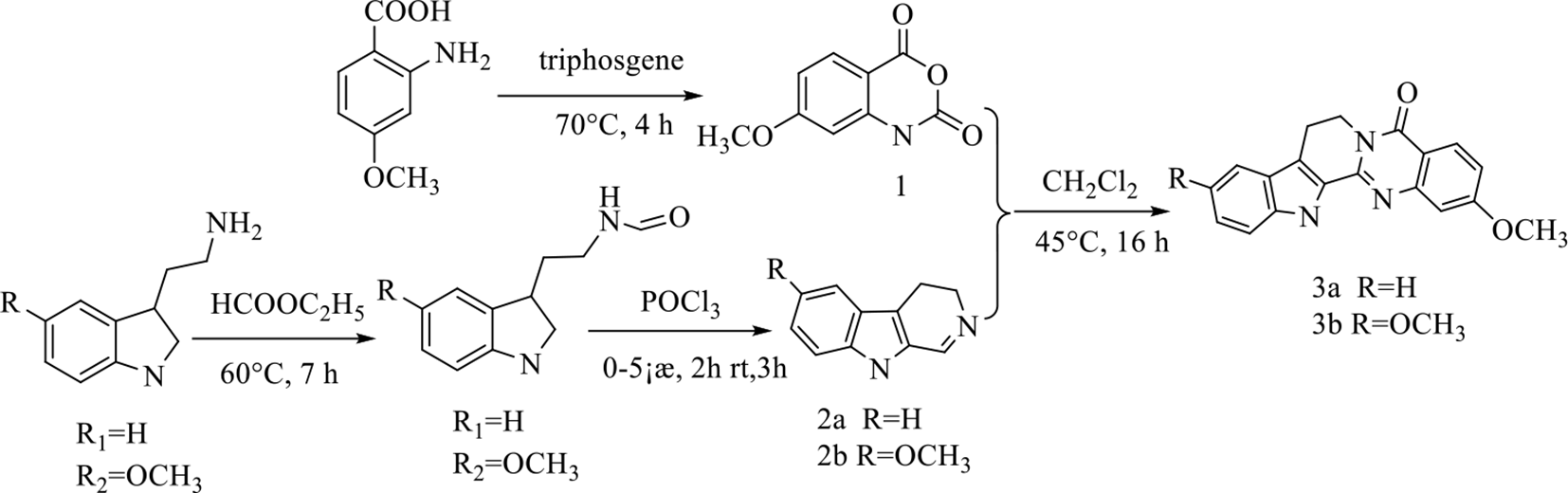
Synthesis of the rutaecarpine derivatives.
Results
Synthesis and Characterization
Although a couple of synthetic procedures for the preparation of rutaecarpine have been reported in the literature,11,13,19 a detailed synthetic procedure for 2-methoxyl rutaecarpine and 2,10-dimethoxy rutaecarpine has not yet been reported. Generally, evodiamine is prepared via condensation of 3,4-dihydro-b-carboline and N-methylisatoic anhydride. Because evodiamine and rutaecarpine have similar structures, we developed a 2-component protocol for a rapid preparation of the 2 methoxy-substituted rutaecarpine (RUT) derivatives using 7-methoxy-1H-benzo[d][1,3]oxazine-2,4-dione and 3,4-dihydro-b-carboline or 6-methoxy-4,9-dihydro-3H-pyrido[3,4-b]indole according to the synthetic method of evodiamine,20 which could avoid tedious purification procedure. The synthetic process is elucidated in Scheme1. The 7-methoxy-1H-benzo[d][1,3]oxazine-2,4-dione (compound 1) was prepared through the cyclization reaction of 2-amino-4-methoxy benzoic acid with triphosgene in dry tetrahydrofuran. The 3, 4-dihydro-b-carboline (compound 2a) or 6-methoxy-4, 9-dihydro-3H-pyrido[3, 4-b]indole (compound 2b) were synthesized according to the method described in Nie et al and Garner and Corminboeuf,4,20 which were then condensed with 7-methoxy-1H-benzo[d][1,3]oxazine-2,4-dione (compound 1) in dichloromethane (CH2Cl2) at 45 °C to produce the desired hybrids of 2-methoxyl rutaecarpine (compounds 3a) and 2,10-dimethoxy rutaecarpine (compounds 3b). The obtained compounds were characterized through 1H and 13C NMR spectroscopic determination to confirm their structures. The spectra were provided in Supplemental Figure S1-S6. These results completely agreed with the desired structures.
Antitumor Activities of Drugs
To evaluate the antiproliferative effects of the derivatives, different human cell lines derived from lung cancer (A549 and H1299), liver cancer (HepG2), and the normal cells (NCM480) were treated with different concentrations (1, 3, 10, 30, 100, 300) of the target compounds, and after 48 hours of incubation, the growth inhibition effect was tested by MTT cytotoxicity assays using paclitaxel as a reference. The results are listed in Figure 1. From Figure 1, for the same compound, show that the inhibition rates of the compounds against the 3 cancer cells increased while elevating the drug concentrations. The inhibition rates of the compounds at various concentrations were different. At drug concentrations (1, 3, 10, 30, 100), the inhibition rates of compounds 3a against those cells were higher than other compounds; the inhibition rate of PTX was higher than that of 3b, while the inhibition rate of RUT was the lowest. At a high drug concentration (300 μM), the inhibition rates of 3a against the cells were still the highest compared with other compounds (RUT, 3b, PTX); however, the inhibition rate of 3b was lower than that of RUT; the inhibition rate of PTX was higher than that of RUT; the inhibition rate of 3b was the lowest. For the normal cells (NCM480), the inhibition rates of the compounds RUT, 3a, 3b were all lower than those of PTX, indicating that the toxicity of these compounds was lower than the positive control (paclitaxel).
The inhibition rate of the rutaecarpine derivatives and control drug (paclitaxel) against different human cell lines derived from lung cancer (A549 and H1299), liver cancer (HepG2), and the normal cells (NCM480) were treated with different concentrations (1, 3, 10, 30, 100, 300) treatment after 48 hours of incubation.
We then further checked the half-maximal inhibitory concentration (IC50) values of the compounds against the 3 cancer cell lines after 48 hours of treatment. For the compounds, the IC50 values of 3a against 3 cancer cells in the range of 87.59‐127.67 μM were obviously smaller than those of RUT, 3b, and PTX (the IC50 values of these compounds all surpassed 200 μM), indicating that the activity of compound 3a was stronger than that of RUT, 3b, and PTX against those 3 cells. For the cells, the IC50 values of RUT, 3a, 3b, and PTX against A549 cells were the lowest compared with other cells (H1299 and HepG2), indicating that the compounds were more sensitive to A549 cells than other cells. In addition, the IC50 values of compounds 3a, 3b against A549 cells (87.59 µM for 3a, 228.60 µM for 3b) were lower than those of RUT (surpassed 300 μM), indicating that the activities of 3a and 3b were stronger than those of RUT, further suggesting that the activities of the compounds could be improved by structural modifications. Additionally, the activity of compound 3a was stronger than that of 3b.
The Oil-Water Partition Coefficient of Compounds
To evaluate the hydrophobicity of the compounds, the oil-water partition coefficients of RUT, 3a, and 3b have been calculated according to the formula , where Co is the concentration of the compound in the oil phase and Cw is the concentration of the compound in the water phase at the partition equilibrium.21,22 Based on the formula, the KP values for the compound RUT, 3a, and 3b were determined to be 121, 1.056, and 5.632, respectively. These results indicate that 3a was relatively hydrophilic, and RUT was hydrophobic, while the hydrophobicity of 3b was between that of the RUT and 3a.23,24 These oil-water partition coefficient results indicated that hydrophobicity affects the physicochemical properties of the molecules, as well as the antitumor activity, which agrees with the antitumor activities in this experiment.
Theoretical Study of RUT, 3a, and 3b
The geometries and electronic structures of compounds were studied by DFT calculations at the B3LYP/6‐31G* level.25 Optimized geometric structure of compounds RUT, 3a, and 3b are shown in Figure 2(A–C). Natural Population and Natural Electron Configuration of some atoms are shown in Supplemental Table S1. From Supplemental Table S1, it can be observed that there is a higher negative charge on the surfaces of oxygen O(15) and nitrogen N(13), N(14) atoms due to the lone pair of electrons and higher electronegativity on these atoms. This makes the linked carbon C(5), C(18), C(19), C(20), C(21) atoms with O(15), N(13), and N(14) atoms have a lower positive charge, while other carbon atoms possess a lower negative charge. Thus, we conclude that the negative charge atoms, such as O(15), N(13), and N(14) will be the aggression sites of the electrophile and C(5), C(18), C(19), C(20), C(21) atoms of the nucleophile. The electrostatic potential (MEP) map of compounds (RUT, 3a, and 3b) were shown in Figure 2(D–E), in which the red regions presented strong repulsion and the blue regions presented the strong attraction. From Figure 2, the red region of compounds RUT, 3a, and 3b is mainly localized on the N13, N14, O23, and O25 atoms, indicating that there are strong repulsions due to the steric effect between N13 and N14 atoms in ring and steric hindrance caused by the methyl group lie in O23 and O25 atoms. The blue region of compounds RUT, 3a, and 3b is mainly localized on the O15, C10, C11, and C12 atoms, indicating that there are strong attractions in these atoms.
Optimized geometric structure (A for RUT, B for 3a, and C for 3b) and electrostatic potential map of compounds (D for RUT, E for 3a, and F for 3b) (red color for strong repulsion and blue color for strong attraction).
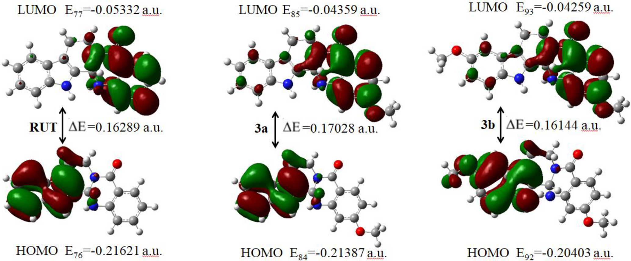
The HOMO-LUMO maps of RUT, 3a, and 3b were presented in Figure 3. Taking HOMO-LUMO calculation results, the HOMO of RUT, 3a, and 3b mainly localized on the A, B, and C rings, while the LUMO mainly on the D and E rings. In addition, RUT, 3a, and 3b possess high HOMO energy levels (−0.21621, 0.21387, and −0.20403 a.u.) and low LUMO energy levels (−0.05332, 0.04359, and −0.04259 a.u.). The value of energy separation between the HOMO and LUMO explains the eventual charge transfer interaction within the molecule, which influences the biological activity of the molecule.26 The bandgaps of RUT, 3a, and 3b are 0.16289, 0.17028, and 0.16144 a.u., respectively. Compared with RUT and 3b, bandgaps of 3a is highest, indicating that the stability of 3a is weakest, so 3a has higher activity than RUT and 3b, which agrees with the results of antitumor activities experiment.
Lowest occupied molecular orbital (LUMO) and highest unoccupied molecular orbital (HOMO) maps for compounds RUT, 3a, and 3b from DFT/B3LYP/6‐31G calculation. The green parts represent positive molecular orbital, and the red parts represent negative molecular orbital.
Experimental
Chemicals and Reagents
The reagents and solvents were purchased from commercial suppliers and used without further purification. The elemental analyses (C, H, and N) were performed using a Vario EL-III instrument. Infrared spectra were recorded on an Equinox55 spectrophotometer within the range of 4000‐400 cm−1 using a potassium bromide pellet. 1H NMR spectra were recorded on a Varian INOVA-400MHz spectrometer with trimethylsilane as an internal standard in deuterated chloroform (CDCl3) at room temperature. Fast atom bombardment-MS were obtained with a JEOL HX-110HF double-focusing spectrometer, operating in the positive-ion detection mode.
Synthesis of Compounds
The synthetic route of the target compounds is depicted in Scheme 1.
2-methoxy rutaecarpine (3a)
The 7-methoxy-1H-benzo[d][1,3]oxazine-2,4-dione (compound 1) was prepared through the cyclization reaction of 2-amino-4-methoxy benzoic acid with triphosgene in dry tetrahydrofuran. The compounds, 3,4-dihydro-β-carboline (2a) and 6-methoxy-4, 9-dihydro-3H-pyrido[3, 4-b]indole (2b), were prepared by refluxing tryptamine or methoxy-tryptamine with ethyl formate and then adding phosphorus oxychloride.4 A solution of 7-methoxy-1H-benzo[d][1,3]oxazine-2,4-dione1 (1.0 g, 5 mmol) and 3,4-dihydro-β-carboline 2a (0.65 g, 5 mmol) in CH2Cl2 (150 mL) was stirred at 45 °C for 16 hours and then cooled to room temperature. The solvent was concentrated under vacuum, and the crude product was purified by column chromatography (hexane/ethylacetate [EtOAc] = 5:1) to give 1.3 g of compound 3a as a white solid. 1H NMR (400 MHz, dimethylsulfoxide [DMSO]-d6) δ 10.89 (s, 1H), 7.67 (d, J = 8.4 Hz, 1H), 7.50 (d, J = 7.6 Hz, 1H), 7.43 (d, J = 7.8 Hz, 1H), 7.12‐7.15 (m, 1H), 7.01-7.05 (m, 1H), 6.95 (s, 1H), 6.42 (d, J = 9.1 Hz, 1H), 6.34 (s, 1H), 6.01 (s, 1H), 4.77 (d, J = 10.9 Hz, 1H), 3.77 (s, 3H), 2.95-2.98 (m, 1H), 2.75‐2.83 (m, 2H). 13C NMR (101 MHz, DMSO-d6) δ 164.06, 163.77, 149.37, 136.72, 131.31, 130.46, 126.32, 122.31, 119.46, 118.89, 112.08, 109.81, 106.61, 98.89, 64.29, 55.67, 38.96, 20.54; IR cm−1: 3302, 1609, 1582, 1448, 733; m/z calcd for C19H17N3O2 (M + H): 319.12; found 318.23.
2,10-dimethoxy rutaecarpine (3b)
The synthetic method of 3b was similar to that of 3a. A solution of 7-methoxy-1H-benzo[d][1,3]oxazine-2,4-dione 1 (1.0 g, 5 mmol) and 6-methoxy-3,4-dihydro-β-carboline 2b (1.3 g, 6.3 mmol) in CH2Cl2 (150 mL) was stirred at 45 °C for 16 hours and then cooled to room temperature. The solvent was concentrated under vacuum, and the crude product was purified by column chromatography (hexane/EtOAc = 5:1) to yield 1.5 g of 3b as a white solid. 1H NMR (400 MHz, DMSO-d6) δ 10.71 (s, 1H), 7.67 (d, J = 8.7 Hz, 1H), 7.32 (d, J = 8.8 Hz, 1H), 6.99 (d, J = 2.4 Hz, 1H), 6.93 (s, 1 H), 6.77 (dd, J = 8.8, 2.5 Hz, 1H), 6.41 (dd, J = 8.7, 2.4 Hz, 1H), 6.33 (d, J = 2.4 Hz, 1 H), 5.98 (s, 1H), 4.76 (dd, J = 12.9, 3.4 Hz, 1H), 3.77 (d, J = 1.8 Hz, 6H), 2.92‐2.99 (m, 1H), 2.67‐2.78 (m, 1H).13CNMR (100 MHz, CDCl3), 166.31, 164.04, 159.70, 153.83, 149.34, 147.89, 144.08, 131.72, 126.65, 112.77, 112.12, 109.79, 106.57, 103.37, 100.67, 98.96, 64.30, 56.41, 55.66, 20.63; IR cm−1: 3310, 1622, 1525, 1461, 744; m/z calcd for C20H19N3O3 (M + H): 349.14; found: 348.13.
Cell Culture and MTT Assay
The cytotoxicity assay of target compounds was evaluated by MTT assay. The compounds were dissolved in DMSO. The stock solution was diluted in the medium before each experiment. The final DMSO concentration did not exceed 0.1% throughout the study. The human tumor cell lines, including A549, H1299, and HepG2 cells were seeded at a density of 5 × 10⁴ cells/mL on a 96-well microplate (180 µL/well) and cultured overnight in a growth medium containing 1% fetal bovine serum. After 24 hours of incubation, 20 µL of fresh culture medium containing varying concentrations of samples was pulsed onto each well to achieve final samples concentration of 1, 3, 10, 30, 100, and 300μM. After 48 hours of incubation, the media were replaced by phosphate-buffered saline medium containing 0.5 mg/mL MTT and incubated for another 4 hours. Then, the medium was removed and 100 mL of DMSO was added into each well to dissolve formazan. The absorbances at 490/630 nm were measured for A549, H1299, and HepG2 cells using a microplate reader (Thermo). The untreated controls were regarded as a cell viability value of 100%. The IC50 values were obtained by following nonlinear regression using GraphPad Prism 4.0. IC50 measurements of the compounds were done in triplicates.
Measurement of the Oil-Water Partition Coefficient of Compounds
The water and 1-octanol, used in the experiment, were saturated with 1-octanol and water, respectively. For water-insoluble compounds, 2 mL of 1-octanol solutions containing compounds (1 mg/mL) were added into test tubes and mixed with 2 mL of water. The test tubes were incubated on a water bath at ambient temperature for 48 hours to achieve equilibrium between the 2 phases. The organic and aqueous phases were separated, and the concentration of compounds was determined at the wavelength of 240 nm by UV spectrophotometry. The oil-water partition coefficient was calculated according to the formula where Co is the concentration of the compound in the oil phase and Cw is the concentration in the water phase at the partition equilibrium.19
Computational Details
The density functional theoretical computations of compounds RUT, 3a, and 3b were performed at DFT/B3LYP/6‐311G level to derive the complete geometry optimization. In addition, natural bond orbitals, HOMO-LUMO, and electrostatic potential were also investigated by B3LYP/6‐311G. All calculations were carried out by Gaussian 16 program.20
Statistical Analysis
All results are presented as mean ± SD. The Student’s t-test was used to analyze the statistically significant differences between the cells exposed to drugs and their controls. The difference between the means was considered statistically significant at P < 0.05.
Conclusions
In this study, the 2 rutaecarpine (RUT) derivatives with methoxy substitutions, 2-methoxyl rutaecarpine (3a), 2,10-dimethoxy rutaecarpine (3b) were synthesized, and the antitumor activities in vitro were studied. For understanding the relationship of antitumor activity and the physicochemical properties of the compounds, the oil-water partition coefficient of RUT and its derivatives were studied by UV spectrometry. The oil-water partition coefficient results indicated that hydrophobicity affects the physicochemical properties of the molecules, as well as the antitumor activities. In addition, in order to discover the relationship of antitumor activity and structures of the compounds, the Natural Electron Configuration, frontier molecular orbital profiles (HOMO, LUMO), and bandgaps of these 3 compounds have been studied based on DFT by means of DFT-B3LYP/6‐31G (d) in Gaussian 16. The calculation results showed that the bandgaps of 3a are highest, indicating that the stability of 3a is weakest compared with RUT and 3b, so 3a has higher activity than RUT and 3b, which agrees with the results of antitumor activities experiment. We believe that our results may help in drug screening and cytotoxicity studies.
Supplemental Material
Supplementary Material 1 - Supplemental material for Synthesis, Antitumor Activity, Oil-Water Partition Coefficient, and Theoretical Calculation of 2 New Rutaecarpine Derivatives With Methoxy Groups
Supplemental material, Supplementary Material 1, for Synthesis, Antitumor Activity, Oil-Water Partition Coefficient, and Theoretical Calculation of 2 New Rutaecarpine Derivatives With Methoxy Groups by Jingjing Ma, Ruolan Yang, Hui Guo, Keyao Zhang, Jingli Liu, Yifan Feng, Jing Zhou, Ruyi Jin, Zhi Li, Dongyan Guo, Yong-gang Yan, Haiyan Zhu and Yuping Tang in Natural Product Communications
Footnotes
Acknowledgments
This work was financially supported by the National Natural Science Foundation of China (81001669), the Natural Science Foundation of Shaanxi Provice (2020JZ-55), the Key Laboratory Research Project of Shaanxi Provice (20J5035), the Postdoctoral Science Foundation of China (2016M592809), the Chinese Medicine Project of Shaanxi Province (ZYMS005) and the Subject Innovation Team of Shaanxi University of Chinese Medicine (2019-PY02).
Declaration of Conflicting Interests
The author(s) declared no potential conflicts of interest with respect to the research, authorship, and/or publication of this article.
Funding
The author(s) disclosed receipt of the following financial support for the research, authorship, and/or publication of this article: This research is funded by the National Natural Science Foundation of China (81001669), the Chinese Medicine Project of Shaanxi Province (ZYMS005), the Key Laboratory Research Project of Shaanxi Provice (20J5035), Subject Innovation Team of Shaanxi University of Chinese Medicine (2019-PY02) and the Key Science Foundation of Shaanxi Provice (2020JZ-55).
ORCID iDs
Hui Guo
Yuping Tang
Supplemental Material
Supplemental material for this article is available online.
References
1.
TianK M.LiJ J.XuS W. Rutaecarpine: a promising cardiovascular protective alkaloid from Evodia rutaecarpa (Wu Zhu Yu). Pharmacol Res. 2019;141:541-550.doi:10.1016/j.phrs.2018.12.01930616017
2.
JiL.WuM.LiZ. Rutacecarpine inhibits angiogenesis by targeting the VEGFR2 and VEGFR2-Mediated Akt/mTOR/p70S6K signaling pathway. Molecules. 2018;23(8):2047.doi:10.3390/molecules2308204730111763
3.
GurungAB.PamayP.TripathyDet al. Bioprospection of anti-inflammatory phytochemicals suggests rutaecarpine and quinine as promising 15-lipoxygenase inhibitors. J Cell Biochem. 2019;120(8):13598-13613.doi:10.1002/jcb.2863430937959
4.
NieL F.WangS S.CaoJ Get al. Straightforward synthesis, characterization, and cytotoxicity evaluation of hybrids of natural alkaloid evodiamine/rutaecarpine and thieno[2,3-d]pyrimidinones. J Asian Nat Prod Res. 2020;22(1):69-82.doi:10.1080/10286020.2018.154059930588834
5.
ChenY C.ZengX Y.HeYet al. Rutaecarpine analogues reduce lipid accumulation in adipocytes via inhibiting adipogenesis/lipogenesis with AMPK activation and UPR suppression. ACS Chem Biol. 2013;8(10):2301-2311.doi:10.1021/cb400389323962138
6.
JinS W.HwangY P.ChoiC Yet al. Protective effect of rutaecarpine against t-BHP-induced hepatotoxicity by upregulating antioxidant enzymes via the CaMKII-Akt and Nrf2/ARE pathways. Food Chem Toxicol. 2017;100:138-148.doi:10.1016/j.fct.2016.12.03128025122
7.
DengJ.QinJ.CaiYet al.Rutaecarpine suppresses proliferation and promotes apoptosis of human pulmonary artery smooth muscle cells in hypoxia possibly through HIF-1α-dependent pathways. J Cardiovasc Pharmacol. 2018;71(5):293-302.doi:10.1097/FJC.000000000000057129438215
8.
GuoH.LiuD.GaoBet al. Antiproliferative activity and cellular uptake of evodiamine and rutaecarpine based on 3D tumor models. Molecules. 2016;21(7):954.doi:10.3390/molecules2107095427455219
9.
FanJ P.YangX M.XuX K,et al. Solubility of rutaecarpine and evodiamine in (ethanol+water) mixed solvents at temperatures from (288.2 to 328.2)K. J Chem Thermodyn. 2015;83:85-89.doi:10.1016/j.jct.2014.12.004
10.
HuangG.DrakopoulosA.SaedtlerMet al. Cytotoxic properties of the alkaloid rutaecarpine and its oligocyclic derivatives and chemical modifications to enhance water-solubility. Bioorg Med Chem Lett. 2017;27(21):4937-4941.doi:10.1016/j.bmcl.2017.08.04528958621
11.
LiQY.ChengSY.TangHTet al. Synthesis of rutaecarpine alkaloids via an electrochemical cross dehydrogenation coupling reaction. Green Chemistry. 2019;21:5517-5520.
12.
LeeD W.KangY.KangM Jet al. Phase I and phase II metabolite identification of rutaecarpine in freshly isolated hepatocytes from male Sprague-Dawley rats. Arch Pharm Res. 2017;40(8):972-979.doi:10.1007/s12272-017-0937-728799086
13.
SonJ K.ChangH W.JahngY. Progress in studies on rutaecarpine. ii.--synthesis and structure-biological activity relationships. Molecules. 2015;20(6):10800-10821.doi:10.3390/molecules20061080026111170
14.
HuangG.RoosD.StadtmüllerP.DeckerMet al. A simple heterocyclic fusion reaction and its application for expeditious syntheses of rutaecarpine and its analogs. Tetrahedron Lett. 2014;55(26):3607-3609.doi:10.1016/j.tetlet.2014.04.120
15.
WenB.RoongtaV.LiuL.MooreD J. Metabolic activation of the indoloquinazoline alkaloids evodiamine and rutaecarpine by human liver microsomes: dehydrogenation and inactivation of cytochrome P450 3A4. Drug Metab Dispos. 2014;42(6):1044-1054.doi:10.1124/dmd.114.05741424696463
16.
KhadkaD B.ChoW J. Topoisomerase inhibitors as anticancer agents: a patent update. Expert Opin Ther Pat. 2013;23(8):1033-1056.doi:10.1517/13543776.2013.79095823611704
JinR Y.ZengC Y.LiangX Het al. Design, synthesis, biological activities and DFT calculation of novel 1,2,4-triazole schiff base derivatives. Bioorg Chem. 2018;80:253-260.doi:10.1016/j.bioorg.2018.06.03029966871
19.
BaoQ Y.LiuA Y.MaYet al. The effect of oil-water partition coefficient on the distribution and cellular uptake of liposome-encapsulated gold nanoparticles. Colloids Surf B Biointerfaces. 2016;146:475-481.doi:10.1016/j.colsurfb.2016.06.04627400242
20.
GarnerM H.CorminboeufC. Correlation between optical activity and the helical molecular orbitals of allene and cumulenes. Dev Growth Differ. 2008;29(2):171-176.
21.
SenadiG C.KudaleV S.WangJ J. Sustainable methine sources for the synthesis of heterocycles under metal- and peroxide-free conditions. Green Chemistry. 2019;21(5):979-985.doi:10.1039/C8GC03839B
22.
DongG.WangS.MiaoZet al. New tricks for an old natural product: discovery of highly potent evodiamine derivatives as novel antitumor agents by systemic structure-activity relationship analysis and biological evaluations. J Med Chem. 2012;55(17):7593-7613.doi:10.1021/jm300605m22867019
23.
BernchouU.IpsenJ H.SimonsenA C. Growth of solid domains in model membranes: quantitative image analysis reveals a strong correlation between domain shape and spatial position. J Phys Chem B. 2009;113(20):7170-7177.doi:10.1021/jp809989t19296622
24.
MetinC O.BaranJ R.NguyenQ P. Adsorption of surface functionalized silica nanoparticles onto mineral surfaces and decane/water interface. J Nanopart Res. 2012;14(11):1246.doi:10.1007/s11051-012-1246-123193372
25.
BouvraisH.PottT.BagatolliL A.IpsenJ H.MéléardP. Impact of membrane-anchored fluorescent probes on the mechanical properties of lipid bilayers. Biochim Biophys Acta. 2010;1798(7):1333-1337.doi:10.1016/j.bbamem.2010.03.02620398624
26.
SuvithaA.PeriandyS.BoomadeviS.GovindarajanM. Vibrational frequency analysis, FT-IR, FT-Raman, ab initio, HF and DFT studies, NBO, HOMO-LUMO and electronic structure calculations on pycolinaldehyde oxime. Spectrochim Acta A Mol Biomol Spectrosc. 2014;117:216-224.doi:10.1016/j.saa.2013.07.08023994677
Supplementary Material
Please find the following supplemental material available below.
For Open Access articles published under a Creative Commons License, all supplemental material carries the same license as the article it is associated with.
For non-Open Access articles published, all supplemental material carries a non-exclusive license, and permission requests for re-use of supplemental material or any part of supplemental material shall be sent directly to the copyright owner as specified in the copyright notice associated with the article.