
Abstract
Tanshinone IIA (Tan IIA) and sinapic acid (SA) are 2 components separately isolated from 2 Asian medicinal plants, Hydnophytum formicarum Jack and Salvia miltiorrhiza Bunge. The antitumor activities of them were worth exploring, therefore, we examined their antitumor activities in A549, HCT116, HeLa, and Colo320 cancer cell lines by means of WST-1 assay. The results show that Tan IIA exerted far higher (IC50 from 1.0 ± 0.0 to 166.3 ± 24.0 µg/mL) antiproliferative activities than SA (IC50 from 2236.3 ± 484.1 to >10 000.0 µg/mL). Of the 4 cell lines, A549 cells were the most sensitive to Tan IIA; thus, we used Western blotting to explore the cytotoxic mechanisms of Tan IIA in A549 cells and found that they rely on simultaneous induction of apoptosis and necroptosis in the cells. Apoptosis was hallmarked by the induction of cleaved caspase-3 by Tan IIA and necroptosis by the necroptotic marker proteins cyclophilin A and high mobility group box 1 (HMGB1), as well as increased lactate dehydrogenase (LDH) activities. The necroptotic effect was confirmed by the necroptosis inhibitor necrostatin-1 (Nec-1), which eliminated these effects and restored cell survival rates. The levels of cyclophilin A decreased in response to the pan-caspase inhibitor z-VAD-fmk, and those of cleaved caspase-3 decreased in response to Nec-1. Conclusively, Tan IIA has the potential to prevent lung cancer and the mechanism seems to be apoptosis and necroptosis, of which the relationship is mutually interdependent. This is the first report of Tan IIA eliciting necroptosis in A549 cells. Tan IIA may be used for necroptosis-based cancer therapy, especially to overcome apoptosis resistance.
Introduction
Tanshinone IIA (Tan IIA, 16,6-trimethyl-6,7,8,9-tetrahydrophenanthro[1,2-b]furan-10,11-dione; Figure 1A) is found in the roots of Salvia miltiorrhiza Bunge, a medicinal plant, and is used to treat cardiovascular disease in East Asia. Tan IIA is an antiangiogenic, antioxidant, antiinflammatory, and antiapoptotic. As reviewed in our previous study, 1 Tan IIA promotes cell apoptosis and exerts anticancer activity in various solid tumor cells of the liver, prostate, bone, esophagus, oral cavity, and cervix and in chronic myeloid leukemia. Tan IIA can stimulate apoptosis in A549 cells, attributable to elevated levels of reactive oxygen species and an increase in mitochondrial membrane potential. 2 The activation of apoptosis is through the cytochrome c-mediated caspase cascade and the c-Jun N-terminal kinase pathway. 3 Accompanying apoptosis, Tan IIA increases the accumulation of S-phase cells. 4 Previous studies have highlighted the apoptotic effects of Tan IIA compared to its necroptotic effects. Tan IIA-triggered necroptosis has not been investigated, except in our study on the HepG2 liver cancer cell line. 5

The chemical structure of Tan IIA and SA are components dominantly present in the root of Salvia miltiorrhiza Bunge (A) and tuber of Hydnophytum formicarum Jack (B). The copyright of the SA image has been released to the public domain by the author of Wikipedia, https://en.wikipedia.org/wiki/Sinapinic_acid#/media/File:Sinapic_acid.png.
Hydnophytum formicarum Jack is a folk medicinal plant used to prevent hepatitis, rheumatism, diarrhea, and lung cancer. The crude extracts of this plant show selective activity against the A549 human lung tumor cell line with EC50 values of 0.78 to 1.03 μg/mL. 6 Sinapic acid (SA, 3,5-dimethoxy-4-hydroxycinnamic acid), an ingredient of H formicarum (Figure 1B), belongs to the phenylpropanoid family. Its antiproliferative activity has been observed in human laryngeal, breast, prostate, and colon cancer cell lines.7–10
Apoptosis or programmed cell death is a tightly regulated mechanism of cell death. Poor regulation of apoptotic cell death results in numerous diseases, including cancer, restenosis, stroke, heart failure, neurodegeneration, and AIDS. 11 Cytotoxic phytochemicals induce tumor cell apoptosis through a series of caspase enzymes. Caspases are functionally classified into 2 types, ie, initiator and executor caspases. Caspase-8 and -9 are initiator caspases and are activated by the extrinsic and intrinsic apoptotic pathways. Caspase-3 and -7 are effector caspases, activated after cleavage by an initiator caspase. The activated caspases subsequently degrade DNA and various proteins to cause cell death. Thus, the cleaved effector caspases can be used as apoptotic markers. 12 Several phytoconstituents that promote apoptosis via the caspase-dependent pathway have been studied. For instance, goniothalamin extracted from Goniothalamus andersonii promotes apoptosis in Jurkat T-cells by activating effector caspase-3 and -7. 13 An aqueous extract of Bryonia dioica can induce apoptosis in BL41 Burkitt's lymphoma cells by upregulating caspase-3 and -9. 14
Necrosis is a form of cell death that occurs through a nonprogrammed pathway and is characterized by increased cell size, enlargement of organelles, breaching of the cell membrane, and the eventual release of intracellular components, for example, lactate dehydrogenase (LDH). However, necroptosis is a programmed form of necrosis. 15 Necroptosis is an important pathogenic mediator of various human diseases including infectious, neurodegenerative, cardiovascular, hepatic, renal, and pulmonary disease and cancer. 16 Necroptosis is initiated by the binding of the extracellular necroptotic ligand to its transmembrane receptor, which subsequently phosphorylates and activates the receptor-interacting serine-threonine protein kinase 1 (RIPK1). A downstream protein, mixed lineage kinase domain-like (MLKL), is in turn phosphorylated by activated RIPK1 and RIPK3 and forms an oligomer complex, called the necrosome. Thereafter, phosphorylated MLKL is translocated from the cytosol to the plasma and intracellular membranes. The oligomerization of MLKL leads to the formation of pores on the membrane, thereby leading to membrane damage and necroptosis.17,18 Thus, activated RIPK1 is a marker for necroptosis, and activators targeting the necrosome have emerged as candidate drugs for cancer treatment. Necroptosis is less studied in cancer cells treated with native compounds. However, shikonin, a phytochemical, can stimulate necroptosis in osteosarcoma, 19 leukemia, 20 glioma, 21 colon cancer, 22 and lung cancer (A549) 23 cell lines. Moreover, a natural phenolic compound, neoalbaconol, induces necroptosis in mouse fibrosarcoma (L929), human amelanotic melanoma (A375), human gastric cancer (AGS-EBV), and human breast cancer (MX-1) 24 cell lines. Recently, cationic peroxidase from proso millet was shown to exert antitumorigenic effect on human colon cancer HCT116 and HT29 cells by inducing necroptosis. 25 In these cells, activated RIPK1 levels were remarkably high, and, therefore, they were determined to undergo RIPK1-dependent necroptosis.
Here, we investigated the anticancer activity of SA and Tan IIA by treating A549 human lung, HCT116 colon, HeLa ovarian, and Colo320 colon cancer cells for 12, 24, and 36 h. All 4 cell lines were more sensitive to the cytotoxic activities of Tan IIA than to those of SA. Moreover, Tan IIA-induced mutually interdependent apoptosis and necroptosis in A549 cells.
Materials and Methods
Cell Line and Culture
The human cancer cell lines A549, HCT116, HeLa, and Colo320 were obtained from Bioresource Collection and Research Center and confirmed to be mycoplasma free. The cells were continuously cultured in Dulbecco's modified Eagle's medium (DMEM) containing phenol red (Gibco) supplemented with 10% fetal bovine serum (Biological Industries) and 100 U/mL penicillin and 100 μg/mL streptomycin (Biological Industries) in a humidified incubator at 37 °C in 5% CO2.
Chemical Compounds
Tan IIA with a purity of 98% (high-performance liquid chromatography) was produced by Kesure Biotechnology Co. SA with purity no less than 98% was procured from Santa Cruz Biotechnology Inc., necrostatin-1 (Nec-1; 99.9% pure) from Enzo Life Sciences, and z-VAD-fmk (99.9% pure) from ApEXBio. All chemicals were prepared in dimethyl sulfoxide (DMSO) at a concentration of 2 mg/mL Tan IIA, 50 mM for Nec-1, and 20 mM for z-VAD-fmk.
Cell Viability Assay
Based on a previous report, 5 1 × 10⁴ cells at the exponential growth stage were seeded in each well of a 96-well tissue culture plate and incubated overnight at 37 °C and 5% CO2 to allow attachment. A549, HCT116, and HeLa cells were exposed to increasing concentrations of either SA (500, 1000, 2000, 5000, and 10 000 μg/mL) or Tan IIA (0.2, 0.5, 1.0, 2.0, and 5.0 μg/mL) for 12, 24, and 36 h. Colo320 cells were treated with 10, 20, 50, 200, and 500 μg/mL of Tan IIA. To study the role of necroptosis and apoptosis in cytotoxicity, either Nec-1 or z-VAD-fmk was added to the phytochemicals. An equal concentration of the solvent vehicle (DMSO) was added to the untreated cells as the negative control. After the treatment was complete, the medium was removed and the cells were rinsed with 200 μL phosphate-buffered saline. Thereafter, the cells were treated with a mixture of 190 μL phenol red-free DMEM and 10 μL cell proliferation reagent 4-[3-[4-iodophenyl]-2-[4-nitrophenyl]-2H-5-tetrazolio]-1,3-benzene disulfonate (WST-1) for 30 min under the same culture conditions, and the absorbance of formazan was determined using a microplate reader (Multiskan Spectrum; Thermo Scientific) at 450 nm. To calculate IC50, the inhibition rate of the phytochemicals was normalized using the formula: absorbance value of formazan influenced by the concentration of each phytochemical/absorbance value of the negative control. The dose-response graph, equation, and IC50 values were plotted and calculated using Microsoft Excel 2010 (Microsoft Inc.). Data are displayed as a mean of IC50 ± standard deviation (SD) of quadruplicates. A 2-tailed Student's t-test was used to analyze the differences between the means of the 2 chosen groups.
LDH Assay
For pretreatment, A549 cells were incubated for 30 min with either 20 μM z-VAD-fmk for the apoptosis inhibition assay or 50 μM Nec-1 for the necroptosis inhibition assay and subsequently exposed to either 5 or 10 μg/mL Tan IIA for 12 h. For necroptosis, the LDH assay (Roche Co.) was prepared according to the manufacturer's instructions. The absorbance of the formazan product was determined at 490 nm using a microplate reader (Thermo Scientific Multiskan). Microsoft Excel 2010 was used for data processing and analysis. Data are displayed as mean ± SD of quadruplicates. The 2-tailed Student's t-test was used to analyze the differences between the means of the 2 groups.
Western Blot Analysis
As described previously, 5 for pretreatment, A549 cells were incubated for 30 min with either 20 μM z-VAD-fmk for the apoptosis inhibition assay or 50 μM Nec-1 for the necroptosis inhibition assay and subsequently treated with either 5 or 10 μg/mL Tan IIA for 1 to 36 h. Thereafter, the cells were collected in 300 μL of radioimmunoprecipitation assay lysis buffer containing the protease inhibitor cocktail (Calbiochem) and homogenized. For Western blot analysis, samples were boiled for 5 min, separated by sodium dodecyl sulfate–polyacrylamide gel electrophoresis, and transferred to polyscreen membranes (polyvinylidene fluoride; Millipore). The membrane was incubated with 5% nonfat milk in Tris-buffered saline buffer with Tween 20 (TBST buffer) (20 mM Tris; pH 7.6, 150 mM NaCl, and 0.3% Tween 20) for 60 min, and washed for 15 min with TBST, twice. For immunoblot analysis, the membrane was incubated with primary antibodies against cyclophilin A, high mobility group box 1 (HMGB1), caspase-3, or β-actin (GeneTex) diluted in the antibody dilution buffer (Ventana Medical Systems), at 4 °C overnight. Thereafter, the membranes were washed for 15 min with TBST twice and incubated with horse-radish peroxidase (HRP) secondary antibodies against either mouse or rabbit IgG (Abcam) in the same buffer for 60 min at room temperature. Subsequently, the membranes were washed for 10 min with the buffer, 3 times, the signal was developed using an HRP-conjugated substrate kit (BIO-RAD), and the results were determined by the Gel Documentation System (BIO-RAD).
Results and Discussion
A549, HCT116, HeLa, and Colo320 Cells are More Sensitive to the Cytotoxicity of Tan IIA Than That of SA
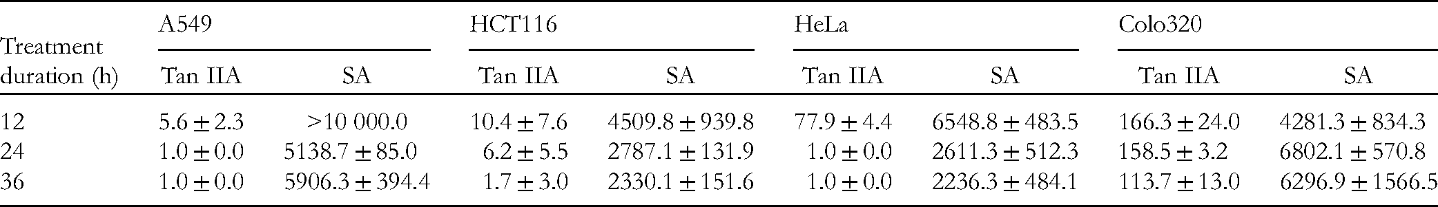
A549, HCT116, HeLa, and Colo320 cells correspond to human lung, colon, cervical, and colorectal cancer, respectively. According to the 2018 report of World Cancer Research Fund International, a total of 2.0 (lung cancer), 1.0 (colon cancer), over 0.5 (cervical cancer), and 1.8 (colorectal cancer) million new cases were reported worldwide. 26 Therefore, we selected these 4 cell lines as the anticancer targets of SA and Tan IIA for 12, 24, and 36 h of treatment. The IC50 values were calculated from the absorbance values obtained from the WST-1 cell proliferation and cytotoxicity assays. The IC50 values of SA were > 2000 μg/mL and those of Tan IIA were <120 μg/mL, indicating that Tan IIA has a higher activity against the 4 cancer cell lines than SA (Table 1). A549 cells contain cancer stem cells, 27 which are characteristically drug-resistant. 28 Cancer stem cells can resist Tan IIA treatment for 36 h compared to that for 24 h, which reflects the insignificant difference between the IC50 values for 24 and 36 h of treatment. Globally, lung cancer has been a fatal disease for several decades. 29 Nonsmall cell lung cancer (NSCLC) accounts for ∼85% of all lung cancers. Although breakthroughs have been achieved in the last decade through screening of high-risk populations and molecular therapies for lung cancer, further progress is expected with the development of next-generation drugs that have more specific target effects. 30 Data indicate that 40% of Food and Drug Administration approved pharmaceutical agents are naturally occurring compounds and their derivatives, 74% of which are used for cancer prevention. Natural products are more biocompatible and less toxic to normal cells than pharmaceuticals. 31 SA had a less antiproliferative effect in NSCLC A549 cells than Tan IIA. Nevertheless, its antimetastatic activity remains to be investigated. H formicarum can suppress A549 cells with relatively high potency 6 ; however, SA displayed lower activity against tumor cells. Several purified ingredients of H formicarum have been tested for their anticancer activities against distinct cell lines7,8,10; however, the potent component remains to be identified. The potent ingredients must be identified or a more effective prescription composed of 2 or more constituents from SA should be developed to treat tumor cells. Thus, Tan IIA inhibits A549 cell growth in a manner distinct from SA.
The IC50 Values (µg/mL) of Tan IIA and SA in 4 Human Cancer Cell Lines.
Tan IIA Induces Caspase-3-Dependent Apoptosis in A549 Cells
As Tan IIA exhibited a high efficiency in inhibiting cellular activities, we focused on its mechanism of action. A549 and HeLa cells were more sensitive to Tan IIA than the other 2 cancer cell lines. To study the mechanism, we chose A549 cells because lung cancer is responsible for the highest number of incidence and deaths globally (1.8 million deaths, 18.4% of the total). 32 The apoptotic potency of Tan IIA has been previously reported in A549 cells using flow cytometry and apoptotic proteins, such as p53, bcl-2, and bax.2,4 Here we have studied apoptosis by determining the cleavage of caspase-3. Caspase-3-mediated apoptosis is marked by the presence of cleaved caspase-3, which can be verified by Western blot analysis. Cells treated with 1 to 20 μg/mL of Tan IIA for 12h were lysed and the lysates were used for Western blotting. Caspase-3 was identified on the blot in a concentration-dependent manner (Figure 2A). Furthermore, the elevated expression of caspase-3 was inhibited by a pan-caspase inhibitor z-VAD-fmk (Figure 3) and the Tan IIA-mediated reduction in cell viability was restored, as shown in our previous study. 1 Thus, Tan IIA-induced apoptosis is caspase-3-dependent.

Tan IIA-induced apoptosis and necroptosis in A549 cells. The apoptotic effect was evidenced by increasing levels of caspase-3 in cleaved form (A). The necroptotic effect was evidenced by increasing levels of cyclophilin A (B), HMGB1 (C), and LDH activities (D). **Indicates the statistical difference (P < .01).

The Tan IIA-induced increase in cyclophilin A was reduced by the pan-caspase inhibitor z-VAD-fmk and the necroptosis inhibitor Nec-1.
Tan IIA Induces Necroptosis in A549 Cells
Cyclophilin A and HMGB1 are biomarkers of necroptosis.33,34 Increased levels of both proteins were detected on a Western blot after 1 to 20 μg/mL Tan IIA treatment for 12 h (Figure 2B and C). LDH assay can directly determine necroptosis. LDH activity increased significantly in response to 5 and 10 μg/mL Tan IIA treatment (Figure 2D). Moreover, increased levels of cyclophilin A were reduced after the addition of the necroptosis inhibitor Nec-1 (Figure 3). Consistently, Tan IIA-induced LDH activities were repressed by Nec-1 (Figure 4). Moreover, Tan IIA-depressed cell proliferation was rescued by Nec-1 at 5, 10, and 20 μg/mL of Tan IIA treatment for 24 and 48 h (Figure 5). Taken together, these results indicate that Tan IIA induces necroptosis in A549 cells. Necroptosis is a double-edged sword. It helps prevent cancer by eliminating cancer cells and inhibiting the initiation, growth, and metastasis of cancer. However, cells killed by necroptosis release certain molecules or provoke inflammatory responses which promote the growth and metastasis of tumor cells.35,36 These disadvantages may be overcome by combining the use of antiinflammatory and antimetastatic phytochemicals in traditional Oriental medicine prescriptions.

Nec-1 (50 µg/mL) suppressed the Tan IIA-induced increase in LDH activities. **Indicates the statistical difference (P < .01).

Nec-1 (50 µg/mL) rescued the Tan IIA-suppressed cell viability. **Indicates the statistical difference (P < .01).
The Induction of Apoptosis and Necroptosis by Tan IIA is Mutually Interdependent
The Tan IIA-induced increase in the necroptotic marker cyclophilin A was reduced by z-VAD-fmk mostly and Nec-1 fully at 5 and 10 μg/mL of Tan IIA treatment for 12, 24, and 36 h (Figure 3). The Tan IIA-induced increase in cleaved caspase-3 was reduced by z-VAD-fmk and Nec-1 under the same conditions (Figure 6). These results suggest that Tan IIA-induced apoptosis and necroptosis are mutually interdependent. Apoptosis and necroptosis are mutually exclusive because the necroptosome, RIPK1, is cleaved by the proapoptotic protease caspase-8, which causes blockage of necroptosis via apoptosis. However, our previous study revealed that both necroptosis and apoptosis are simultaneously induced by Tan IIA in HepG2 cells. 5 In this study, both necroptosis and apoptosis occurred in A549 cells. Moreover, in our previous study with HepG2 cells caspase inhibition increased the rate of necroptosis caused by Tan IIA. 5 However, here we report that caspase suppression by z-VAD-fmk results in lowering the level of cyclophilin A, suggesting that the reduction of Tan IIA-induced apoptosis decreases the level of necroptosis. The mechanism remains to be studied. Nec-1 is reported to have no influence on apoptosis. Here we report that Nec-1 reduced the levels of cleaved caspase-3, suggesting a reduction of apoptosis. This is consistent with previous findings. 5

The Tan IIA-induced increase in the cleaved form of caspase-3 was reduced by the pan-caspase inhibitor z-VAD-fmk and the necroptosis inhibitor Nec-1.
Conclusions
We compared the antiproliferative activities of SA and Tan IIA on A549 human lung cancer cells and found that Tan IIA is more suppressive of cell growth than SA, with a 25.7-fold difference in their IC50 values. The cytotoxic mechanism relies on the simultaneous occurrence of apoptosis and necroptosis. The cytotoxic effect of Tan IIA was higher than that of SA on all 4 cancer cell lines, which may be attributed to the simultaneous induction of apoptosis and necroptosis. This is the first report of necroptosis in A549 cells. The study of this form of cell death is currently very popular and necroptosis-based cancer therapy has been suggested as an alternative method to overcome apoptosis resistance. Indeed, advanced lung cancer is frequently resistant to apoptosis-based anticancer drugs, and A549 cells are usually selected as a model to evaluate drug resistance. 37 Tan IIA shows great potential for lung cancer therapy, especially in avoiding apoptotic resistance.
Footnotes
Acknowledgments
We are grateful to Professor Dr Jye-Siung Fang for his support and encouragement, Chung-Wei Lin for preparing the manuscript, and Professor Dr. Peng Yeong Woon and Ruth Wang for providing language help.
Author Contributions
CYL designed and performed part of the experiments. MTL carried out the experiments. SHY, SCL, and TWC conceived the presented idea and verified the analytical methods. IHL and YJT conceived the study and were in charge of overall direction and planning. YJT wrote the manuscript with input from all authors. All authors discussed the results and contributed to the final manuscript.
Declaration of Conflicting Interests
The authors declared no potential conflicts of interest with respect to the research, authorship, and/or publication of this article.
Funding
The authors disclosed receipt of the following financial support for the research, authorship, and/or publication of this article: This study was supported by grants from Tzu Chi University (TCMRC-P-106008-01); the Ministry of Science and Technology of the Executive Yuan in Taiwan (MOST 103-2633-B-320-001); the Buddhist Tzu Chi Medical Foundation (TCMMPSP104-09 and TCMMP-108-02-02); and the Intramural Research Project of Buddhist Tzu Chi General Hospital (TCRD107-53, TCRD108-35, and TCRD109-42). The research was also supported by Deutsche Akademische Austauschdienst (DAAD). The funding bodies provided material costs and personnel expenses needed in the study.
Ethical Approval
Ethical approval is not applicable for this article.
Statement of Human and Animal Rights
This article does not contain any studies with human or animal subjects.
Statement of Informed Consent
There are no human subjects in this article and informed consent is not applicable.